PT-141
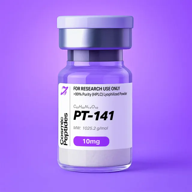
PT-141
Commonly researched with these items


Patent-pending lot tracking: Complete transparency from production to vial.
| CAS Number | 189691-06-3 |
|---|---|
| Molecular Formula | C50H68N14O10 |
| Molecular Weight | 1025.18 g/Mol |
| Purity | 99.8% |
| Lot Number | 25056 |
| Quantity | 10.14mg |
| Sequence | Ac-Nle-cyclo[Asp-His-D-Phe-Arg-Trp-Lys]-OH |
PT-141 (Bremelanotide): A Cyclic Lactam α-Melanotropin Heptapeptide and Melanocortin Receptor Agonist
Disclaimer
Products described here are supplied for research use only and are not intended for diagnostic, therapeutic, or clinical application. All statements regarding biological activity reflect preclinical and in vitro findings exclusively and have not been evaluated by the U.S. Food and Drug Administration. These materials and related content are provided for educational and investigational purposes only and are intended solely for qualified researchers in laboratory and academic settings.
Key Facts
- Compound class
- Synthetic cyclic lactam heptapeptide; side-chain-to-side-chain macrocyclic analogue of α-melanocyte-stimulating hormone (α-MSH) residues 4–10
- Sequence / structure
Ac-Nle-cyclo[Asp-His-D-Phe-Arg-Trp-Lys]-OH; acetylated N-terminus, 23-membered lactam bridge between the aspartate β-carboxyl and the lysine ε-amine, free C-terminal carboxylic acid on lysine- Molecular formula
- C50H68N14O10
- Molecular weight
- 1025.18 g/mol (average, free base); monoisotopic mass 1024.5243 Da
- CAS number
- 189691-06-3
- PubChem CID
- 9941379
- Stereochemistry
- Seven defined stereocentres; six L-configured residues plus D-phenylalanine at the position corresponding to Phe7 of α-MSH
- Physical form as supplied
- Lyophilized powder
- Lot traceability
- Every unit is tied to a tracked lot; the lot number, recorded purity and verified quantity of the lot on sale are shown live on this page, with its certificate of analysis.
Molecular Identity and Classification
PT-141, assigned the International Nonproprietary Name bremelanotide, is a synthetic macrocyclic heptapeptide written Ac-Nle-cyclo[Asp-His-D-Phe-Arg-Trp-Lys]-OH. It belongs to the melanotropin family: ligands built around the recognition sequence of α-melanocyte-stimulating hormone (α-MSH), the tridecapeptide Ac-Ser-Tyr-Ser-Met-Glu-His-Phe-Arg-Trp-Gly-Lys-Pro-Val-NH2 produced by processing of pro-opiomelanocortin (POMC). PT-141 is not a literal fragment of that hormone but a conformationally constrained redesign of residues 4 through 10, in which two side chains are joined into a covalent ring and two residues are replaced to remove chemical liabilities.
The molecular formula is C50H68N14O10, giving an average molecular weight of 1025.18 g/mol and a monoisotopic mass of 1024.5243 Da. The compound is registered under CAS number 189691-06-3 and catalogued as PubChem compound 9941379, InChIKey FFHBJDQSGDNCIV-MFVUMRCOSA-N. Systematic nomenclature renders the macrocycle as a cyclotricosane ring system: (3S,6S,9R,12S,15S,23S)-15-[[(2S)-2-acetamidohexanoyl]amino]-9-benzyl-6-[3-(diaminomethylideneamino)propyl]-12-(1H-imidazol-5-ylmethyl)-3-(1H-indol-3-ylmethyl)-2,5,8,11,14,17-hexaoxo-1,4,7,10,13,18-hexazacyclotricosane-23-carboxylic acid. The 9R descriptor is the D-phenylalanine stereocentre; every other stereocentre is S (L).
Two substitutions distinguish the scaffold from the parent sequence. Methionine 4 of α-MSH is replaced by norleucine, (2S)-2-aminohexanoic acid (C6H13NO2, 131.17 g/mol), an isosteric non-proteinogenic residue that keeps the straight-chain hydrophobic side chain while deleting the thioether sulfur; PT-141 therefore contains no sulfur at all, unlike α-MSH (C77H109N21O19S, 1664.9 g/mol), and cannot form a methionine sulfoxide. The L-phenylalanine of the recognition core is inverted to D-phenylalanine, a change introduced by Sawyer and colleagues in 1980 and retained in essentially every potent melanotropin analogue since.
By chemotype, PT-141 is an N-acetylated, side-chain-to-side-chain lactam-bridged macrocycle with one exocyclic residue: six of its seven residues lie inside the ring and only the N-acetyl-norleucine hangs outside it, the same architecture as MT-II (its C-terminal carboxamide congener) and SHU9119.
Structure and Chemical Features
Each residue contributes a distinct chemical function, and because six of them are locked inside a macrocycle their spatial relationships are far more tightly defined than in a linear peptide of the same length.
Norleucine gives an unbranched butyl side chain and, through its acetylated α-amine, caps the N-terminus, so the molecule has no free primary amine. Aspartate commits its β-carboxyl to the lactam, leaving no free aspartate carboxylate. Histidine supplies an imidazole of pKa near 6.0, the only group whose ionization state changes appreciably across the ordinary laboratory pH range. D-Phenylalanine presents a benzyl group on the face opposite an L-residue. Arginine supplies a guanidinium of pKa near 12.5, protonated under all practical aqueous conditions and the dominant source of positive charge. Tryptophan supplies the indole ring, the only strong chromophore above 250 nm and the most oxidation-sensitive element. Lysine commits its ε-amine to the lactam and retains a free α-carboxylic acid, the single titratable acid.
Computed descriptors reflect this composition: a topological polar surface area of 379 Å2, 13 hydrogen-bond donors, 12 hydrogen-bond acceptors, 17 rotatable bonds, 74 heavy atoms and a calculated XLogP3 of 0.7. XLogP3 is an atom-additive estimate for the neutral structure and ignores ionization, so it should not be read as a measured partition coefficient. The comparison is still informative: the linear parent α-MSH computes at −4.3, whereas PT-141 sits near zero because indole, benzyl and butyl side chains make up a large share of a smaller molecule, its N-terminus is capped, and two polar side chains are consumed by the ring. That is consistent with its comfortable retention on C18 columns.
The 23-Membered Lactam Ring
The macrocycle is formed by condensing the aspartate β-carboxyl with the lysine ε-amine. Counting round the ring — the aspartate α- and β-carbons and bridging carbonyl, the lysine ε-nitrogen, its four side-chain carbons and its α-carbon, then the five backbone amide units running back through tryptophan, arginine, D-phenylalanine and histidine — gives exactly 23 ring atoms.
That number is not incidental. In the series of cyclic lactam melanotropins reported by Al-Obeidi, Castrucci, Hadley and Hruby in 1989, ring size was varied systematically by changing the bridging residues (Glu or Asp on one side; Lys, Orn, Dab or Dpr on the other). The 23-membered Asp-to-Lys macrocycles were the most potent members of the set, about two orders of magnitude above the linear parent hormone in the melanotropic bioassays of the time, and both enlarging and contracting the ring from 23 atoms reduced potency: the Asp/Orn (22-atom), Asp/Dab (21-atom) and Asp/Dpr (20-atom) analogues fell 4- to 18-fold below the Asp/Lys compound. The design rationale, derived from molecular dynamics, was that a ring of this size holds the His-D-Phe-Arg-Trp segment in a turn geometry while still closing without strain.
The Melanotropin Message Sequence
Structure–activity dissection of α-MSH converged on His-Phe-Arg-Trp as the minimal recognition, or "message", sequence (Hruby and co-workers, 1987), later mapped in detail for the capped tetrapeptide Ac-His-D-Phe-Arg-Trp-NH2 by Holder, Xiang, Bauzo and Haskell-Luevano (2003). PT-141 presents this tetrapeptide, D-inverted at phenylalanine, on a ring rather than a flexible chain; the norleucine, aspartate and lysine exist to build and anchor the macrocycle.
Receptor structural biology has made this logic concrete. Yu and colleagues reported a 2.8 Å crystal structure of MC4R bound to the antagonist SHU9119 in 2020, identifying a divalent calcium ion as a cofactor coordinated jointly by receptor carboxylates (Glu1002.60, Asp1223.25, Asp1263.29) and by main-chain carbonyls of the bound macrocyclic ligand; in the same work, extracellular Ca2+ raised the affinity and potency of α-MSH at MC4R 37-fold and 600-fold respectively, a reminder that assay-buffer calcium is a pharmacological variable for this receptor. Zhang and colleagues then reported cryo-electron microscopy structures of the full-length receptor in complex with heterotrimeric Gs and four agonists, bremelanotide among them, establishing a conserved binding mode in which the ligand's aromatic and guanidinium groups occupy a compact orthosteric pocket. Together these explain why ring size and the stereochemistry of one aromatic residue dominate the structure–activity relationships of the whole series.
Synthesis and Manufacturing Chemistry
PT-141 is assembled by solid-phase peptide synthesis, introduced by Merrifield in 1963, using base-labile 9-fluorenylmethoxycarbonyl (Fmoc) α-amine protection. Because the target carries a free C-terminal acid, lysine is loaded through its α-carboxyl onto an acid-labile support such as 2-chlorotrityl chloride or Wang resin, and the chain is extended towards the N-terminus: Trp, Arg, D-Phe, His, Asp, Nle.
The defining synthetic problem is the lactam. Closing a side-chain-to-side-chain amide requires the aspartate β-carboxyl and the lysine ε-amine to be unmasked while every other protecting group stays in place — a three-dimensional orthogonal strategy in the sense of Kates, Solé, Albericio, Barany and colleagues (1993). In practice an allyl ester on aspartate is paired with allyloxycarbonyl (Alloc) on the lysine ε-amine, the approach Grieco, Gitu and Hruby evaluated in 2001 for automated synthesis of seven-residue lactam-bridged peptides: both are removed together on-resin by Pd(PPh3)4 with phenylsilane as allyl scavenger in dichloromethane under neutral conditions, leaving acid-labile side-chain protections and base-labile Fmoc chemistry untouched. The remaining side chains carry Arg(Pbf), Trp(Boc) and His(Trt) or His(Boc).
Three sequence-specific hazards shape the process. Histidine epimerisation is the most serious: histidine is the most racemisation-prone common residue under carbodiimide or uronium activation, because its own imidazole can deprotonate the activated α-carbon, so the His coupling is run cold with an additive such as Oxyma or HOAt and minimal base. Aspartimide formation threatens the Asp-His junction, since repeated piperidine exposures can cyclise a side-chain-protected aspartate into a five-membered imide with epimerisation; backbone-amide protection of the following residue, or a bulkier ester with a less basic deprotection cocktail, suppresses it. Tryptophan alkylation during final acidolysis is controlled by the Boc group on the indole nitrogen and by cleavage-mixture scavengers.
Once the fully protected, resin-bound heptapeptide is complete, the N-terminus is acetylated with acetic anhydride and a tertiary base, the allyl and Alloc groups are removed with palladium(0), and macrolactamisation is performed on-resin, where pseudo-dilution favours intramolecular closure over oligomerisation; HATU, PyBOP or DIC with HOAt are typical reagents. The macrocycle is then released with trifluoroacetic acid and scavengers, which simultaneously remove Pbf, Boc and Trt, precipitated into cold ether, purified by preparative reversed-phase HPLC and lyophilized.
Because trifluoroacetic acid is used throughout acidolysis and purification, the isolated solid is normally a trifluoroacetate salt unless deliberately exchanged into acetate or chloride. With one guanidinium and one imidazole available for salt formation, PT-141 can carry one or two counterion equivalents, with the arithmetic consequences set out below.
Analytical Characterization
Reversed-phase HPLC is the primary purity method. PT-141 contains one tryptophan and one phenylalanine, so unlike aliphatic peptides it can be monitored both near 214 nm, in the peptide-bond region, and at 280 nm, where the indole absorbs. Using the residue coefficients of Pace and colleagues (1995), a single tryptophan gives a molar absorption coefficient of about 5,500 M−1cm−1 at 280 nm; dividing by the molecular weight, a 1 mg/mL solution of the free base has a calculated A280 near 5.4 in a 1 cm cell, so an absorbance of 0.5 corresponds to roughly 93 µg/mL — a spectrophotometric cross-check on concentration that is independent of weighing.
Retention behaviour follows from the charge arithmetic. In a mobile phase containing 0.1% trifluoroacetic acid (approximately pH 2), the calculated net charge is close to +1.9: guanidinium and imidazole are both protonated and the single carboxyl is neutral. Raising the eluent pH towards neutrality titrates the imidazole and the carboxyl and drops the net charge to roughly +0.1, shifting retention markedly. A C18 column with a gradient of roughly 15–45% acetonitrile against 0.1% trifluoroacetic acid places the peak mid-gradient; an unexpected retention shift is more often eluent pH than column failure.
Mass spectrometry confirms identity. The two basic sites generate a prominent doubly charged ion from acidic mobile phases, and the macrocycle suppresses the clean fragment series a linear peptide of this mass would give.
| Ion | Calculated m/z | Origin and diagnostic value |
|---|---|---|
| [M+H]+ | 1025.5316 | Protonated molecule; neutral monoisotopic mass 1024.5243 Da |
| [M+2H]2+ | 513.2694 | Usually the base peak from acidic eluent; guanidinium plus imidazole |
| [M+3H]3+ | 342.5154 | Minor; appears only with strong charge-enhancing conditions |
| b1 (Ac-Nle acylium, C8H14NO2+) | 156.1019 | Cleavage of the single exocyclic amide bond |
| y6 (protonated amino-macrocycle) | 870.4369 | Complement of b1; b1 + y6 = [M+H]+ + 1.0073 |
| Trp immonium (C10H11N2+) | 159.0917 | Confirms tryptophan; often accompanied by the 3-methyleneindoleninium ion C9H8N+ at 130.0651 |
| His immonium (C5H8N3+) | 110.0713 | Confirms histidine |
| Ring-opened (+H2O) product | 1043.5421 (2+: 522.2747) | +18.0106 Da; hydrolysis of any ring amide, including the lactam |
| Mono-oxidised tryptophan | 1041.5265 (2+: 521.2669) | +15.9949 Da; oxidative degradation marker |
| MT-II carry-over, [M+H]+ | 1024.5475 (2+: 512.7774) | C-terminal carboxamide congener; sits one unit below the PT-141 monoisotopic ion |
The fragmentation behaviour deserves emphasis, because it is the commonest source of confusion when a laboratory first runs tandem MS on this compound. A linear heptapeptide gives a readable b/y ladder; PT-141 has only one amide bond outside the macrocycle, so low-energy collision-induced dissociation produces the complementary pair b1 at m/z 156.10 and y6 at m/z 870.44 (their masses sum to [M+H]+ plus one proton, the usual check that two ions are true complements) and then stalls. Sequencing the ring requires a ring-opening event followed by a second fragmentation, generating a superposition of product ions from several opening sites rather than one interpretable series. MS/MS is therefore excellent for confirming identity against a reference spectrum and poor for de novo sequence confirmation.
Distinguishing PT-141 from Its Carboxamide Congener
PT-141 differs from MT-II, the other widely used member of this lactam series, by a single functional group: MT-II terminates in a lysine carboxamide (C50H69N15O9, average 1024.20 g/mol, monoisotopic 1023.5403 Da) where PT-141 terminates in the free acid, so the two differ by only 0.984 Da.
That near-unit difference creates a specific analytical trap. Detecting MT-II is easy even at unit resolution: its own monoisotopic [M+H]+ falls at m/z 1024.5475 (512.7774 for the 2+ ion), one mass unit below the PT-141 monoisotopic peak, where pure PT-141 gives no signal at all. Quantifying it is harder, because the first 13C isotopologue of MT-II (1024.5436 Da) lies just 0.0193 Da above the 1024.5243 Da of PT-141 and adds silently to the target's monoisotopic peak. Resolving that overlap at m/z 1025.5 needs a resolving power of about 53,000 (1025.53 / 0.0193): within reach of an orbital-trapping or Fourier-transform instrument, out of reach for a quadrupole, on which a contaminated sample reads as a distorted isotope envelope with an extra peak below it. Chromatography solves the problem more simply, since the acid and the carboxamide differ in net charge at intermediate pH and separate under a pH-adjusted gradient.
A second trap concerns stereochemistry. The D-Phe to L-Phe epimer is exactly isobaric with the target — identical formula, identical exact mass, indistinguishable by any mass measurement. Only chromatography or chiral analysis after hydrolysis reveals it, which is why chiral amino-acid analysis is a meaningful part of a characterization package here rather than a formality.
Amino-acid analysis after acid hydrolysis carries two caveats specific to this molecule. Hydrolysis opens the lactam, so the method reports composition but cannot confirm cyclicity: it gives the same answer for the macrocycle and for its linear precursor. Expected molar ratios after 6 M HCl hydrolysis are Asp:His:Phe:Arg:Lys:Nle of 1:1:1:1:1:1, with tryptophan largely destroyed unless an alkaline or mercaptoethanesulfonic-acid hydrolysis is substituted. And norleucine is itself a standard internal marker in amino-acid analysis, precisely because it is absent from natural proteins; here it is an analyte, so using it as the internal standard gives a silently wrong result and α-aminobutyric acid should be substituted.
Finally, purity and peptide content are different quantities. HPLC purity is the fraction of ultraviolet-absorbing material that is the target peak; peptide content is the fraction of the weighed powder that is peptide at all, as against counterions, water and salts. For PT-141 the arithmetic is substantial: one trifluoroacetate equivalent (114.02 g/mol) on the 1025.18 g/mol free base gives a salt mass of 1139.21 g/mol and 90.0% peptide content by weight; two equivalents give 1253.23 g/mol and 81.8%. Acetate is lighter: 1085.23 g/mol and 94.5% for one equivalent, 1145.29 g/mol and 89.5% for two. A powder 99% pure by HPLC can therefore be 82% peptide by mass. Ion chromatography or 19F NMR quantifies the trifluoroacetate, Karl Fischer titration the water, and quantitative NMR or amino-acid analysis the peptide content.
Physicochemical Properties
| Property | Value | Notes |
|---|---|---|
| Molecular formula | C50H68N14O10 | Free base, no counterions; contains no sulfur |
| Average molecular weight | 1025.18 g/mol | Used for gravimetric preparation of solutions |
| Monoisotopic mass | 1024.5243 Da | Used for high-resolution MS |
| Macrocycle size | 23-membered lactam ring | Asp β-carboxyl to Lys ε-amine |
| Defined stereocentres | 7 | Six L, one D (D-phenylalanine) |
| Heavy atom count | 74 | Structural complexity index 1,950 |
| XLogP3 | 0.7 | Near-neutral lipophilicity, unusual for a peptide |
| Topological polar surface area | 379 Å2 | Dominated by backbone amides and guanidinium |
| Hydrogen-bond donors / acceptors | 13 / 12 | Computed |
| Rotatable bonds | 17 | Fewer effective degrees of freedom than the count implies, owing to the ring |
| Ionizable groups | Arg guanidinium (pKa ≈ 12.5), His imidazole (≈ 6.0), Lys α-COOH (≈ 3.2) | N-terminus acetylated; Asp and Lys side chains consumed by the ring |
| Calculated net charge, pH 7.0 | ≈ +0.1 | Near-neutral; zwitterionic character |
| Calculated net charge, pH 2.0 | ≈ +1.9 | Explains the dominant 2+ ion from acidic eluents |
| UV absorbance maxima | ≈ 214 nm (amide), ≈ 280 nm (indole) | ε280 ≈ 5,500 M−1cm−1 from one Trp |
| Fluorescence | Excitation ≈ 280 nm, emission ≈ 350 nm | Tryptophan; usable for sensitive detection |
| Solubility | Soluble in water and aqueous buffers; soluble in dimethyl sulfoxide | Mild acidification aids dissolution of the solid |
| Principal chemical liabilities | Tryptophan oxidation; lactam hydrolysis at alkaline pH | No Met, Cys or Asn, so no sulfoxide or classic deamidation pathway |
The two most ionizable side chains are built into the ring and the N-terminus is blocked, leaving one guanidinium, one imidazole and one carboxyl. Net charge therefore falls from about +1.9 at pH 2 to close to zero by pH 7 and stays there, the guanidinium balancing the carboxylate, until the guanidinium begins to titrate above pH 11. That profile predicts good C18 retention, strong positive-mode electrospray response and solubility that improves with a trace of acid.
In solution the chemistry is comparatively clean: no methionine to oxidise to a sulfoxide, no cysteine to form disulfides, no asparagine or glutamine to deamidate. The two pathways that matter are oxidation of the indole ring, which proceeds through hydroperoxide and oxindole intermediates and can fragment the side chain under forced conditions (Schöneich 2018), and base-catalysed amide hydrolysis, which opens the macrocycle and adds 18 Da. In the solid state the hygroscopic amorphous lyophilisate is most sensitive to moisture, which lowers its glass transition temperature and accelerates both pathways.
Research Context
PT-141 is studied as a melanocortin receptor agonist. The melanocortin receptors are a five-member subfamily of class A G protein-coupled receptors, MC1R through MC5R, cloned in the early 1990s and reviewed by Cone in 2006. Four of the five recognise α-MSH and its analogues; MC2R is selective for adrenocorticotropic hormone. All couple principally to Gs and are read out in vitro through cyclic AMP accumulation. The subfamily also has an endogenous inverse agonist, agouti-related protein, which suppresses MC4R constitutive activity in cell-based assays.
Because the five receptors share a recognition pharmacophore, subtype selectivity is the central ligand-design problem of the field, and PT-141 serves as a reference agonist in that work.
Reported In Vitro Affinity and Potency
The table below gathers published in vitro values from independent laboratories. The spread is instructive rather than contradictory: competition-binding constants depend on the radioligand, membrane preparation, carrier protein, receptor construct and expression level, while agonist potency in a cyclic-AMP assay shifts with receptor reserve.
| Receptor | Ki, BHK570 cells, filtration binding (Conde-Frieboes 2012) | Ki, HEK293 membranes, scintillation proximity (Durek 2018, 2021) | EC50, cyclic AMP, doxycycline-inducible HEK293 cells (Durek 2018, 2021) |
|---|---|---|---|
| MC1R | 6.4 nM | 1.9 nM | 0.078–0.096 nM |
| MC3R | 53 nM | 191–200 nM | 0.68–2.4 nM |
| MC4R | 0.25 nM (cyclic AMP EC50 0.2 nM) | 20–25 nM | 0.12–4.1 nM |
| MC5R | 17 nM | 195–200 nM (IC50) | Not reported in these series |
| MC2R | Not applicable | Not applicable | Does not recognise α-MSH-type ligands |
Two features are consistent across sources. PT-141 is a potent, broadly acting agonist at MC1R, MC3R, MC4R and MC5R rather than a subtype-selective ligand, and its highest affinity in every data set is at MC1R or MC4R. The roughly hundred-fold difference in reported MC4R affinity between the two binding columns illustrates assay dependence. Both groups displaced an iodinated [Nle4,D-Phe7]-α-MSH radioligand, but the first used receptors expressed in BHK570 cells, incubations of one to two hours and ovalbumin as carrier, while the second used a scintillation proximity format on HEK293 membranes incubated for 16–23 hours; host cell, receptor density, separation method and equilibration time all differ. Receptor reserve matters as much for function: in the 2021 cyclic AMP series, under identical 45-minute incubations, the MC4R EC50 was 0.12 nM in cells induced to high receptor expression with doxycycline and 1.45 nM at low expression, a twelve-fold shift from expression level alone. Potency ratios, not absolute potencies, are therefore the meaningful currency across publications, and a comparator should be re-measured in the reader's own system.
Signalling Read-Outs and Mechanistic Questions
Standard in vitro read-outs for this receptor family are cyclic AMP accumulation by competitive immunoassay or luminescent biosensor, reporter-gene assays driven by a cyclic-AMP response element, β-arrestin recruitment by enzyme complementation or resonance energy transfer, and receptor internalisation followed by imaging or surface labelling. Signalling is not confined to Gs: Breit and colleagues reviewed alternative G protein coupling and biased agonism at MC4R in 2011, and in the crystallographic study that identified the calcium cofactor the crystallised receptor construct still coupled to the inwardly rectifying potassium channel Kir7.1 while lacking cyclic AMP stimulation, pointing to a G protein-independent signalling mode.
The mechanistic questions for which PT-141 is a useful tool compound are accordingly structural and biochemical. How does a 23-membered macrocycle position the His-D-Phe-Arg-Trp pharmacophore relative to the calcium-bridged orthosteric site, and how far can the ring be perturbed before recognition fails? Does the free C-terminal acid, versus the carboxamide of MT-II, alter the balance between G protein coupling and arrestin recruitment? Grieco and colleagues mapped the systematic side of this in 2003 across a series of lactam derivatives of MT-II and SHU9119, showing how bridge length and bridging-residue identity partition activity among MC3R, MC4R and MC5R. Typical designs are concentration–response series from roughly 0.01 nM to 1 µM, Schild analysis against SHU9119, side-by-side comparison of the acid and carboxamide congeners, and parallel cyclic AMP and arrestin end points in one cell background to detect pathway bias.
Design History as Chemistry
The scaffold is the product of four sequential chemical decisions, easier to remember as a sequence than as a formula.
| Step | Chemical change | Consequence |
|---|---|---|
| 1. Substitution and inversion (Sawyer 1980) | Met4 → Nle; L-Phe7 → D-Phe, giving NDP-α-MSH | Thioether removed; greater potency and resistance to proteolysis |
| 2. Truncation to the message (Hruby 1987) | His-Phe-Arg-Trp identified as the minimal recognition sequence | Residues outside 6–9 modulate potency but are not required; KPV (Lys-Pro-Val) lies wholly outside the core |
| 3. Macrocyclisation (Al-Obeidi 1989) | Side-chain lactam from position 5 to position 10, ring sizes 20–24 atoms | 23-membered Asp/Lys optimum; its C-amidated 4–10 member is MT-II |
| 4. C-terminal acid | Carboxamide replaced by a free carboxylic acid, built on a C-terminal-acid resin | PT-141: +0.984 Da; altered charge at intermediate pH, retention and hydrogen bonding, with the pharmacophore untouched |
The same laboratory's 1995 finding that swapping D-phenylalanine for the bulkier D-2-naphthylalanine converts the agonist scaffold into the MC3R/MC4R antagonist SHU9119 completes the picture: within one macrocyclic framework a single aromatic side chain governs whether the ligand activates the receptor, and the C-terminal group governs its physicochemical behaviour.
Related Compounds and Comparative Chemistry
PT-141 belongs to a tight family of melanotropin analogues used together in the laboratory, differing from one another almost entirely by one functional group or one side chain.
| Compound | Formula and average mass | Relationship to PT-141 |
|---|---|---|
| PT-141 (bremelanotide) | C50H68N14O10, 1025.18 g/mol | Reference compound; C-terminal free acid |
| MT-II | C50H69N15O9, 1024.20 g/mol | Same macrocycle; C-terminal carboxamide; −0.984 Da |
| SHU9119 | C54H71N15O9, 1074.2 g/mol | D-Phe replaced by D-2-naphthylalanine; antagonist at MC3R and MC4R |
| NDP-α-MSH ([Nle4,D-Phe7]-α-MSH) | C78H111N21O19, 1646.8 g/mol | Linear tridecapeptide bearing the same Nle/D-Phe substitutions (Sawyer 1980); its 125I-labelled form is the standard competition-binding radioligand |
| α-MSH | C77H109N21O19S, 1664.9 g/mol | Parent hormone; contains methionine, hence the only sulfur |
For macrocyclisation chemistry rather than pharmacology, oxytocin is the classic contrast: a ring closed by a disulfide rather than a lactam, and therefore reducible where PT-141 is not.
Laboratory Handling and Storage
Storage of the solid. Keep lyophilized PT-141 tightly closed at −20 °C, protected from light and moisture. Let a cold vial equilibrate to room temperature in a desiccator before opening it, so moisture does not condense onto the hygroscopic solid and inflate the weighed mass.
Preparing solutions. PT-141 dissolves in water and aqueous buffers, faster and more completely with a trace of acid, and dimethyl sulfoxide suits concentrated stocks for later dilution into buffer. For accurate molar concentrations, correct the weighed mass for peptide content rather than assuming pure free base: 10 mg of the free base is 9.75 µmol, giving 4.88 mM in 2 mL, whereas the same mass of a di-trifluoroacetate salt delivers only about 82% of that. Where the counterion content is unknown, the A280 measurement above gives an independent estimate.
Stock solutions. Prepare stocks at 0.1–10 mM, pass them through a 0.22 µm membrane when destined for cell culture, divide into single-use aliquots and store at −20 °C or −80 °C, avoiding repeated freeze–thaw cycles. Store in buffers near pH 4–6 rather than alkaline buffers, which hydrolyse the lactam and open the macrocycle, and dilute into assay buffer immediately before use.
Adsorption and carrier proteins. At low nanomolar concentrations, peptides of this size adsorb appreciably to polypropylene and glass. Low-binding labware and a carrier protein in the assay buffer limit these losses; the published work cited above used ovalbumin in competition binding and 0.1% human serum albumin in cyclic AMP assays for exactly this reason, which is also why affinity values are not transferable between buffers with and without carrier.
Counterion controls. Where the material is a trifluoroacetate salt, include a vehicle control matched for trifluoroacetate in cell-based work: Cornish and colleagues showed in 1999 that trifluoroacetate alone can affect cell proliferation in culture, and at the ratios calculated above the accompanying trifluoroacetate is not negligible.
Re-verification. After long storage, a short LC-MS run confirms integrity: the [M+2H]2+ ion at m/z 513.27 should dominate, with no significant +18 Da ring-opened product at m/z 1043.54 or +16 Da oxidised species at m/z 1041.53. Each lot's certificate of analysis, shown live on this page, records the results for the material on sale.
Frequently Asked Research Questions
- What is PT-141?
- PT-141, also named bremelanotide, is a synthetic cyclic lactam heptapeptide, Ac-Nle-cyclo[Asp-His-D-Phe-Arg-Trp-Lys]-OH: a conformationally constrained analogue of residues 4–10 of α-melanocyte-stimulating hormone that acts in vitro as an agonist at MC1R, MC3R, MC4R and MC5R. Formula C50H68N14O10, average molecular weight 1025.18 g/mol, CAS number 189691-06-3, PubChem CID 9941379 (InChIKey FFHBJDQSGDNCIV-MFVUMRCOSA-N).
- How is PT-141 structurally different from MT-II?
- They share an identical 23-membered macrocycle and the same His-D-Phe-Arg-Trp sequence. PT-141 ends in a free carboxylic acid on the lysine α-carbon, MT-II in a carboxamide, so PT-141 is 0.984 Da heavier (C50H68N14O10 against C50H69N15O9) and the two differ in net charge at intermediate pH and in chromatographic retention.
- Which ions should I expect for PT-141 in electrospray mass spectrometry?
- The monoisotopic neutral mass is 1024.5243 Da, giving [M+H]+ at m/z 1025.5316, [M+2H]2+ at m/z 513.2694 and a weak [M+3H]3+ at m/z 342.5154. The doubly charged ion usually dominates from acidic mobile phases, because both the guanidinium and the imidazole are protonated.
- Why does tandem mass spectrometry of PT-141 give such a sparse fragment spectrum?
- Only one amide bond lies outside the macrocycle, the Ac-Nle–Asp linkage. Low-energy collision-induced dissociation cleaves it to the complementary pair b1 at m/z 156.10 and y6 at m/z 870.44, then stalls, because sequencing the ring needs a ring-opening event plus a second fragmentation. MS/MS therefore suits identity confirmation against a reference spectrum, not de novo sequencing.
- How do I detect MT-II (carboxamide) contamination in PT-141 by mass spectrometry?
- Look one mass unit below the PT-141 monoisotopic ion: MT-II gives [M+H]+ at m/z 1024.5475 and [M+2H]2+ at m/z 512.7774, where pure PT-141 has no signal. To resolve the MT-II 13C isotopologue (1024.5436 Da) from PT-141's monoisotopic mass (1024.5243 Da), a difference of 0.0193 Da, requires a resolving power of about 53,000 at m/z 1025.5, so on a quadrupole the two are best separated chromatographically.
- Can PT-141 be quantified by ultraviolet absorbance?
- Yes. The single tryptophan gives a molar absorption coefficient near 5,500 M−1cm−1 at 280 nm, so a 1 mg/mL solution of the free base reads about A280 5.4 in a 1 cm cell and an absorbance of 0.5 corresponds to roughly 93 µg/mL — a concentration check independent of the powder's counterion content.
- What is the difference between HPLC purity and peptide content for this compound?
- HPLC purity is the fraction of ultraviolet-absorbing material that is the target peak; peptide content is the fraction of the weighed powder that is peptide rather than counterion, water or salt. One trifluoroacetate equivalent gives a salt mass of 1139.21 g/mol and 90.0% peptide content, two give 1253.23 g/mol and 81.8% — so a powder 99% pure by HPLC can be 82% peptide by mass.
- Why should norleucine not be used as the internal standard in amino-acid analysis of PT-141?
- Norleucine is a standard internal marker precisely because it is absent from natural proteins, but it is residue 1 of PT-141. Using it here co-adds analyte and standard and gives a wrong result with no visible warning; α-aminobutyric acid or another non-constituent amino acid should be substituted.
- Which degradation products should be monitored in stored PT-141 solutions?
- Two markers cover the principal pathways. Lactam hydrolysis, accelerated by alkali, opens the macrocycle and adds 18.011 Da, giving [M+H]+ at m/z 1043.54; indole oxidation adds 15.995 Da per oxygen, giving m/z 1041.53. With no methionine, cysteine, asparagine or glutamine, sulfoxide formation, disulfide scrambling and classical deamidation are not pathways here.
- Is PT-141 selective for one melanocortin receptor subtype?
- No. Published in vitro data from several laboratories agree that it is a potent agonist across MC1R, MC3R, MC4R and MC5R, with highest affinity at MC1R or MC4R depending on the assay system; MC2R recognises adrenocorticotropic hormone rather than α-MSH-type ligands. This breadth is why PT-141 serves as a benchmark for candidate subtype-selective ligands.
- Why do published affinity values for PT-141 at MC4R differ by more than an order of magnitude?
- Competition-binding constants depend on the radioligand, membrane preparation, carrier protein, receptor construct and receptor density, and cyclic-AMP potency additionally on receptor reserve. Reported Ki values span 0.25 nM to about 25 nM, and in one cyclic-AMP series the MC4R EC50 moved twelve-fold (0.12 to 1.45 nM) with doxycycline-induced receptor expression level alone. Comparative work should generate the reference value in the same system as the test compound.
- How should lyophilized PT-141 be stored, and what limits solution stability?
- Store the solid tightly closed at −20 °C, protected from light and moisture, and warm it in a desiccator before opening, because the powder is hygroscopic. For solutions, avoid alkaline buffers, which hydrolyse the lactam, and prolonged exposure to air and light, which promotes tryptophan oxidation; aliquot and freeze rather than freeze–thaw repeatedly.
Sources & Further Reading
- National Center for Biotechnology Information. PubChem Compound Summary for CID 9941379, Bremelanotide. PubChem CID 9941379.
- Sawyer TK, Sanfilippo PJ, Hruby VJ, et al. 4-Norleucine, 7-D-phenylalanine-α-melanocyte-stimulating hormone: a highly potent α-melanotropin with ultralong biological activity. Proceedings of the National Academy of Sciences 1980;77(10):5754–5758. doi:10.1073/pnas.77.10.5754
- Hruby VJ, Wilkes BC, Hadley ME, et al. α-Melanotropin: the minimal active sequence in the frog skin bioassay. Journal of Medicinal Chemistry 1987;30(11):2126–2130. doi:10.1021/jm00394a033
- Al-Obeidi F, Castrucci AM, Hadley ME, Hruby VJ. Potent and prolonged-acting cyclic lactam analogs of α-melanotropin: design based on molecular dynamics. Journal of Medicinal Chemistry 1989;32(12):2555–2561. doi:10.1021/jm00132a010
- Hruby VJ, Lu D, Sharma SD, et al. Cyclic lactam α-melanotropin analogs of Ac-Nle4-cyclo[Asp5,D-Phe7,Lys10]-α-melanocyte-stimulating hormone-(4-10)-NH2 with bulky aromatic amino acids at position 7 show high antagonist potency and selectivity at specific melanocortin receptors. Journal of Medicinal Chemistry 1995;38(18):3454–3461. doi:10.1021/jm00018a005
- Grieco P, Gitu PM, Hruby VJ. Preparation of 'side-chain-to-side-chain' cyclic peptides by Allyl and Alloc strategy: potential for library synthesis. The Journal of Peptide Research 2001;57(3):250–256. doi:10.1111/j.1399-3011.2001.00816.x
- Holder JR, Xiang Z, Bauzo RM, Haskell-Luevano C. Structure–activity relationships of the melanocortin tetrapeptide Ac-His-DPhe-Arg-Trp-NH2 at the mouse melanocortin receptors. Peptides 2003;24(1):73–82. doi:10.1016/S0196-9781(02)00278-4
- Grieco P, Balse-Srinivasan P, Han G, et al. Extensive structure–activity studies of lactam derivatives of MT-II and SHU-9119: their activity and selectivity at human melanocortin receptors 3, 4, and 5. The Journal of Peptide Research 2003;62(5):199–206. doi:10.1034/j.1399-3011.2003.00087.x
- Cone RD. Studies on the physiological functions of the melanocortin system. Endocrine Reviews 2006;27(7):736–749. doi:10.1210/er.2006-0034
- Breit A, Büch TRH, Boekhoff I, et al. Alternative G protein coupling and biased agonism: new insights into melanocortin-4 receptor signalling. Molecular and Cellular Endocrinology 2011;331(2):232–240. doi:10.1016/j.mce.2010.07.007
- Conde-Frieboes K, Thøgersen H, Lau JF, et al. Identification and in vivo and in vitro characterization of long acting and melanocortin 4 receptor (MC4-R) selective α-MSH analogues. Journal of Medicinal Chemistry 2012;55(5):1969–1977. doi:10.1021/jm201489a
- Durek T, Cromm PM, White AM, et al. Development of novel melanocortin receptor agonists based on the cyclic peptide framework of sunflower trypsin inhibitor-1. Journal of Medicinal Chemistry 2018;61(8):3674–3684. doi:10.1021/acs.jmedchem.8b00170
- Schöneich C. Novel chemical degradation pathways of proteins mediated by tryptophan oxidation: tryptophan side chain fragmentation. Journal of Pharmacy and Pharmacology 2018;70(5):655–665. doi:10.1111/jphp.12688
- Yu J, Gimenez LE, Hernandez CC, et al. Determination of the melanocortin-4 receptor structure identifies Ca2+ as a cofactor for ligand binding. Science 2020;368(6489):428–433. doi:10.1126/science.aaz8995
- Zhang H, Chen LN, Yang D, et al. Structural insights into ligand recognition and activation of the melanocortin-4 receptor. Cell Research 2021;31(11):1163–1175. doi:10.1038/s41422-021-00552-3
- Durek T, Kaas Q, White AM, et al. Melanocortin 1 receptor agonists based on a bivalent, bicyclic peptide framework. Journal of Medicinal Chemistry 2021;64(14):9906–9915. doi:10.1021/acs.jmedchem.1c00095
- Merrifield RB. Solid phase peptide synthesis. I. The synthesis of a tetrapeptide. Journal of the American Chemical Society 1963;85(14):2149–2154. doi:10.1021/ja00897a025
- Kates SA, Solé NA, Johnson CR, Hudson D, Barany G, Albericio F. A novel, convenient, three-dimensional orthogonal strategy for solid-phase synthesis of cyclic peptides. Tetrahedron Letters 1993;34(10):1549–1552. doi:10.1016/0040-4039(93)85003-F
- Pace CN, Vajdos F, Fee L, Grimsley G, Gray T. How to measure and predict the molar absorption coefficient of a protein. Protein Science 1995;4(11):2411–2423. doi:10.1002/pro.5560041120
- Cornish J, Callon KE, Lin CQX, et al. Trifluoroacetate, a contaminant in purified proteins, inhibits proliferation of osteoblasts and chondrocytes. American Journal of Physiology-Endocrinology and Metabolism 1999;277(5):E779–E783. doi:10.1152/ajpendo.1999.277.5.E779
Last reviewed:
In the research library
Everything the library holds on PT-141, and the methods behind the numbers on its certificate.
- PT-141 reference entry Identity, calculated properties, the mass spectrometry ions to expect and analytical notes, from the structure.
- Check a certificate against this sequence Paste the masses from a spectrum or a certificate; the mass-match tool compares them with what this sequence should give.
- Peptide calculator, with this sequence Formula, average and monoisotopic mass, charge across pH, isoelectric point and absorbance, recalculated as you edit.
- Melanocortin-related peptides The family side by side: sequences aligned, each member's relation to the reference peptide, and how analysis tells them apart.
- How to read a certificate of analysis What a peptide certificate of analysis should contain, how to check that it belongs to the material in hand, how to read its identity and purity results, and the signs that it deserves a second look.
- HPLC purity analysis of peptides How reversed-phase HPLC measures the chromatographic purity of a peptide, what the figure includes, and what it leaves out.
- Mass spectrometry for peptide identity How mass spectrometry confirms that a peptide is the intended sequence: electrospray and MALDI, working out the expected mass, reading charge states and adducts, sequence confirmation, and the limits of the method.
- Net peptide content and purity: two different numbers Why a peptide that is 99% pure by HPLC can be only 75–85% peptide by weight: what the powder contains, how each part is measured, and how the two figures combine.
- Preparing peptide solutions in the laboratory From a freeze-dried vial to a solution of known concentration: the arithmetic, the solvent, the charge chemistry and the handling, with the errors each step can introduce and the article that covers it.
- Peptide stability and storage How peptides degrade, chemically and physically, why the dry solid lasts far longer than a solution, which sequences are most at risk, and the storage and handling that slow it down.