AOD-9604
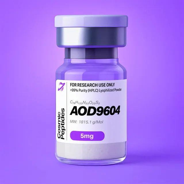
AOD-9604
Commonly researched with these items
Patent-pending lot tracking: Complete transparency from production to vial.
AOD-9604: A Disulfide-Cyclized Tyr-hGH(177–191) Peptide from the C-Terminus of Human Growth Hormone
Disclaimer
Products described here are supplied for research use only and are not intended for diagnostic, therapeutic, or clinical application. All statements regarding biological activity reflect preclinical and in vitro findings exclusively and have not been evaluated by the U.S. Food and Drug Administration. These materials and related content are provided for educational and investigational purposes only and are intended solely for qualified researchers in laboratory and academic settings.
Key Facts
- Compound class
- Synthetic cyclic hexadecapeptide (one intramolecular disulfide); N-terminally tyrosine-modified analogue of residues 176–191 of human growth hormone (hGH, somatotropin)
- Sequence / structure
H-Tyr-Leu-Arg-Ile-Val-Gln-Cys-Arg-Ser-Val-Glu-Gly-Ser-Cys-Gly-Phe-OH, disulfide Cys7–Cys14 (one-letter code: YLRIVQ[CRSVEGSC]GF); all chiral residues L- Molecular formula
- C78H123N23O23S2 (oxidized, disulfide form; free peptide without counterions)
- Molecular weight
- 1815.1 g/mol (average); monoisotopic mass 1813.8604 Da
- CAS number
- 221231-10-3
- PubChem CID
- 71300630
- Other identifiers
- AOD9604, Tyr-hGH(177–191), UNII 7UP768IP4M
- Physical form as supplied
- Lyophilized powder
- Lot traceability
- Every unit is tied to a tracked lot; the lot number, recorded purity and verified quantity of the lot on sale are shown live on this page, with its certificate of analysis.
Molecular Identity and Classification
AOD-9604 is a sixteen-residue synthetic peptide whose sequence, H-Tyr-Leu-Arg-Ile-Val-Gln-Cys-Arg-Ser-Val-Glu-Gly-Ser-Cys-Gly-Phe-OH, reproduces the last fifteen residues of human growth hormone (residues 177–191 of the mature 191-residue protein) with one tyrosine added at the N-terminus. In the native hormone the residue immediately before Leu177 is Phe176, so the molecule can be described in two equivalent ways: as Tyr-hGH(177–191), the name used by the group that developed it, or as [Tyr176]hGH(176–191), a 176–191 fragment in which phenylalanine has been replaced by tyrosine. The two cysteines, at positions 7 and 14 of the peptide (Cys182 and Cys189 in hGH numbering), are joined by a disulfide bond, exactly as they are in the intact hormone. AOD-9604 is therefore a cyclic peptide with a linear N-terminal tail: six residues outside the ring, an eight-residue disulfide-bridged loop, and a two-residue C-terminal tail.
The molecular formula of the disulfide form is C78H123N23O23S2, the average molecular weight 1815.1 g/mol and the monoisotopic mass 1813.8604 Da. The compound is registered under CAS number 221231-10-3 and as PubChem compound 71300630, whose IUPAC name describes the ring as a 1,2-dithia-5,8,11,14,17,20,23-heptazacyclohexacosane: a 26-membered macrocycle made of two sulfur atoms, two cysteine β-carbons and 22 backbone atoms. PubChem records 15 defined stereocentres, which matches the composition: fourteen α-carbons (the two glycines are achiral) plus the β-carbon of isoleucine. Note for literature and database searches: one synonym attached to the PubChem record reads "Tyr-somatostatin (177–191)", a mislabelling of somatotropin; somatostatin is an unrelated 14-residue peptide.
By composition AOD-9604 is a small, moderately amphipathic peptide. It carries two arginines and a free N-terminal amine as its basic groups, one glutamic acid and the C-terminal carboxylate as its acidic groups, and a cluster of hydrophobic side chains (Leu, Ile, two Val, Phe, Tyr). It contains no methionine, tryptophan, histidine, lysine, aspartic acid or asparagine, which, as discussed below, simplifies both its analytics and its stability profile.
In proportion to its parent, AOD-9604 is a minimal piece: sixteen of the 191 residues of hGH, or roughly 8% of the chain and about 8% of the hormone's mass of approximately 22.1 kDa. Its interest as a research tool lies precisely in that minimalism. Human growth hormone recognizes its receptor through a large, discontinuous surface assembled from three separate stretches of the chain, and the fragment approach asks which properties, if any, survive when one contiguous stretch is isolated from the folded protein.
Structure and Chemical Features
Human growth hormone is a four-helix bundle with an up-up-down-down topology, held together by two disulfide bonds: Cys53–Cys165, which links the first long loop to helix 4, and Cys182–Cys189, which closes a small loop at the C-terminus. In the crystal structure of the hormone bound to the extracellular domain of its receptor, solved by de Vos, Ultsch and Kossiakoff in 1992, helix 4 runs to about residue 183 and the final eight residues form a short excursion pinned back by that second disulfide. The AOD-9604 sequence therefore spans the C-terminal turns of helix 4 (residues 177–183) and the whole of the disulfide-closed C-terminal loop (184–191).
Walking along the peptide from the N-terminus shows how each part contributes chemically:
- Tyr1. The added phenol provides a 275–280 nm chromophore, a site for iodination or other aromatic labelling, and an ionizable side chain (pKa near 10). Its substitution for the native Phe176 adds exactly one oxygen atom (+15.9949 Da).
- Leu2-Arg3-Ile4-Val5-Gln6. A short, largely hydrophobic stretch broken by a guanidinium group (pKa about 12.5). In the folded hormone these residues are helical; in the isolated peptide this tail has no partner helices to pack against and is conformationally mobile.
- Cys7 to Cys14. The disulfide-closed loop,
C-R-S-V-E-G-S-C, containing the second arginine, the only carboxylic acid side chain (Glu11), two serines and a glycine. The loop constrains backbone motion and shields its peptide bonds from proteases. - Gly15-Phe16. A flexible C-terminal dipeptide ending in a free carboxylic acid, as in the native hormone.
Charge and Polarity
At neutral pH AOD-9604 carries three positive charges (the N-terminal α-amine and two arginines) and two negative charges (Glu11 and the C-terminal carboxylate), for a net charge of about +1. Under the acidic conditions of reversed-phase HPLC (0.1% TFA, pH about 2) the carboxylates are protonated and the net charge approaches +3. The calculated isoelectric point lies between about 9.4 and 9.9 depending on the pKa set used, because above pH 9 the N-terminal amine begins to deprotonate while both arginines remain charged. The Kyte–Doolittle grand average of hydropathy (GRAVY) is +0.30, mildly hydrophobic for a peptide, which reflects the six hydrophobic residues; the PubChem-computed XLogP3 of −4.8 and topological polar surface area of 815 Å2 instead reflect the large number of polar backbone groups (28 hydrogen-bond donors and 28 acceptors). Both descriptors are correct in their own terms: the molecule is water-soluble overall, yet has a hydrophobic face that governs its retention on C18 phases and its tendency to adsorb to surfaces.
From hGH(177–191) to AOD-9604: A Chemical History
The molecule grew out of a long series of fragment studies on human growth hormone at Monash University in Melbourne. The primary structure of the hormone had been revised by Niall in 1971, fixing the C-terminal sequence that ends in ...Cys-Arg-Ser-Val-Glu-Gly-Ser-Cys-Gly-Phe. In the late 1970s, solid-phase syntheses were reported for three C-terminal peptides, hGH(177–191), hGH(178–191) and hGH(179–191), together with a reduced and S-carbamidomethylated form of hGH(177–191) in which both cysteines are capped as carboxamidomethyl thioethers. That work established two chemical themes that run through all later research on this region: how much N-terminal extension the fragment needs, and whether the disulfide ring itself is essential, a question the alkylated analogue was designed to answer.
Around 2000 the 15-residue disulfide peptide hGH(177–191) was described under the code AOD9401, and the tyrosine-extended analogue under the code AOD9604. The added residue is chemically the most conservative way to put a phenol on the N-terminus of the native sequence: it replaces Phe176 with its hydroxylated counterpart, keeps the native chain length of the 176–191 segment, and gives the peptide a tyrosine for spectrophotometric quantification and for radioiodination, a standard labelling route for peptides at the time.
| Molecule | Sequence | Formula | Average / monoisotopic mass |
|---|---|---|---|
| hGH, mature 22 kDa form | 191 residues, two disulfides (C53–C165, C182–C189) | C990H1528N262O300S7 | about 22,125 g/mol |
| hGH(176–191), native fragment | FLRIVQ[CRSVEGSC]GF | C78H123N23O22S2 | 1799.1 / 1797.8654 Da |
| hGH(177–191) (AOD9401) | LRIVQ[CRSVEGSC]GF | C69H114N22O21S2 | 1651.9 / 1650.7970 Da |
| AOD-9604, Tyr-hGH(177–191) | YLRIVQ[CRSVEGSC]GF | C78H123N23O23S2 | 1815.1 / 1813.8604 Da |
The table makes one analytical point plain: AOD-9604 and the native 176–191 fragment differ by a single oxygen atom, 15.9949 Da. Material described generically as an "hGH fragment 176–191" should therefore always be checked by mass spectrometry, which distinguishes the Phe and Tyr forms unambiguously at the [M+2H]2+ level (899.94 versus 907.94).
Synthesis and Manufacturing Chemistry
AOD-9604 is made by solid-phase peptide synthesis (SPPS), the method introduced by Merrifield in 1963, using the base-labile 9-fluorenylmethoxycarbonyl (Fmoc) α-amine protection described by Carpino and Han in 1972. Because the C-terminus is a free acid, assembly starts from phenylalanine loaded on an acid-labile resin such as Wang or 2-chlorotrityl chloride resin, and proceeds residue by residue towards Tyr1, each cycle consisting of Fmoc removal (typically 20% piperidine in DMF) and coupling of the next activated Fmoc amino acid.
The side chains that need protection, and the standard choices in Fmoc/tert-butyl chemistry, are:
| Position | Residue | Building block | Comment |
|---|---|---|---|
| 16 | Phe | Fmoc-Phe-Wang or Fmoc-Phe-2-ClTrt resin | Loaded first; free acid after cleavage |
| 14, 7 | Cys | Fmoc-Cys(Trt)-OH (or Cys(Acm) for directed oxidation) | Activate under low-base conditions (e.g. DIC/Oxyma) to limit racemization |
| 13, 9 | Ser | Fmoc-Ser(tBu)-OH | Ether protection removed by TFA |
| 11 | Glu | Fmoc-Glu(OtBu)-OH | Only acidic side chain |
| 8, 3 | Arg | Fmoc-Arg(Pbf)-OH | Pbf removal needs sufficient TFA time; residual Pbf adds 252.1 Da |
| 6 | Gln | Fmoc-Gln(Trt)-OH | Trityl prevents side-chain dehydration to a nitrile |
| 1 | Tyr | Fmoc-Tyr(tBu)-OH | Last residue; N-terminus left free |
| 2, 4, 5, 10, 12, 15 | Leu, Ile, Val, Gly | Fmoc-AA-OH, no side-chain protection | The Ile-Val pair is a sterically demanding coupling |
Cleavage from the resin and global deprotection use a trifluoroacetic acid cocktail containing scavengers. Because the peptide contains two cysteines and a tyrosine, a thiol scavenger such as 1,2-ethanedithiol or 3,6-dioxa-1,8-octanedithiol is normally included alongside water and triisopropylsilane, to trap trityl and tert-butyl cations that would otherwise alkylate the free thiols or the phenol ring. The product of cleavage is the linear, reduced dithiol peptide, C78H125N23O23S2 (monoisotopic mass 1815.8760 Da).
Forming the Disulfide
The ring is closed in a separate oxidation step, reviewed in detail by Postma and Albericio (2014). With Cys(Trt) protection, the crude dithiol is dissolved at high dilution, typically 0.1–1 mg/mL, in a mildly basic aqueous buffer (pH about 7.5–8.5) and oxidized by air, by dimethyl sulfoxide, or by a glutathione redox pair. Dilution matters because thiolate anions can react either within the same molecule, closing the 26-membered ring, or with a second molecule, giving covalent dimers and oligomers; intramolecular closure wins at low concentration. An alternative is to carry the cysteines through synthesis as acetamidomethyl (Acm) thioethers and close the ring with iodine, which removes the Acm groups and forms the disulfide in one step. Reaction progress is followed by analytical HPLC and by Ellman's reagent (5,5′-dithiobis(2-nitrobenzoic acid)), which reports remaining free thiol through the 412 nm absorbance of the released thionitrobenzoate anion.
The oxidized peptide is purified by preparative reversed-phase HPLC with water/acetonitrile gradients containing 0.1% TFA and lyophilized. As with most synthetic peptides, the product is isolated as a salt of its basic groups, a trifluoroacetate unless it is exchanged into acetate or chloride; acetate salts of AOD-9604 are common, and "AOD9604 acetate" appears among the PubChem synonyms. The certificate of analysis for each lot states the results that apply to that material.
Sequence-specific impurities a laboratory may encounter include: the reduced (open-chain) peptide (+2.016 Da); disulfide-linked dimers (exactly twice the monomer mass); deletion sequences such as des-Arg (−156.1011 Da) or des-Val (−99.0684 Da) from incomplete couplings; incompletely deprotected species carrying Pbf (+252.10 Da) or tert-butyl (+56.06 Da) groups; and D-Cys diastereomers from racemization during cysteine activation, which have the same mass and are detected only chromatographically. The sequence contains no Asp-Gly or Asp-Ser motif, so aspartimide formation is not a concern, and no methionine or tryptophan, so the common oxidation impurities of those residues do not arise.
Analytical Characterization
Reversed-phase HPLC. AOD-9604 is retained well on C18 or C8 columns with water/acetonitrile gradients containing 0.1% TFA; a gradient of roughly 10–50% acetonitrile over 20–30 minutes is a sensible starting point, as outlined in general terms for peptides by Mant and colleagues. Detection at 210–220 nm (peptide bond) gives the purity figure; a second channel at 275–280 nm, where only the tyrosine phenol and the cystine absorb appreciably, helps confirm that a peak is peptide-derived. Purity is reported as the percentage of total integrated peak area. The oxidized and reduced forms, and disulfide-linked dimers, usually separate on a standard gradient, so a single chromatogram also reports the redox state of the material.
Mass Spectrometry: Expected Ions
Three basic sites make AOD-9604 ionize readily in positive electrospray, with the doubly and triply protonated ions usually most abundant. The values below were calculated from the elemental composition with monoisotopic atomic masses and a proton mass of 1.00728 Da.
| Species | Formula / description | m/z |
|---|---|---|
| [M+H]+, disulfide form | C78H124N23O23S2+ | 1814.8676 |
| [M+2H]2+, disulfide form | Usually the base peak | 907.9375 |
| [M+3H]3+, disulfide form | Abundant at low pH | 605.6274 |
| [M+4H]4+, disulfide form | Minor | 454.4724 |
| [M+2H]2+, reduced dithiol | +2.016 Da on the neutral | 908.9453 |
| [M+2H]2+, reduced and bis-carbamidomethylated | +2 × 57.0215 Da after iodoacetamide | 965.9667 |
| b2 / b3 / b6 fragments | Tyr-Leu / Tyr-Leu-Arg / Tyr…Gln | 277.1547 / 433.2558 / 773.4668 |
| y1 / y2 fragments | Phe / Gly-Phe | 166.0863 / 223.1077 |
| y10 fragment | Intact ring plus Gly-Phe | 1042.4081 |
Several features of these numbers are worth knowing. First, the isotope envelope of a C78 peptide is broad: calculated relative intensities are about 100 : 97 : 60 : 28 for the monoisotopic, M+1, M+2 and M+3 peaks. The monoisotopic peak of the reduced peptide falls exactly on the M+2 isotope of the oxidized one, so partial reduction cannot be read off a low-resolution spectrum by eye. It is quantified instead by alkylating free thiols (iodoacetamide adds 57.0215 Da per cysteine) or by Ellman's assay. Second, a covalent disulfide-linked dimer has a neutral mass of 3627.7207 Da, exactly twice that of the monomer, so its [2M+2H]2+ ion coincides with the monomer's [M+H]+ at m/z 1814.87. The two are told apart by the isotope spacing (0.5 versus 1.0 m/z units) and by HPLC retention. Third, in tandem MS, fragment ions are named by the convention of Roepstorff and Fohlman. Backbone cleavage inside the disulfide ring does not release a fragment, because the two halves remain joined through sulfur, so collision-induced spectra of the intact peptide are dominated by b ions from the linear Tyr1–Gln6 tail and y ions from the C-terminal Gly-Phe. Full sequence coverage of the ring requires reduction and alkylation before MS/MS.
A diagnostic digest. Trypsin cleaves C-terminal to Arg3 and Arg8. Without reduction this releases the tripeptide YLR ([M+H]+ 451.2663) and a two-chain, disulfide-linked product IVQCR/SVEGSCGF (monoisotopic 1399.6224 Da, [M+2H]2+ 700.8185), because the Arg8–Ser9 bond lies inside the ring. After reduction and alkylation the two chains separate (carbamidomethylated IVQCR [M+H]+ 675.3607; SVEGSCGF 842.3349). The appearance of the 700.82 ion only under non-reducing conditions is direct evidence of the Cys7–Cys14 connectivity, which is the only possible pairing for a peptide with two cysteines but is still a useful identity test for the cyclic form.
Amino-acid analysis. After 6 M HCl hydrolysis the expected molar composition is Arg 2, Glx 2 (Gln6 is converted to glutamic acid and counted with Glu11), Gly 2, Ser 2, Val 2, Ile 1, Leu 1, Phe 1, Tyr 1 and cystine 1. Two cautions apply: the Ile-Val bond is hydrolysed slowly and can give low Ile and Val recoveries at 24 hours, and serine is partly destroyed during hydrolysis. Cysteine is best measured as cysteic acid after performic acid oxidation.
Purity versus peptide content. HPLC purity is the fraction of UV-absorbing material that is AOD-9604. Peptide content is the fraction of the weighed powder that is peptide at all, as opposed to counterions, water and residual salts. With three protonatable groups, a fully protonated tris-trifluoroacetate salt has a formula weight of about 2157.2 g/mol (peptide fraction 84.1% by weight), and a tris-acetate about 1995.3 g/mol (91.0%), before any water is counted. Amino-acid analysis, quantitative NMR or nitrogen determination give the peptide content when exact molar concentrations are required.
Physicochemical Properties
| Property | Value | Notes |
|---|---|---|
| Molecular formula | C78H123N23O23S2 | Without counterions; PubChem CID 71300630 |
| Average molecular weight | 1815.1 g/mol | PubChem |
| Monoisotopic mass | 1813.8604 Da | PubChem exact mass 1813.86036 |
| Reduced (dithiol) form | C78H125N23O23S2; 1817.1 g/mol; 1815.8760 Da | Calculated |
| Residues / heavy atoms | 16 / 126 | Ring of 26 atoms closed by Cys7–Cys14 |
| Defined stereocentres | 15 | 14 α-carbons plus Ile β-carbon; all L |
| Net charge | about +1 at pH 7; about +3 at pH 2 | N-terminus, Arg3, Arg8 versus Glu11, C-terminus |
| Isoelectric point | about 9.4–9.9 | Calculated; depends on the pKa set |
| Molar absorption coefficient, 280 nm | about 1,615 M−1 cm−1 | 1 Tyr (1,490) + 1 cystine (125), Pace method |
| A280 of a 1 mg/mL solution | about 0.89 (1 cm path) | Peptide basis; calculated from the coefficient above |
| GRAVY (Kyte–Doolittle) | +0.30 | Calculated; mildly hydrophobic |
| XLogP3 | −4.8 | PubChem, computed |
| Topological polar surface area | 815 Å2 | PubChem, computed |
| Hydrogen-bond donors / acceptors | 28 / 28 | PubChem, computed |
| Rotatable bonds | 45 | PubChem, computed |
| Chromophores | Peptide bond (about 214 nm), Tyr (275 nm), Phe (257 nm), cystine (weak, broad) | No tryptophan |
The composition explains most of the practical behaviour. With a net positive charge at neutral pH and a strongly positive one in acid, AOD-9604 dissolves in water and dilute aqueous acids; its hydrophobic Leu-Ile-Val and Phe/Tyr side chains give it clear retention on reversed-phase media and a measurable tendency to adsorb to plastic and glass at low concentrations. Because the only absorbing side chains at 280 nm are one tyrosine and one cystine, the molar absorption coefficient is small. A 0.1 mg/mL solution has an A280 of only about 0.09, so 280 nm absorbance is a usable but insensitive concentration check, and the far-UV region or a colorimetric peptide assay is preferable for dilute solutions.
Chemically the principal liabilities are the disulfide and the peptide bonds at extremes of pH. Above about pH 8, traces of thiolate catalyse disulfide exchange and can generate dimers, and strongly alkaline conditions promote β-elimination at cystine. Reducing agents such as dithiothreitol, 2-mercaptoethanol or TCEP open the ring quantitatively, which is useful analytically but must be avoided when the cyclic form is the object of study. The absence of methionine, tryptophan, asparagine and aspartic acid removes the most common oxidation and deamidation pathways; Gln6 is internal and so cannot cyclize to pyroglutamate.
Research Context
AOD-9604 is studied as a defined structural fragment of human growth hormone (hGH). The crystal structure of hGH bound to the extracellular domain of its receptor, solved by de Vos, Ultsch and Kossiakoff in 1992, shows that the hormone engages two receptor molecules through two distinct binding sites assembled from residues scattered across its four-helix bundle. The C-terminal segment that AOD-9604 reproduces, residues 177–191, lies outside the main receptor-binding helices and includes the Cys182–Cys189 disulfide loop that closes the hormone's C-terminus.
Because high-affinity receptor binding requires the folded hormone, an isolated C-terminal peptide is not expected to reproduce it. That makes AOD-9604 useful as a structural probe: researchers compare the conformation of the excised, disulfide-closed segment in solution with the same residues in the intact hormone, and use receptor-signalling counter-screens, such as STAT5 phosphorylation in receptor-expressing cell lines, to confirm that the fragment does not act through the full-length hormone's receptor. Its N-terminal tyrosine also makes it a convenient, radioiodinatable reference peptide for analytical and binding-assay method development.
Related Compounds
Other compact, disulfide-containing or analytically well-defined peptides in the catalogue include Oxytocin, a cyclic nonapeptide closed by a disulfide bridge, which shares with AOD-9604 the analytical questions of disulfide verification and reduction–alkylation mapping. For comparison of fragment design strategies, CJC-1295 (No DAC) is a substituted fragment of growth hormone-releasing hormone.
Laboratory Handling and Storage
Storage of the solid. Lyophilized AOD-9604 is best kept tightly closed at −20 °C or below, protected from light and moisture. Like most peptide salts it is hygroscopic, so a container taken from the freezer should reach room temperature in a desiccator before opening, to avoid condensation on the cold powder.
Dissolution. The peptide dissolves in high-purity water and in dilute acetic acid; for stock solutions, slightly acidic conditions (for example 0.1–1% acetic acid, or water followed by dilution into buffer) are preferable to basic ones because they suppress thiolate formation and therefore disulfide exchange. If a concentrated stock is slow to dissolve, a small proportion of acetonitrile or dimethyl sulfoxide can be used, provided it is compatible with the downstream assay. Avoid buffers containing reducing agents unless the reduced form is intended.
A worked concentration example. A 1.0 mM solution of AOD-9604 requires 1.815 mg of peptide per mL. If the certificate of analysis or an amino-acid analysis gives a peptide content of, say, 85%, the mass of powder to weigh is 1.815 / 0.85 = 2.14 mg per mL. Ignoring peptide content in this example would make the solution about 15% weaker than labelled.
Solutions. Prepare stocks at 0.5–5 mM, divide into single-use aliquots in low-binding polypropylene tubes, and store at −20 °C or −80 °C; avoid repeated freeze–thaw cycles. Filter through a 0.22 µm low-protein-binding membrane when solutions are destined for cell culture. At sub-micromolar working concentrations, adsorption to surfaces can remove a meaningful fraction of the peptide; low-binding ware and, where the design allows, a carrier protein in the assay buffer reduce these losses. For cell-based experiments, a vehicle control matched for counterion is advisable, since Cornish and colleagues showed that trifluoroacetate alone can alter proliferation of cultured cells.
Re-verification. Before critical experiments, or after long storage of a solution, a short LC-MS run confirms identity and redox state: the [M+2H]2+ ion at m/z 907.94 for the cyclic peptide, the absence of a +2 Da shift (reduced form at 908.95) and of a chromatographically distinct dimer, and, in biological media, monitoring of the stable ring fragment at m/z 448.17. Each lot's certificate of analysis, linked on this page, records the analytical results for that lot.
Frequently Asked Research Questions
- What is AOD-9604?
- AOD-9604 is a synthetic 16-residue peptide, H-Tyr-Leu-Arg-Ile-Val-Gln-Cys-Arg-Ser-Val-Glu-Gly-Ser-Cys-Gly-Phe-OH with a Cys7–Cys14 disulfide. It reproduces residues 177–191 of human growth hormone with an added N-terminal tyrosine, and is also written Tyr-hGH(177–191).
- Is AOD-9604 the same as hGH fragment 176–191?
- Not exactly. The native 176–191 sequence begins with phenylalanine (Phe176); AOD-9604 has tyrosine in that position. The two differ by one oxygen atom (15.9949 Da): monoisotopic masses are 1813.8604 Da for AOD-9604 and 1797.8654 Da for the native fragment.
- What are the CAS number, formula and molecular weight of AOD-9604?
- CAS 221231-10-3; formula C78H123N23O23S2 for the disulfide form; average molecular weight 1815.1 g/mol; monoisotopic mass 1813.8604 Da; PubChem CID 71300630.
- Which ions should I see for AOD-9604 by ESI-MS?
- For the cyclic peptide: [M+H]+ at m/z 1814.868, [M+2H]2+ at 907.938 (usually most intense) and [M+3H]3+ at 605.627. The reduced dithiol appears 2.016 Da heavier ([M+2H]2+ 908.945).
- How can I confirm the disulfide is intact?
- Compare non-reduced and reduced samples. The intact ring gives [M+2H]2+ 907.94, shifts by +1.008 m/z on reduction and by +58.02 m/z (2+) after reduction and iodoacetamide alkylation. Ellman's assay of the untreated sample should show negligible free thiol, and a non-reducing tryptic digest gives the disulfide-linked IVQCR/SVEGSCGF ion at m/z 700.82 (2+).
- Why do MS/MS spectra of intact AOD-9604 show few fragments from the middle of the sequence?
- Residues 7–14 form a disulfide-closed ring, so a single backbone cleavage inside the ring does not release a fragment. Spectra are dominated by b ions from the linear Tyr–Gln tail (b2 277.155, b3 433.256, b6 773.467) and y ions of Gly-Phe; reduce and alkylate for full coverage.
- Where can I see the purity of the lot I am buying?
- This page shows the lot number, recorded purity and verified quantity of the lot currently on sale, taken live from the shop's lot-tracking database, with that lot's certificate of analysis.
Sources & Further Reading
- National Center for Biotechnology Information. PubChem Compound Summary for CID 71300630, AOD-9604. PubChem CID 71300630.
- Niall HD. Revised primary structure for human growth hormone. Nature New Biology 1971;230(11):90–91. doi:10.1038/newbio230090a0
- de Vos AM, Ultsch M, Kossiakoff AA. Human growth hormone and extracellular domain of its receptor: crystal structure of the complex. Science 1992;255(5042):306–312. doi:10.1126/science.1549776
- Andreu D, Albericio F, Solé NA, Munson MC, Ferrer M, Barany G. Formation of disulfide bonds in synthetic peptides and proteins. In: Peptide Synthesis Protocols. Humana Press; 1994:91–170. doi:10.1385/0-89603-273-6:91
- Postma TM, Albericio F. Disulfide formation strategies in peptide synthesis. European Journal of Organic Chemistry 2014;2014(17):3519–3530. doi:10.1002/ejoc.201402149
- Ellman GL. Tissue sulfhydryl groups. Archives of Biochemistry and Biophysics 1959;82(1):70–77. doi:10.1016/0003-9861(59)90090-6
- Merrifield RB. Solid phase peptide synthesis. I. The synthesis of a tetrapeptide. Journal of the American Chemical Society 1963;85(14):2149–2154. doi:10.1021/ja00897a025
- Carpino LA, Han GY. 9-Fluorenylmethoxycarbonyl amino-protecting group. The Journal of Organic Chemistry 1972;37(22):3404–3409. doi:10.1021/jo00795a005
- Mant CT, Chen Y, Yan Z, et al. HPLC analysis and purification of peptides. Methods in Molecular Biology 2007;386:3–55. doi:10.1007/978-1-59745-430-8_1
Last reviewed:
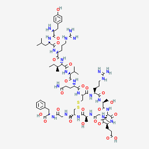
| CAS Number | 221231-10-3 |
|---|---|
| Molecular Formula | C78H123N23O23S2 |
| Molecular Weight | 1815.1 g/Mol |
| Purity | 99.7% |
| Lot Number | 26072 |
| Quantity | 2.21mg |
| Sequence | H-Tyr-Leu-Arg-Ile-Val-Gln-Cys(1)-Arg-Ser-Val-Glu-Gly-Ser-Cys(1)-Gly-Phe-OH |
Cosmic Peptides Certificate of Analysis
Unable to load certificate. Download COA