NAD+
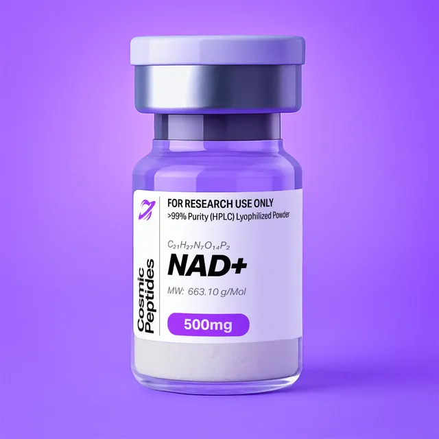
NAD+
Commonly researched with these items
Patent-pending lot tracking: Complete transparency from production to vial.
| CAS Number | 53-84-9 |
|---|---|
| Molecular Formula | C21H27N7O14P2 |
| Molecular Weight | 664.0 g/Mol |
| Purity | 99.8% |
| Lot Number | 26096 |
| Quantity | 503mg |
NAD+ (Nicotinamide Adenine Dinucleotide): The Pyridine Dinucleotide Coenzyme of Hydride Transfer and ADP-Ribosylation
Disclaimer
Products described here are supplied for research use only and are not intended for diagnostic, therapeutic, or clinical application. All statements regarding biological activity reflect preclinical and in vitro findings exclusively and have not been evaluated by the U.S. Food and Drug Administration. These materials and related content are provided for educational and investigational purposes only and are intended solely for qualified researchers in laboratory and academic settings.
Key Facts
- Compound class
- Dinucleotide coenzyme (pyridine nucleotide); not a peptide. Two ribonucleotides, nicotinamide mononucleotide and adenosine 5′-monophosphate, joined 5′ to 5′ through a pyrophosphate bridge
- Structure
- β-Nicotinamide adenine dinucleotide, oxidized form (β-NAD+), as the inner salt: a 3-carbamoylpyridinium (nicotinamide) cation balanced by one ionized phosphate. Synonyms: nadide, coenzyme I, diphosphopyridine nucleotide (DPN), cozymase
- Molecular formula
- C21H27N7O14P2 (free acid, inner salt)
- Molecular weight
- 663.43 g/mol (average); monoisotopic mass 663.1091 Da
- CAS number
- 53-84-9
- PubChem CID
- 5892
- Physical form as supplied
- White to off-white solid powder
- Lot traceability
- Every unit is tied to a tracked lot; the lot number, recorded purity and verified quantity of the lot on sale are shown live on this page, with its certificate of analysis.
Molecular Identity and Classification
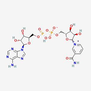
NAD+ is nicotinamide adenine dinucleotide in its oxidized form. Although it appears in a catalogue of research peptides, it is not a peptide: it contains no amino acids and no amide backbone. It is a dinucleotide, built from two nucleotides that share a pyrophosphate (diphosphate) linkage. One nucleotide is nicotinamide mononucleotide (NMN), in which the pyridine base nicotinamide is attached to C1′ of a D-ribose through a β-N-glycosidic bond; the other is adenosine 5′-monophosphate (AMP). The 5′-phosphate of each ribose is joined to the other through a phosphoanhydride bond, giving the sequence nicotinamide–ribose–phosphate–phosphate–ribose–adenine.
The PubChem IUPAC name, [[(2R,3S,4R,5R)-5-(6-aminopurin-9-yl)-3,4-dihydroxyoxolan-2-yl]methoxy-hydroxyphosphoryl] [(2R,3S,4R,5R)-5-(3-carbamoylpyridin-1-ium-1-yl)-3,4-dihydroxyoxolan-2-yl]methyl phosphate, spells out the eight stereocentres of the two D-ribofuranose rings and the positive charge on the pyridinium nitrogen. The neutral, isolable compound registered under CAS 53-84-9 is the inner salt (sometimes called the free acid): the cationic pyridinium ring is balanced by one ionized phosphate, while the second phosphate carries a proton. Its formula is C21H27N7O14P2, its average molecular weight 663.43 g/mol and its monoisotopic mass 663.1091 Da (PubChem CID 5892).
The "+" in NAD+ refers to the formal charge on the nicotinamide ring, not to the charge of the whole molecule. At pH 7 both phosphates are ionized, so the species in solution carries a net charge of −1 (one positive pyridinium, two negative phosphates). The reduced form, NADH (C21H29N7O14P2 as the free acid, PubChem CID 439153), carries an uncharged 1,4-dihydronicotinamide ring and therefore a net charge of −2 at pH 7. The phosphorylated relative NADP+ (CAS 53-59-8, PubChem CID 5885) carries a third phosphate on the 2′-hydroxyl of the adenosine ribose.
The natural and enzymatically active compound is the β-anomer, in which the nicotinamide sits on the same face of its ribose as the C5′ hydroxymethyl group. The α-anomer (α-NAD+) is a known impurity of older preparations and a separate research reagent; most dehydrogenases do not accept it. Commercial material is also offered as salts, of which the monosodium salt C21H26N7NaO14P2 (CAS 20111-18-6, 685.41 g/mol) is the most common; molar calculations must use the molecular weight of the form actually in the vial.
Structure and Chemical Features
The nicotinamide ring is the reactive end. It is a pyridinium cation carrying a carboxamide at C3. The positive charge on N1 makes C4 (para to the ring nitrogen) electron-poor, so it can accept a hydride ion (H−, one proton and two electrons) from a substrate. Accepting a hydride converts the aromatic pyridinium into the non-aromatic 1,4-dihydropyridine of NADH. The carboxamide is essential: it is held in a defined orientation by hydrogen bonds in dehydrogenase active sites and tunes the redox potential of the ring. The N-glycosidic bond that links the pyridinium N1 to ribose C1′ is unusually reactive, because the departing nicotinamide is a neutral, good leaving group. That reactivity is what allows ADP-ribosyl transferases and NAD+ glycohydrolases to cleave it. The free energy of hydrolysis of this bond is commonly quoted at about −34 kJ/mol (roughly −8 kcal/mol), comparable to the phosphoanhydride bonds of ATP.
The adenosine end carries no redox chemistry. It serves as a binding handle. The adenine ring, the adenosine ribose and the pyrophosphate make most of the contacts that hold the coenzyme in the nucleotide-binding domains of dehydrogenases, and the 2′-hydroxyl of the adenosine ribose is the position that distinguishes NAD+ from NADP+, which is why enzymes can discriminate between the two coenzymes with high selectivity. Adenine is also the main chromophore: it is responsible for the strong absorbance band of NAD+ at 259–260 nm.
The pyrophosphate bridge provides two negative charges, considerable water solubility and flexibility. Combined with the rotatable bonds around the two riboses (11 rotatable bonds by PubChem's count), it allows NAD+ to adopt both extended and folded conformations. In free solution the molecule samples a population of conformers, some in which the two rings stack face to face; bound to dehydrogenases, it is held in an extended conformation with the two rings well apart.
Stereochemistry of Hydride Transfer
When NAD+ is reduced, C4 of the nicotinamide ring changes from an sp2 ring carbon into a CH2 group whose two hydrogens are no longer equivalent: they lie on opposite faces of the ring and are designated pro-R (the A-side) and pro-S (the B-side). Deuterium-labelling experiments by Westheimer, Vennesland and colleagues in the early 1950s, with yeast alcohol dehydrogenase and deuterated substrates, showed that the enzyme transfers hydrogen directly between substrate and coenzyme, with no exchange with solvent, and always to and from the same face of the ring. Every dehydrogenase since characterized is either A-specific or B-specific. Stereospecifically labelled [4-2H]NADH remains a tool of mechanistic enzymology.
Redox Chemistry
The NAD+/NADH couple is a two-electron, one-proton system: NAD+ + 2 e− + H+ ⇌ NADH. Its standard reduction potential at pH 7 and 25 °C is tabulated as −0.32 V, so NADH is a strong biological reductant and NAD+ a comparatively weak oxidant. Chemically, NADH is a hydride donor rather than a source of free electrons; it does not react rapidly with molecular oxygen without a catalyst, which is why the couple is kinetically stable in air-saturated buffers and is useful as a reagent in coupled enzyme assays.
Synthesis and Manufacturing Chemistry
NAD+ is made by biochemical and chemical routes unrelated to solid-phase peptide synthesis. Historically it was extracted from baker's yeast and fractionated by precipitation and ion-exchange chromatography.
The first enzymatic synthesis was reported by Arthur Kornberg in 1950. He showed that an enzyme now called nicotinamide mononucleotide adenylyltransferase (NMNAT) condenses NMN with ATP to give NAD+ and inorganic pyrophosphate, and that the reaction is reversible. The same reaction underlies modern chemoenzymatic manufacture: NMN and ATP are combined with a recombinant NMNAT, and the pyrophosphate by-product is often removed with an inorganic pyrophosphatase to drive the equilibrium towards the product.
The first total chemical synthesis was reported by Hughes, Kenner and Todd in 1957, in the series of papers in which Alexander Todd's group worked out the chemistry of nucleotide coenzymes. The key step is the formation of an unsymmetrical pyrophosphate: one nucleotide's phosphate is activated and coupled with the phosphate of the other, here NMN and AMP. Building an unsymmetrical pyrophosphate without forming the two symmetrical by-products, and without damaging the labile glycosidic bond, remains the central problem in making NAD+ analogues.
Purification typically relies on anion-exchange or reversed-phase chromatography followed by precipitation or freeze-drying. The related substances that analysts look for follow directly from the bonds that can break or the steps that can be incomplete, and each has a distinct mass, which makes an LC-MS impurity screen straightforward.
| Species | Formula | Monoisotopic mass (Da) | [M−H]− m/z | Origin |
|---|---|---|---|---|
| Nicotinamide | C6H6N2O | 122.0480 | Seen as [M+H]+ 123.0553 | Glycosidic bond hydrolysis |
| Nicotinamide mononucleotide (NMN) | C11H15N2O8P | 334.0566 | 333.0493 | Unreacted starting material; pyrophosphate cleavage |
| AMP | C10H14N5O7P | 347.0631 | 346.0558 | Pyrophosphate cleavage |
| ADP | C10H15N5O10P2 | 427.0294 | 426.0221 | Synthesis side product |
| ADP-ribose | C15H23N5O14P2 | 559.0717 | 558.0644 | Glycosidic bond hydrolysis (with nicotinamide) |
| α-NAD+ | C21H27N7O14P2 | 663.1091 | 662.1018 | Anomer; same mass, resolved only by chromatography or enzymatic assay |
| NADH | C21H29N7O14P2 | 665.1248 | 664.1175 | Reduction; also flagged by A340 |
The certificate of analysis for each lot states the analytical results that apply to that material.
Analytical Characterization
Because NAD+ is a nucleotide, its analytical chemistry follows nucleotide practice rather than peptide practice. Four approaches are used, and each answers a different question.
UV spectroscopy. NAD+ absorbs maximally at 259–260 nm, from the adenine ring, with a molar absorption coefficient commonly tabulated at about 17.8–18.0 × 10³ M−1 cm−1 at pH 7. It has no absorbance at 340 nm. NADH keeps an adenine band near 259 nm (about 14.4 × 10³ M−1 cm−1) and gains a second band at 340 nm from the dihydronicotinamide ring, with ε340 = 6.22 × 10³ M−1 cm−1, the value established by Horecker and Kornberg in 1948 and still used in almost every dehydrogenase assay. An A340 reading of an NAD+ solution is therefore a quick test for contaminating NADH, and the A260/A340 ratio of about 2.3 is a classic purity criterion for NADH itself.
Enzymatic assay. Chromatographic purity does not by itself prove that the material is biochemically active. The classical active-content assay reduces NAD+ with ethanol and alcohol dehydrogenase at mildly alkaline pH, with a carbonyl-trapping agent such as semicarbazide to pull the reaction to completion, and reads the NADH formed at 340 nm. Because the enzyme accepts only the β-anomer, it reports active β-NAD+ specifically. For very small amounts, the enzymatic cycling methods of Lowry and colleagues (1961) recycle the coenzyme repeatedly between two dehydrogenases, amplifying the signal so that picomole quantities can be measured.
HPLC. NAD+ is too polar and too charged to be retained well by ordinary reversed-phase chromatography. Laboratories use ion-pair reversed-phase HPLC (with a tetraalkylammonium ion-pairing agent in phosphate buffer), anion-exchange chromatography, porous graphitic carbon or hydrophilic interaction chromatography (HILIC), with detection at 260 nm. These methods separate NAD+ from NADH, NADP+, NMN, AMP, ADP, ADP-ribose and nicotinamide, the principal related substances.
Mass spectrometry. Electrospray ionization detects NAD+ in both polarities. The table below gives calculated monoisotopic m/z values for the main ions and diagnostic fragments, which allow a laboratory to confirm identity independently.
| Ion | m/z | Notes |
|---|---|---|
| NAD+ [M+H]+ | 664.1164 | Positive mode; the intact pyridinium cation with the phosphate protonated |
| NAD+ [M−H]− | 662.1018 | Negative mode; commonly used in metabolomics |
| NAD+ [M+Na]+ | 686.0983 | Sodium adduct, prominent with sodium salt forms |
| NAD+ [M+2H]2+ | 332.5618 | Doubly charged; weak under most conditions |
| [M+H − nicotinamide]+ | 542.0684 | Loss of neutral nicotinamide (122.0480 Da) at the labile glycosidic bond |
| [ADP+H]+ | 428.0367 | Adenosine diphosphate fragment |
| [adenine+H]+ | 136.0618 | Diagnostic adenine fragment |
| [nicotinamide+H]+ | 123.0553 | Diagnostic nicotinamide fragment |
| [M−H − nicotinamide]− | 540.0538 | Negative-mode equivalent of the 542 fragment |
| NADH [M−H]− | 664.1175 | Two mass units above NAD+ in negative mode |
One pitfall deserves emphasis. In negative mode the [M−H]− ion of NADH (m/z 664.1175) falls within about 0.01 m/z of the second isotope peak (M+2) of NAD+ (about m/z 664.108), and the M+1 isotope of NAD+ is roughly a quarter of its monoisotopic peak, because of its 21 carbon atoms. Unless the two coenzymes are separated chromatographically, a small amount of NADH can be hidden in, or invented by, the isotope envelope of an NAD+ peak. In-source fragmentation and interconversion during sample preparation are further sources of error, which is why Lu and colleagues (2018) recommended cold, acidic organic solvent extraction and chromatographic separation of all four redox species when NAD(P)(H) is measured.
Complementary techniques include 1H and 31P NMR (the two phosphorus atoms of the pyrophosphate appear as an AB pattern of coupled signals, and the nicotinamide ring protons resonate far downfield, between about 8 and 9.5 ppm, because of the ring's positive charge), Karl Fischer titration for water content, and ion chromatography or elemental analysis for sodium content in salt forms.
Physicochemical Properties
| Property | Value | Notes |
|---|---|---|
| Molecular formula | C21H27N7O14P2 | Inner salt (free acid), CAS 53-84-9 |
| Average molecular weight | 663.43 g/mol | PubChem CID 5892 (663.4) |
| Monoisotopic mass | 663.1091 Da | For high-resolution MS |
| Monosodium salt | C21H26N7NaO14P2, 685.41 g/mol | CAS 20111-18-6; hydrates are heavier still |
| Net charge at pH 7 | −1 | Pyridinium +1, pyrophosphate −2 |
| Adenine N1 pKa | about 3.5–4 | Protonated only in acidic solution |
| Standard reduction potential (NAD+/NADH) | −0.32 V | pH 7, 25 °C; shifts about −30 mV per pH unit |
| UV maximum | 259–260 nm, ε about 17.8–18.0 × 10³ M−1 cm−1 | No absorbance at 340 nm |
| Fluorescence | Non-fluorescent | NADH fluoresces (excitation 340 nm, emission about 450–460 nm) |
| XLogP3 | −6.0 | Computed (PubChem); extremely hydrophilic |
| Topological polar surface area | 321 Å2 | Computed (PubChem) |
| Hydrogen-bond donors / acceptors | 7 / 18 | Computed (PubChem) |
| Rotatable bonds | 11 | Computed (PubChem) |
| Phosphorus content | 9.34% by mass | Two P atoms; basis of phosphate-based quantification |
| Solubility | Freely soluble in water | Practically insoluble in ethanol, acetone and ether |
The physicochemical profile of NAD+ is dominated by its charge: a polar surface area above 300 Å2, eighteen hydrogen-bond acceptors and a computed partition coefficient of −6 rule out passive diffusion across lipid bilayers.
The compound's characteristic instability is pH-dependent and opposite to that of NADH. NAD+ is reasonably stable in mild acid but is destroyed by alkali, which attacks the nicotinamide–ribose glycosidic bond and the pyridinium ring; NADH is stable in alkali but decomposes rapidly in acid. Lowry, Passonneau and Rock characterized these kinetics in 1961, and the asymmetry is still exploited deliberately: heating an extract in acid destroys the reduced coenzymes while sparing NAD+, and heating in alkali does the reverse, so that each form can be measured separately. The inner salt dissolves to give a distinctly acidic solution, so buffering to the intended pH is part of preparing any stock.
| Property | NAD+ | NADH | NADP+ |
|---|---|---|---|
| Formula (free acid) | C21H27N7O14P2 | C21H29N7O14P2 | C21H28N7O17P3 |
| Average MW (g/mol) | 663.43 | 665.45 | 743.4 |
| Monoisotopic mass (Da) | 663.1091 | 665.1248 | 743.0755 |
| PubChem CID | 5892 | 439153 | 5885 |
| Absorbance at 340 nm | None | ε = 6.22 × 10³ M−1 cm−1 | None |
| Fluorescence | No | Yes (ex 340 / em about 460 nm) | No |
| Stable in | Mild acid | Alkali | Mild acid |
| Destroyed by | Alkali, especially with heat | Acid; phosphate and low pH accelerate | Alkali, especially with heat |
Research Context
NAD+ plays two chemically distinct roles, and research on it divides accordingly. As a coenzyme, it accepts and donates hydride in hundreds of oxidoreductase reactions and is regenerated each time, so the total pool is conserved. As a substrate, it is consumed: sirtuins, poly(ADP-ribose) polymerases and NAD+ glycohydrolases break the nicotinamide glycosidic bond and use the ADP-ribose part, releasing free nicotinamide. Pollak, Dölle and Ziegler (2007) reviewed both roles. For the enzymologist, the distinction defines the experiment: in a dehydrogenase assay NAD+ is a co-substrate whose reduction is read at 340 nm; in a sirtuin or PARP assay it is a substrate that is irreversibly cleaved.
Dehydrogenases and the Rossmann Fold
Dehydrogenases that use NAD+, such as lactate, malate, alcohol and glyceraldehyde-3-phosphate dehydrogenases, share a nucleotide-binding domain first described by Rossmann, Moras and Olsen in 1974 from crystal structures of several of these enzymes. The "Rossmann fold" is an alternating arrangement of β-strands and α-helices with a glycine-rich loop that wraps the pyrophosphate; a conserved acidic residue at the end of the second β-strand hydrogen-bonds to the 2′- and 3′-hydroxyls of the adenosine ribose and is a main reason these enzymes prefer NAD+ over NADP+. NAD+ is used in steady-state kinetics, coenzyme-specificity engineering, kinetic isotope-effect studies and crystallography of ternary complexes. In practical biochemistry, NAD+-linked dehydrogenases are also the readout for a large family of coupled spectrophotometric assays: any reaction that can be connected to the formation or consumption of NADH can be followed continuously at 340 nm.
A worked example shows how directly this works. With ε340 = 6.22 mM−1 cm−1 and a 1 cm path length, an absorbance change of 0.100 at 340 nm corresponds to 0.100 / 6.22 = 0.0161 mM, or 16.1 µM NADH formed. In a 1.0 mL cuvette that is 16.1 nmol of hydride transferred, and an initial rate of 0.050 absorbance units per minute is 8.0 µM per minute. An absorbance of 1.5, near the linear limit of most instruments, is about 240 µM NADH.
Sirtuins: NAD+-Dependent Deacylases
In 2000, Landry and colleagues showed with purified recombinant proteins that the yeast silencing protein Sir2 and several of its homologues from other organisms are NAD+-dependent protein deacetylases, a finding reported at the same time by other groups. Tanner and colleagues showed in the same year that the reaction consumes one NAD+ for every acetyl group removed and generates an unusual product, O-acetyl-ADP-ribose, together with nicotinamide and the deacetylated lysine. Jackson and Denu (2002) then established that the enzymatic product is 2′-O-acetyl-ADP-ribose, which equilibrates non-enzymatically with the 3′-isomer by intramolecular acyl transfer.
Mechanistically, as reviewed by Sauve, Wolberger, Schramm and Boeke (2006), the acetyl-lysine carbonyl oxygen attacks C1′ of the nicotinamide ribose, displacing nicotinamide and forming a covalent ADP-ribose-peptidyl imidate intermediate. That intermediate can be captured again by free nicotinamide (base exchange), which regenerates NAD+ and explains why nicotinamide is an inhibitor of sirtuins; Jackson, Schmidt, Oppenheimer and Denu (2003) dissected this inhibition and the accompanying transglycosidation kinetically with purified enzymes. For assay design, this means that nicotinamide accumulation can slow a sirtuin reaction over time, and that reported Michaelis constants for NAD+, which differ by more than an order of magnitude across the seven mammalian sirtuins (SIRT1–SIRT7) and with the acyl-peptide used, must be compared under matched conditions. Activity is measured with acetylated peptides by HPLC or MS, by coupled assays for released nicotinamide, or with fluorogenic peptides, which do not always behave like native sequences.
Poly(ADP-Ribose) Polymerases
PARPs are ADP-ribosyl transferases. PARP1, the best-studied member, binds DNA strand breaks through its zinc-finger domains, which activates its catalytic domain to transfer ADP-ribose units from NAD+ onto acceptor proteins, including itself. Successive units are joined by ribose–ribose glycosidic bonds into linear and branched chains of poly(ADP-ribose), releasing one nicotinamide per unit. Gibson and Kraus (2012) reviewed the mammalian PARP family of 17 members, most of which transfer only a single ADP-ribose (mono-ADP-ribosylation). In vitro, PARP activity is measured by incorporation of radiolabelled or biotinylated NAD+, by antibody detection of poly(ADP-ribose), by consumption of NAD+ (converted into a fluorescent derivative or measured by cycling) or by release of nicotinamide. The chains are hydrolysed by poly(ADP-ribose) glycohydrolase (PARG), making the system a model of a reversible, NAD+-fuelled modification.
NAD+ Glycohydrolases and Calcium-Mobilizing Messengers
CD38 and its homologue CD157 are multifunctional ectoenzymes that hydrolyse NAD+ into ADP-ribose and nicotinamide and, as a minor reaction, cyclize it into cyclic ADP-ribose (cADPR), in which C1′ of the nicotinamide ribose is bonded to N1 of adenine. Under acidic conditions and in the presence of nicotinic acid, the same enzyme family exchanges the nicotinamide of NADP+ for nicotinic acid to make NAADP. Lee (2001) reviewed both molecules as calcium-mobilizing messengers in cell systems. Analogues of NAD+ such as nicotinamide guanine dinucleotide (NGD+), whose cyclic product is fluorescent, are used to separate the cyclase and hydrolase activities in vitro.
History of the Molecule
NAD+ was the first coenzyme to be recognized. In 1906, Arthur Harden and William John Young found that yeast juice lost its ability to ferment glucose when it was dialysed or filtered through gelatin, and regained it when the boiled, filtered fraction was added back. They called the heat-stable, dialysable factor the "coferment". Hans von Euler-Chelpin purified it, worked out that it was a nucleotide containing adenine, sugar and phosphate, and named it cozymase; Harden and von Euler-Chelpin shared the 1929 Nobel Prize in Chemistry for work on fermentation. In the mid-1930s Otto Warburg and Walter Christian identified nicotinamide as the part of the molecule that carries hydrogen, and established the spectroscopic difference between the oxidized and reduced forms that is still the basis of NAD+-linked assays.
Since the 2000 sirtuin discoveries it has been studied as a substrate as well as a coenzyme. Older names (DPN, DPNH, coenzyme I, cozymase) matter when searching pre-1960s papers.
Related Compounds
Common companion reagents are NADH as a standard, NADP+ for coenzyme-specificity comparisons, NMN as a biosynthetic intermediate and nicotinamide as a reaction product and inhibitor. Nicotinamide itself is a substrate of nicotinamide N-methyltransferase (NNMT), which transfers a methyl group from S-adenosylmethionine to form 1-methylnicotinamide; the quinolinium compound 5-Amino-1MQ is studied as an inhibitor of this enzyme in biochemical assays, which makes it a natural companion reagent in studies of nicotinamide metabolism.
Laboratory Handling and Storage
Storage of the solid. Solid NAD+ is hygroscopic. It is best kept tightly closed, desiccated and protected from light at −20 °C. Allow a container taken from the freezer to reach room temperature, ideally in a desiccator, before opening it, so that moisture does not condense on the cold powder and add to the weighed mass.
Preparing solutions. NAD+ dissolves readily in high-purity water. Because the inner salt gives an acidic solution, stocks for enzyme work are usually prepared in, or adjusted with, a buffer near pH 6.5–7.5; avoid alkaline buffers, in which the nicotinamide glycosidic bond is hydrolysed. As a reference calculation, 6.63 mg of the inner salt (663.43 g/mol) dissolved to 1.00 mL gives a 10.0 mM solution, and 500 mg of the inner salt is 0.754 mmol; for the monosodium salt the corresponding figures are 6.85 mg and 0.729 mmol. Because powders contain variable water, the concentration of a stock is best confirmed by absorbance at 260 nm (using ε260 ≈ 18.0 mM−1 cm−1 at pH 7) or by an enzymatic assay rather than assumed from the weighed mass. Stocks intended for cell culture are filtered through a 0.22 µm membrane, divided into single-use aliquots and frozen at −20 °C or −80 °C; repeated freeze–thaw cycles should be avoided.
Working with NADH alongside NAD+. Because the reduced and oxidized forms degrade under opposite conditions, they should be stored as separate stocks: NAD+ in neutral to slightly acidic solution, NADH in slightly alkaline solution (for example pH 8–9). Phosphate buffers and low pH accelerate the breakdown of NADH, so assay buffers such as Tris or HEPES are often preferred when NADH is held for long periods. Check NAD+ stocks occasionally for A340, which indicates reduction or contamination.
Verification. After long storage, an LC-MS check for [M−H]− at m/z 662.10, with the related substances tabulated above absent, confirms identity and integrity.
Frequently Asked Research Questions
- Is NAD+ a peptide?
- No. NAD+ is a dinucleotide coenzyme: nicotinamide mononucleotide and adenosine monophosphate joined by a pyrophosphate bridge. It contains no amino acids. Its analytical chemistry (UV at 260 nm, ion-pair or HILIC chromatography, enzymatic assay) is that of a nucleotide.
- What is the molecular formula and molecular weight of NAD+?
- The inner salt (CAS 53-84-9, PubChem CID 5892) has the formula C21H27N7O14P2, an average molecular weight of 663.43 g/mol and a monoisotopic mass of 663.1091 Da. The monosodium salt (CAS 20111-18-6) is C21H26N7NaO14P2, 685.41 g/mol.
- Why is it written NAD+ if the molecule is neutral or negatively charged?
- The "+" denotes the formal positive charge on the nitrogen of the oxidized nicotinamide ring. The whole molecule is neutral as the inner salt and carries a net charge of −1 at pH 7, because both phosphates are ionized.
- What is the difference between NAD+ and NADH?
- NADH is the reduced form, carrying an extra hydride at C4 of the nicotinamide ring (C21H29N7O14P2, 665.45 g/mol). Only NADH absorbs at 340 nm (ε = 6.22 × 10³ M−1 cm−1) and fluoresces; the two forms are stable under opposite pH conditions.
- What mass should I see for NAD+ by mass spectrometry?
- In negative electrospray ionization, [M−H]− at m/z 662.1018; in positive mode, [M+H]+ at m/z 664.1164. Characteristic fragments include loss of nicotinamide (m/z 542.0684 in positive mode), adenine at m/z 136.0618 and nicotinamide at m/z 123.0553.
- How do I convert a change in A340 into an amount of NADH?
- Divide by the molar absorption coefficient and the path length: with ε340 = 6.22 mM−1 cm−1 and a 1 cm cuvette, ΔA340 = 0.100 equals 16.1 µM NADH.
- How do sirtuins use NAD+?
- Sirtuins consume one NAD+ for each acyl group removed from a lysine, releasing nicotinamide and producing 2′-O-acetyl-ADP-ribose, which equilibrates with the 3′-isomer. Free nicotinamide inhibits the reaction by base exchange with an enzyme-bound intermediate.
- Why is NAD+ unstable in alkaline solution?
- Hydroxide attacks the electron-poor pyridinium ring and the reactive N-glycosidic bond between nicotinamide and ribose. NAD+ is reasonably stable in mild acid, whereas NADH is stable in alkali and destroyed by acid, an asymmetry used to measure the two forms separately.
- Why doesn't extracellular NAD+ simply enter cultured cells?
- NAD+ is large (663 Da), carries a net negative charge and has a computed XLogP of about −6, so it cannot diffuse across lipid membranes. In culture medium it is commonly reported to be cleaved by cell-surface enzymes such as CD38 and nucleotide pyrophosphatases into smaller nucleotides and nucleosides, which must be considered when interpreting such experiments.
- How should solid NAD+ be stored?
- Tightly closed, desiccated and protected from light at −20 °C, warmed to room temperature before opening (it is hygroscopic). Solutions: near neutral pH, aliquoted, frozen.
- Where can I see the purity of the NAD+ I am buying?
- This page shows the lot number, recorded purity and verified quantity of the lot currently on sale, taken live from the shop's lot-tracking database, together with that lot's certificate of analysis.
Sources & Further Reading
- National Center for Biotechnology Information. PubChem Compound Summary for CID 5892, Nadide (NAD+). PubChem CID 5892. Related records: NADH, CID 439153; NADP+, CID 5885.
- Harden A, Young WJ. The alcoholic ferment of yeast-juice. Part II. The coferment of yeast-juice. Proceedings of the Royal Society of London. Series B 1906;78(526):369–375. doi:10.1098/rspb.1906.0070
- Horecker BL, Kornberg A. The extinction coefficients of the reduced band of pyridine nucleotides. Journal of Biological Chemistry 1948;175(1):385–390. doi:10.1016/S0021-9258(18)57268-9
- Kornberg A. Reversible enzymatic synthesis of diphosphopyridine nucleotide and inorganic pyrophosphate. Journal of Biological Chemistry 1950;182(2):779–793. doi:10.1016/S0021-9258(18)56513-3
- Fisher HF, Conn EE, Vennesland B, Westheimer FH. The enzymatic transfer of hydrogen. Journal of Biological Chemistry 1953;202(2):687–697. doi:10.1016/S0021-9258(18)66181-2
- Hughes NA, Kenner GW, Todd A. Codehydrogenases. Part III. A synthesis of diphosphopyridine nucleotide (cozymase), and some observations on the synthesis of triphosphopyridine nucleotide. Journal of the Chemical Society 1957:3733. doi:10.1039/JR9570003733
- Lowry OH, Passonneau JV, Rock MK. The stability of pyridine nucleotides. Journal of Biological Chemistry 1961;236(10):2756–2759. doi:10.1016/S0021-9258(19)61730-8
- Lowry OH, Passonneau JV, Schulz DW, Rock MK. The measurement of pyridine nucleotides by enzymatic cycling. Journal of Biological Chemistry 1961;236(10):2746–2755. doi:10.1016/S0021-9258(19)61729-1
- Rossmann MG, Moras D, Olsen KW. Chemical and biological evolution of a nucleotide-binding protein. Nature 1974;250(5463):194–199. doi:10.1038/250194a0
- Landry J, Sutton A, Tafrov ST, Heller RC, Stebbins J, Pillus L, Sternglanz R. The silencing protein SIR2 and its homologs are NAD-dependent protein deacetylases. Proceedings of the National Academy of Sciences USA 2000;97(11):5807–5811. doi:10.1073/pnas.110148297
- Tanner KG, Landry J, Sternglanz R, Denu JM. Silent information regulator 2 family of NAD-dependent histone/protein deacetylases generates a unique product, 1-O-acetyl-ADP-ribose. Proceedings of the National Academy of Sciences USA 2000;97(26):14178–14182. doi:10.1073/pnas.250422697
- Jackson MD, Denu JM. Structural identification of 2′- and 3′-O-acetyl-ADP-ribose as novel metabolites derived from the Sir2 family of β-NAD+-dependent histone/protein deacetylases. Journal of Biological Chemistry 2002;277(21):18535–18544. doi:10.1074/jbc.M200671200
- Jackson MD, Schmidt MT, Oppenheimer NJ, Denu JM. Mechanism of nicotinamide inhibition and transglycosidation by Sir2 histone/protein deacetylases. Journal of Biological Chemistry 2003;278(51):50985–50998. doi:10.1074/jbc.M306552200
- Sauve AA, Wolberger C, Schramm VL, Boeke JD. The biochemistry of sirtuins. Annual Review of Biochemistry 2006;75:435–465. doi:10.1146/annurev.biochem.74.082803.133500
- Pollak N, Dölle C, Ziegler M. The power to reduce: pyridine nucleotides – small molecules with a multitude of functions. Biochemical Journal 2007;402(2):205–218. doi:10.1042/BJ20061638
- Gibson BA, Kraus WL. New insights into the molecular and cellular functions of poly(ADP-ribose) and PARPs. Nature Reviews Molecular Cell Biology 2012;13(7):411–424. doi:10.1038/nrm3376
- Lee HC. Physiological functions of cyclic ADP-ribose and NAADP as calcium messengers. Annual Review of Pharmacology and Toxicology 2001;41:317–345. doi:10.1146/annurev.pharmtox.41.1.317
- Lu W, Wang L, Chen L, Hui S, Rabinowitz JD. Extraction and quantitation of nicotinamide adenine dinucleotide redox cofactors. Antioxidants & Redox Signaling 2018;28(3):167–179. doi:10.1089/ars.2017.7014
Last reviewed:
In the research library
The methods behind the numbers on a certificate of analysis.
- How to read a certificate of analysis What a peptide certificate of analysis should contain, how to check that it belongs to the material in hand, how to read its identity and purity results, and the signs that it deserves a second look.
- HPLC purity analysis of peptides How reversed-phase HPLC measures the chromatographic purity of a peptide, what the figure includes, and what it leaves out.
- Mass spectrometry for peptide identity How mass spectrometry confirms that a peptide is the intended sequence: electrospray and MALDI, working out the expected mass, reading charge states and adducts, sequence confirmation, and the limits of the method.
- Net peptide content and purity: two different numbers Why a peptide that is 99% pure by HPLC can be only 75–85% peptide by weight: what the powder contains, how each part is measured, and how the two figures combine.
- Preparing peptide solutions in the laboratory From a freeze-dried vial to a solution of known concentration: the arithmetic, the solvent, the charge chemistry and the handling, with the errors each step can introduce and the article that covers it.
- Peptide stability and storage How peptides degrade, chemically and physically, why the dry solid lasts far longer than a solution, which sequences are most at risk, and the storage and handling that slow it down.