MOTS-c
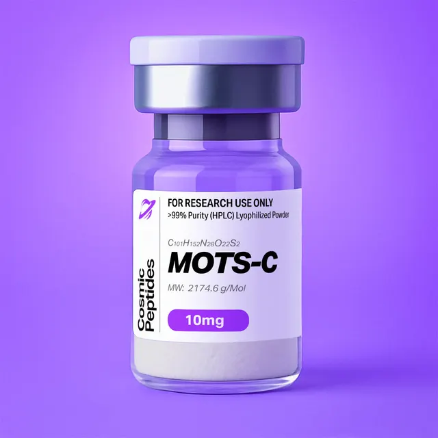
MOTS-c
Commonly researched with these items

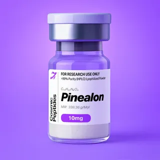

Patent-pending lot tracking: Complete transparency from production to vial.
| CAS Number | 1627580-64-6 |
|---|---|
| Molecular Formula | C101H152N28O22S2 |
| Molecular Weight | 2174.62 g/Mol |
| Purity | 99.87% |
| Lot Number | 26118 |
| Quantity | 10.22mg |
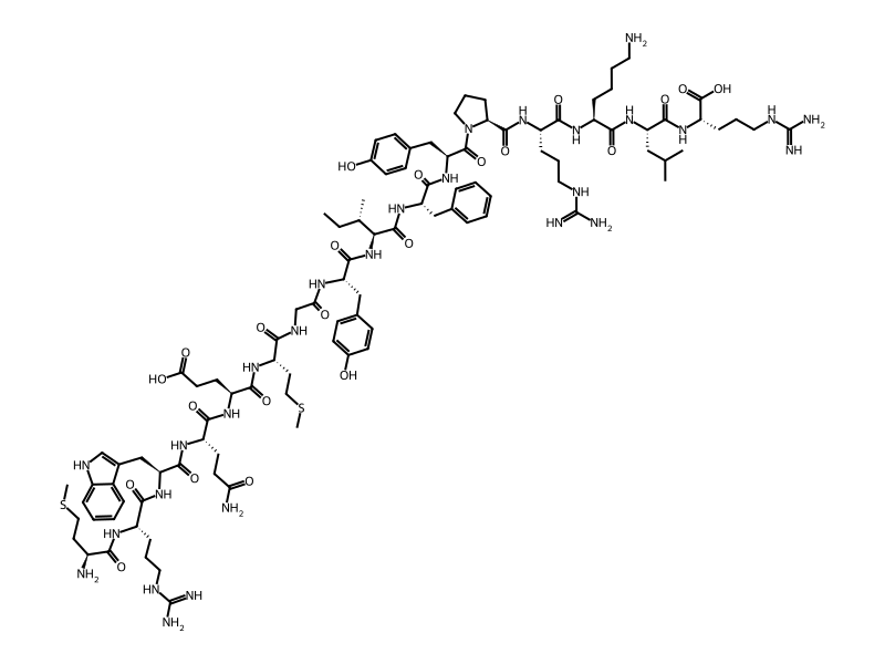
| Sequence | H-Met-Arg-Trp-Gln-Glu-Met-Gly-Tyr-Ile-Phe-Tyr-Pro-Arg-Lys-Leu-Arg-OH |
MOTS-c: The 16-Residue Peptide Encoded Inside the Mitochondrial 12S rRNA Gene
Disclaimer
Products described here are supplied for research use only and are not intended for diagnostic, therapeutic, or clinical application. All statements regarding biological activity reflect preclinical and in vitro findings exclusively and have not been evaluated by the U.S. Food and Drug Administration. These materials and related content are provided for educational and investigational purposes only and are intended solely for qualified researchers in laboratory and academic settings.
Key Facts
- Compound class
- Linear 16-residue mitochondrial-derived peptide (MDP), encoded by a short open reading frame inside the mitochondrial 12S rRNA gene MT-RNR1
- Sequence
H-Met-Arg-Trp-Gln-Glu-Met-Gly-Tyr-Ile-Phe-Tyr-Pro-Arg-Lys-Leu-Arg-OH(one-letter codeMRWQEMGYIFYPRKLR); all-L, free N-terminal amine and free C-terminal acid- Molecular formula
- C101H152N28O22S2
- Molecular weight
- 2174.62 g/mol (average); monoisotopic mass 2173.108 Da
- CAS number
- 1627580-64-6
- PubChem CID
- 146675088
- Genomic coordinates
- Nucleotides 1343–1390 of the revised Cambridge Reference Sequence (rCRS, NC_012920.1), in-frame TAG terminator at 1391–1393
- Physical form as supplied
- Lyophilized powder
- Lot traceability
- Every unit is tied to a tracked lot; the lot number, recorded purity and verified quantity of the lot on sale are shown live on this page, with its certificate of analysis.
Molecular Identity and Classification
MOTS-c — mitochondrial open reading frame of the twelve S rRNA type-c — is a hexadecapeptide, sequence MRWQEMGYIFYPRKLR. It belongs to the mitochondrial-derived peptides (MDPs): short polypeptides encoded inside mitochondrial genes long annotated only as ribosomal RNA. The first described was humanin, from a frame inside the 16S rRNA gene MT-RNR2; MOTS-c, reported by Lee and colleagues in 2015, comes from the 12S rRNA gene MT-RNR1. Further frames in the region have since been catalogued as the SHLP series.
The free peptide has the molecular formula C101H152N28O22S2, an average molecular weight of 2174.62 g/mol and a monoisotopic mass of 2173.108 Da. It is indexed under CAS number 1627580-64-6 and PubChem CID 146675088, with the systematic name L-methionyl-L-arginyl-L-tryptophyl-L-glutaminyl-L-α-glutamyl-L-methionylglycyl-L-tyrosyl-L-isoleucyl-L-phenylalanyl-L-tyrosyl-L-prolyl-L-arginyl-L-lysyl-L-leucyl-L-arginine.
MOTS-c is a medium-length linear peptide with no disulfide bond, no post-translational modification in its canonical form and no terminal capping: the N-terminal methionine retains a free α-amino group and the C-terminal arginine a free carboxylate. That matters when literature reports are compared, since amidated or acetylated analogues differ in net charge, chromatographic retention and observed mass by predictable increments (−0.984 Da for C-terminal amidation, +42.011 Da for N-terminal acetylation).
Encoding in the Mitochondrial Genome
The frame can be located exactly. In the rCRS (NC_012920.1), nucleotides 1343–1390 read ATG AGG TGG CAA GAA ATG GGC TAC ATT TTC TAC CCC AGA AAA CTA CGA — sixteen codons followed by a TAG terminator at 1391–1393. The frame sits inside MT-RNR1 (nucleotides 648–1601), nested within a structural RNA gene — one reason it escaped annotation for decades.
A chemical curiosity follows from which genetic code is applied. Vertebrate mitochondria use a variant code in which AGA and AGG are terminators rather than arginine codons; read with that code, the frame terminates at codon 2 (AGG) and again at codon 13 (AGA). The 16-residue sequence emerges only under the standard cytoplasmic code, consistent with the reported translation of the transcript outside the organelle. The arginines at positions 2, 13 and 16 are therefore not incidental: they are where the two codes disagree, and they carry most of the peptide's positive charge.
Structure and Chemical Features
MOTS-c is best read by walking along the chain. The N-terminal half, Met-Arg-Trp-Gln-Glu-Met, is the most reactive segment: both methionine thioethers and the single tryptophan indole sit here, the three side chains most prone to oxidation and alkylation, along with the only acidic side chain, Glu5. The centre, Gly-Tyr-Ile-Phe-Tyr, is a compact aromatic and aliphatic cluster behind a glycine hinge. The C-terminal segment, Pro-Arg-Lys-Leu-Arg, is strongly basic and constrained by the proline.
Four of the sixteen side chains are aromatic — one Trp, one Phe, two Tyr — high for a peptide of this length, which gives MOTS-c a usable UV chromophore (see Analytical Characterization), intrinsic tryptophan fluorescence and a tendency toward π-stacking and cation–π contacts. Against that core stand three arginines and one lysine: an aromatic centre with flanking cationic residues is the signature shared by many cationic, membrane-associating peptides, and the feature most often invoked when researchers ask how a cytoplasmically translated peptide reaches other compartments.
The proline at position 12 interrupts the chain's hydrogen-bonding capacity: its ring nitrogen bears no amide hydrogen, so the Tyr11–Pro12 bond is a tertiary amide and cannot donate a hydrogen bond. Because the cis/trans energy gap of an X–Pro bond is small, a measurable cis population persists in solution and exchanges slowly on the NMR timescale, doubling some resonances near the proline. The proline restricts the backbone locally while the Gly7 hinge does the opposite, so the molecule is flexible overall: in water, circular dichroism of peptides of this length and composition typically shows the random-coil signature — a strong negative band near 198 nm, little ellipticity between 208 and 222 nm — rather than helical minima.
Charge, Polarity and Isoelectric Behaviour
Protonation sites are the N-terminal α-amine (pKa ≈ 9), the ε-amine of Lys14 (≈ 10.5) and the guanidinium groups of Arg2, Arg13 and Arg16 (≈ 12.5); deprotonation sites are the C-terminal carboxylate (≈ 2), the γ-carboxylate of Glu5 (≈ 4.1) and, at high pH, the two tyrosine phenols (≈ 10.1). Summing these titration curves gives a net charge near +3 at pH 7.0 and a calculated isoelectric point near 10.5. At the pH of a typical reversed-phase mobile phase (0.1% trifluoroacetic acid, pH ≈ 2) the net charge rises to about +4.5, which is why MOTS-c ionizes efficiently in positive-mode electrospray and appears predominantly as triply and quadruply charged ions.
Hydropathy tells the complementary story. The Kyte–Doolittle grand average of hydropathicity (GRAVY) is −0.94, the three arginines and the lysine outweighing the Ile, Leu, Phe and Met side chains, and the computed XLogP is −3.9. With 31 hydrogen-bond donors, 29 acceptors, 73 rotatable bonds and a topological polar surface area near 890 Å2, MOTS-c is a highly polar, highly flexible solute: good aqueous solubility, no passive permeation of lipid bilayers, and handling questions that centre on adsorption and oxidation rather than dissolution.
Synthesis and Manufacturing Chemistry
MOTS-c is assembled by solid-phase peptide synthesis, the technique Merrifield introduced in 1963, in the Fmoc/tBu variant systematized for peptide work by Fields and Noble. Chain growth runs from the C-terminus: Arg16 is anchored to the resin and the remaining fifteen residues are added in reverse sequence order. A free C-terminal acid requires an acid-labile linker, so 2-chlorotrityl chloride or Wang resin is the usual choice; 2-chlorotrityl also suppresses racemization and diketopiperazine formation at the first residue.
The side-chain protection scheme is heavy: the three arginines as Fmoc-Arg(Pbf)-OH, the two tyrosines as Tyr(tBu), the lysine as Lys(Boc), the glutamate as Glu(OtBu) and the glutamine usually as Gln(Trt), which limits dehydration of the side-chain amide to a nitrile during activation. Tryptophan is best introduced as Fmoc-Trp(Boc)-OH, since an unprotected indole is readily alkylated by tert-butyl cations released during final deprotection. The two methionines carry no protecting group; their thioethers are defended instead by scavengers in the cleavage cocktail.
Three steps deserve the attention the literature on "difficult peptides" gives them. First, acylation of the proline nitrogen: coupling Tyr11 onto the secondary amine of Pro12 is intrinsically slower than onto a primary amine and benefits from a stronger activator (HATU or COMU rather than DIC/Oxyma) or a repeat coupling. Second, the arginine couplings: the bulky Pbf group slows acylation and Fmoc-Arg(Pbf)-OH is prone to δ-lactam formation on activation, so short pre-activation times and double couplings are routine. Third, the aromatic stretch Tyr8-Ile9-Phe10-Tyr11 drives on-resin aggregation through interchain hydrogen bonding; elevated-temperature coupling, polar aprotic co-solvents or a low-loading polyethylene-glycol resin are the standard countermeasures.
Final cleavage uses trifluoroacetic acid with scavengers chosen for Met and Trp: water plus a thiol or thioether such as 1,2-ethanedithiol, thioanisole or dimethyl sulfide traps the tert-butyl and Pbf cations that would otherwise alkylate the indole and thioethers, while triisopropylsilane serves as a hydride donor. The crude peptide is precipitated into cold diethyl ether, purified by preparative reversed-phase HPLC and lyophilized. Because trifluoroacetic acid is present throughout, the isolated solid is normally a trifluoroacetate salt unless exchanged for acetate or chloride — a choice that changes the mass of a weighed sample appreciably, as the worked calculations show.
Analytical Characterization
Identity and purity are established with a short panel of orthogonal methods. Reversed-phase HPLC on C18 with a water/acetonitrile gradient containing 0.1% trifluoroacetic acid is the workhorse; roughly 15–45% acetonitrile over 20–30 minutes places the main peak in a well-resolved region. Unlike very short peptides, MOTS-c has a genuine UV chromophore beyond the amide bond, so two wavelengths are informative: 214–220 nm for the backbone and 280 nm for the aromatic side chains. Comparing peak-area ratios between them is a quick way to spot a non-aromatic by-product hiding under the main peak.
The molar absorption coefficient follows the Pace additivity rule, ε280 = 5500 (Trp) + 2 × 1490 (Tyr) = 8480 M−1cm−1, so a 1 mg/mL solution of the free peptide gives A280 ≈ 3.90 in a 1 cm cell and a 100 µM solution A280 ≈ 0.85. Spectrophotometry is thus an independent concentration check — unavailable for sequences lacking Trp and Tyr — provided the sample is free of nucleic acid or scattering particulates. The single tryptophan also gives intrinsic fluorescence (excitation ≈ 280 nm, emission ≈ 350 nm in water, blue-shifting in less polar environments), making MOTS-c tractable for binding and partitioning work without a label.
Expected Mass Spectral Signatures
In positive-mode electrospray, five basic sites spread the peptide across several charge states. Calculated monoisotopic m/z values are 2174.115 [M+H]+, 1087.561 [M+2H]2+, 725.377 [M+3H]3+, 544.284 [M+4H]4+ and 435.629 [M+5H]5+, with 3+ and 4+ usually dominant from an acidified mobile phase. Isotope spacing confirms the assignment: 1.003 m/z for 1+, 0.502 for 2+, 0.334 for 3+, 0.251 for 4+.
One subtlety trips up newcomers to peptides of this size: with 101 carbon atoms, the monoisotopic peak is not the tallest in the envelope. Convolving natural isotope abundances over the formula gives A+0 81%, A+1 100%, A+2 72% and A+3 38% of the base peak, so the most abundant isotopologue sits one neutron above the monoisotopic ion, at 2174.111 Da for the neutral molecule — a number not to be mistaken for the average mass of 2174.62 g/mol, which it resembles only by coincidence. Where the envelope is unresolved, the monoisotopic mass has to be recovered from the intensity pattern rather than read off the tallest peak.
Tandem mass spectrometry gives a clean ladder. Because collision-induced dissociation preferentially cleaves the bond N-terminal to proline, the y5 ion at m/z 669.452 (PRKLR) is characteristically intense, with the complementary b11 at 1505.670. Other diagnostic singly charged fragments are b2 288.149, b3 474.228, y2 288.203 (LR), y3 416.298 (KLR) and y4 572.399 (RKLR). The near-coincidence of b2 and y2 — 0.054 Da apart, about 190 ppm — is a reminder to work at high resolution, since the two are easily conflated on a low-resolution instrument.
The modification masses to watch for are methionine oxidation, +15.995 Da per sulfoxide (monoisotopic 2189.103 for the mono-, 2205.098 for the bis-sulfoxide); tryptophan oxidation, also +15.995 Da but chromatographically distinguishable and often accompanied by +31.990 and +3.995 Da (kynurenine) species; and deamidation of Gln4, +0.984 Da. With two methionines and one tryptophan, an oxidized shoulder eluting slightly earlier than the main peak on C18 is the most likely purity defect in an aged sample, and the mass shift confirms it unambiguously. Integrating parent and oxidized peaks at 280 nm gives a rough extent of oxidation; integration at 214 nm is less reliable, because the oxidized indole's absorbance differs from the parent's.
Complementary methods complete the picture. Amino-acid analysis after 6 M HCl hydrolysis should return Arg 3, Met 2, Tyr 2 and one each of Gly, Ile, Leu, Phe, Pro and Lys, with Glx 2 rather than separate Gln and Glu because the glutamine side chain hydrolyses to glutamate, and with tryptophan absent because the indole is destroyed; a Trp determination needs alkaline hydrolysis or a spectrophotometric estimate. Ion chromatography or 19F NMR quantifies trifluoroacetate, Karl Fischer titration or thermogravimetry gives water content, and chiral amino-acid analysis confirms that no D-epimers arose during the arginine couplings. HPLC purity and peptide content are different quantities — the first is the fraction of UV-absorbing material that is the target, the second the fraction of the weighed powder that is peptide — and only the second converts milligrams into moles.
Physicochemical Properties
| Property | Value | Notes |
|---|---|---|
| Molecular formula | C101H152N28O22S2 | Free peptide; PubChem CID 146675088 |
| Average molecular weight | 2174.62 g/mol | From the formula, standard atomic weights |
| Monoisotopic mass | 2173.108 Da | Residue masses + H2O (18.0106 Da) |
| Most abundant isotopologue | 2174.111 Da (A+1) | A+0 81%, A+1 100%, A+2 72% |
| [M+2H]2+ / [M+3H]3+ / [M+4H]4+ | m/z 1087.561 / 725.377 / 544.284 | Monoisotopic, positive ESI |
| Residue count | 16 | Arg 3, Met 2, Tyr 2; one each of Gly, Gln, Glu, Ile, Leu, Lys, Phe, Pro, Trp |
| Net charge at pH 7.0 | approximately +3 | Five basic sites, two acidic sites |
| Net charge at pH 2.0 | approximately +4.5 | TFA-containing mobile phase |
| Isoelectric point (calculated) | ≈ 10.5 | Strongly basic |
| ε280 | 8480 M−1cm−1 | 1 Trp + 2 Tyr, Pace additivity rule |
| A280 of 1 mg/mL | ≈ 3.90 (1 cm path) | Free peptide basis |
| Trp fluorescence | λex ≈ 280 nm, λem ≈ 350 nm | Single Trp3; label-free |
| GRAVY (Kyte–Doolittle) | −0.94 | Net hydrophilic |
| XLogP / TPSA | −3.9 / ≈ 890 Å2 | PubChem computed descriptors |
| Hydrogen-bond donors / acceptors | 31 / 29 | PubChem computed descriptors |
| Rotatable bonds / heavy atoms | 73 / 153 | Conformationally flexible |
| Secondary structure in water | Predominantly random coil | Expected CD minimum near 198 nm |
| Solubility | Readily soluble in water and aqueous buffers | Consistent with +3 net charge, GRAVY −0.94 |
| Chemical liabilities | Met and Trp oxidation, Gln deamidation | +15.995, +15.995/+31.990, +0.984 Da |
Two entries in this table shape every experiment. At neutral pH MOTS-c is a polycation, so it adsorbs to negatively charged surfaces — borosilicate glass, untreated polystyrene, anionic filter membranes — and losses at low micromolar concentrations are real. And two thioethers plus an indole in sixteen residues make it far more sensitive to dissolved oxygen, trace metals and light than a peptide of similar size without them.
Research Context
Interest in MOTS-c rests on an idea rather than on any single assay: that the mitochondrial genome encodes small polypeptides acting as signalling molecules in the cell that hosts them. Before 2001 the thirteen protein-coding genes of mitochondrial DNA were taken to be a closed list of oxidative-phosphorylation subunits. Finding humanin within MT-RNR2, and then MOTS-c within MT-RNR1, reframed the two ribosomal RNA genes as dual-function loci and created the research category of mitochondrial-derived peptides, called "mitokines" in reviews of the field.
AMPK and One-Carbon Metabolism
The mechanism reported in the original 2015 description runs through folate-dependent one-carbon metabolism. In cultured cells, metabolomic profiling after exposure to the peptide showed accumulation of 5-methyltetrahydrofolate and of de novo purine-synthesis intermediates, consistent with inhibition at the folate-cycle step feeding the purine pathway. One of those intermediates, 5-aminoimidazole-4-carboxamide ribonucleotide (AICAR, also written ZMP), is an endogenous AMP mimetic: it binds the γ-subunit of AMP-activated protein kinase (AMPK) and increases phosphorylation of the activation-loop threonine, Thr172, on the catalytic α-subunit. That chain — folate-cycle inhibition, AICAR accumulation, AMPK activation — is why MOTS-c is of interest in cell-based energy-sensing work rather than as a receptor ligand.
AMPK is a heterotrimeric αβγ kinase whose γ-subunit binds AMP, ADP and ATP competitively, with further control by the upstream kinases LKB1 and CaMKK2. The standard in vitro read-outs follow from that architecture: immunoblotting for phospho-AMPKα Thr172 and for the canonical substrate acetyl-CoA carboxylase at Ser79, cellular AMP:ATP and ADP:ATP ratios by LC-MS, and extracellular-flux measurement of oxygen-consumption and acidification rates in cultured monolayers. Concentration–response series of roughly 0.1–10 µM with matched vehicle and counterion controls, plus time courses separating early phosphorylation from later transcriptional changes, are the usual experimental shape.
Nuclear Localisation Under Metabolic Stress
A second strand concerns where the peptide goes inside a cell. Kim and colleagues reported in 2018 that in cultured cells under metabolic stress — glucose restriction or an oxidative challenge — MOTS-c accumulates in the nucleus and associates with stress-responsive transcription-factor complexes, with changes in the expression of nuclear genes carrying antioxidant-response elements. Structurally this is unsurprising for a peptide whose C-terminal region is Pro-Arg-Lys-Leu-Arg: a short basic cluster of that form resembles the classical monopartite nuclear-localisation signal consensus K(K/R)X(K/R), and the +3 net charge is concentrated there. Immunofluorescence or fractionation with immunoblotting, chromatin immunoprecipitation and reporter assays follow this behaviour in cell culture, with basic-residue substitution variants as the structural control.
Both strands make MOTS-c useful chiefly as a tool compound for in vitro cell biology and biophysics: a defined, synthetically accessible peptide with intrinsic tryptophan fluorescence, a documented route to AMPK activation and a documented nuclear distribution under stress, against which analogues and variants are compared.
Sequence Variation and Its Analytical Consequences
Because the reading frame lies inside a well-sequenced region of mitochondrial DNA, nucleotide substitutions map onto the peptide predictably. The most-discussed is m.1382A>C: codon 14 occupies nucleotides 1382–1384 and reads AAA, encoding Lys14, and the A-to-C substitution converts it to CAA, encoding glutamine. The resulting peptide, written K14Q, has the sequence MRWQEMGYIFYPRQLR and differs from the reference peptide in two chemically meaningful ways.
First, charge: replacing lysine with glutamine removes one protonatable amine, lowering the net charge at pH 7 from about +3 to about +2 and shifting the calculated isoelectric point downward, which changes retention on reversed-phase and ion-exchange columns and alters the charge-state distribution in electrospray. Second, mass: lysine and glutamine are near-isobaric. The reference peptide is 2173.108 Da monoisotopic and the K14Q variant 2173.071 Da — 0.0364 Da, or 16.7 ppm, apart. Accurate mass better than 5 ppm distinguishes them comfortably, but baseline-resolving two co-eluting species that close at the 3+ charge state requires a resolving power near 60,000. Where the distinction matters, tandem MS is safer: the y3 ion shifts from 416.298 (KLR) to 416.262 (QLR), with y4 and b14 shifting correspondingly while fragments outside position 14 are unchanged. MOTS-c is a textbook illustration of the Lys/Gln near-isobaricity that is a standing caution in peptide mass spectrometry.
Worked Calculations for the Bench
Molar mass from the sequence. Adding the residue masses of MRWQEMGYIFYPRKLR and one water reproduces the catalogue figures: the monoisotopic residue sum of 2155.097 Da plus 18.011 Da gives 2173.108 Da, and the same arithmetic with average residue masses gives 2174.62 g/mol. A disagreement beyond about 0.1 g/mol points to a different salt form, different terminal chemistry, or an error in the record.
Counterion burden. MOTS-c has five basic sites, so a fully paired trifluoroacetate salt is the peptide with five equivalents of trifluoroacetic acid: the proton sits on the peptide and the counterion is CF3CO2− (113.01 g/mol), together adding 114.02 g/mol per equivalent. Five equivalents give a formula weight of 2744.7 g/mol, 79.2% of it peptide; four give 2630.7 g/mol and 82.7%; three give 2516.7 g/mol and 86.4%. Exchanged to acetate (60.05 g/mol per equivalent) the same stoichiometries give 87.9%, 90.1% and 92.3%. Salt form alone can move the peptide fraction of a weighed powder by more than ten percentage points, before water content is considered.
Molar concentrations. On a free-peptide basis, 1 mg is 459.9 nmol and a 1 mM solution contains 2.175 mg/mL. A 10 mg portion of a lot whose peptide content is 85% by weight holds 8.5 mg of peptide, or 3.91 µmol; in 3.91 mL that is exactly 1.00 mM, whereas assuming 100% peptide gives 0.85 mM — a 15% systematic error carried into every downstream dilution. Where exact molarity matters, prepare by weight and verify spectrophotometrically with ε280 = 8480 M−1cm−1.
Oxidation accounting. A stored stock whose LC-MS trace shows a second species 15.995 Da heavier at every charge state — for example m/z 730.708 alongside 725.377 at 3+ — carries one methionine or the tryptophan as its oxide.
Related Research Compounds
In cell culture, MOTS-c is often studied alongside other defined reagents that approach the same chemistry from different directions. The mitochondria-targeted tetrapeptide SS-31 is an instructive comparison: also short, strongly cationic and aromatic, but its cationic–aromatic alternation was designed for accumulation at the inner mitochondrial membrane and association with cardiolipin, whereas MOTS-c is a natural gene product whose cationic cluster is confined to its C-terminus.
Because the reported pathway runs through folate-dependent one-carbon chemistry and nucleotide pools, reagents acting on adjacent nodes are frequent companions in experimental design. NAD is the central redox cofactor whose pools these pathways intersect, and 5-amino-1MQ inhibits nicotinamide N-methyltransferase, which consumes S-adenosylmethionine to methylate nicotinamide and so sits at the junction of methyl-group and nicotinamide metabolism. Together they let a researcher perturb methyl-donor supply, nucleotide synthesis and redox pools independently.
Laboratory Handling and Storage
The solid. Lyophilized MOTS-c should be kept tightly closed at −20 °C or colder, protected from light and moisture. Peptide lyophilisates are hygroscopic, and a container taken straight from a freezer condenses atmospheric water on the cold powder; let it equilibrate to room temperature in a desiccator before opening, then close and return it promptly. With two methionines and a tryptophan, headspace oxygen matters: for material opened repeatedly, backfilling with argon or nitrogen and storing single-use aliquots is worth the effort.
Dissolution. The peptide dissolves readily in high-purity water and aqueous buffers; its basic character means mildly acidic water (0.01–0.1% acetic acid, say) gives the fastest, cleanest dissolution and the best stock stability. Avoid stocks above about pH 8, where tyrosine and methionine oxidation accelerate and the peptide's charge is reduced. Dimethyl sulfoxide is not required for this sequence and is best avoided, since it can drive thioether oxidation on standing.
Stocks and aliquots. Prepare stocks at 1–10 mM, correcting the weighed mass for peptide content; filter through a low-protein-binding 0.22 µm membrane for cell-culture work; divide into single-use aliquots in low-binding polypropylene; freeze at −80 °C. Repeated freeze–thaw cycles and prolonged storage of dilute working solutions are the commonest causes of drifting results with cationic peptides, so prepare very low concentrations fresh from a concentrated stock, and consider a carrier protein in the assay buffer where the design permits: adsorptive losses at nanomolar levels can exceed the peptide intended to be present.
Oxidation and counterion controls. Degassed buffers, a chelator such as 0.1 mM EDTA for trace transition metals, amber tubes or foil and minimal headspace all reduce methionine and tryptophan oxidation; reducing agents suppress sulfoxide formation but add their own interferences. Trifluoroacetate carried over from purification is not inert in cell culture, as Cornish and colleagues showed for cultured cell proliferation, so a vehicle control matched for counterion concentration is good practice and acetate-exchanged material is preferable for sensitive assays.
Re-verification. A short LC-MS check of a stored stock — the 3+ ion at m/z 725.38, no +15.995 Da satellite — confirms identity and integrity in minutes. Each lot's certificate of analysis, shown on this page, records the analytical results for that material.
Frequently Asked Research Questions
- What is MOTS-c?
- MOTS-c is a 16-residue peptide, sequence
MRWQEMGYIFYPRKLR, encoded by a short open reading frame inside the mitochondrial 12S rRNA gene MT-RNR1. It is a mitochondrial-derived peptide with molecular formula C101H152N28O22S2 and an average molecular weight of 2174.62 g/mol. - What are the CAS number and PubChem CID for MOTS-c?
- CAS 1627580-64-6 and PubChem CID 146675088, whose record lists the same formula, C101H152N28O22S2, and an average mass of 2174.6 g/mol.
- Where exactly is MOTS-c encoded in mitochondrial DNA?
- At nucleotides 1343–1390 of the revised Cambridge Reference Sequence (NC_012920.1): sixteen codons followed by a TAG terminator at 1391–1393, entirely within MT-RNR1 (nucleotides 648–1601).
- Why does the reading frame only make sense with the standard genetic code?
- Codon 2 is AGG and codon 13 is AGA. Both are terminators in the vertebrate mitochondrial code, so the frame would stop at residue 2; under the standard cytoplasmic code they encode arginine, which is how the full 16-residue sequence arises — consistent with translation of the transcript outside the organelle.
- What masses should MOTS-c give by mass spectrometry?
- Monoisotopic mass 2173.108 Da; monoisotopic m/z 1087.561 [M+2H]2+, 725.377 [M+3H]3+, 544.284 [M+4H]4+. The most abundant isotopologue of the neutral molecule is the A+1 peak at 2174.111 Da, not the monoisotopic peak, because the molecule has 101 carbon atoms.
- Can I determine the concentration of a MOTS-c solution by UV absorbance?
- Yes. One tryptophan and two tyrosines give a calculated ε280 of 8480 M−1cm−1 by the Pace additivity rule. A 1 mg/mL solution of the free peptide reads about A280 = 3.90 in a 1 cm cell, and A280 = 0.85 is roughly 100 µM.
- What is the net charge and isoelectric point of MOTS-c?
- Three arginines, one lysine and the N-terminal amine give five basic sites against one glutamate side chain and the C-terminal carboxylate: net charge about +3 at pH 7, calculated isoelectric point near 10.5, and about +4.5 at the pH of a TFA-containing mobile phase.
- Which degradation products are most likely in a stored sample?
- Oxidation products. Each methionine sulfoxide or tryptophan oxide adds 15.995 Da — monoisotopic 2189.103 for one oxidation, 2205.098 for two — and deamidation of Gln4 adds 0.984 Da. Oxidized species typically elute slightly earlier than the parent peak on C18.
- What is the difference between HPLC purity and peptide content for this peptide?
- HPLC purity is the fraction of UV-absorbing material that is MOTS-c; peptide content is the fraction of the weighed powder that is peptide rather than counterion, water or salt. With five basic sites, a fully trifluoroacetate-paired salt is only about 79% peptide by mass, so molar concentrations calculated without that correction can be low by fifteen percent or more.
- Does MOTS-c have a defined secondary structure?
- Not in plain water. With 73 rotatable bonds, a glycine hinge at position 7 and a proline at position 12 it is conformationally flexible, and circular dichroism of peptides of this length and composition typically shows the random-coil signature, a strong negative band near 198 nm. Ordered conformations would be sought in membrane-mimetic or crowded environments.
- Why is the Lys14-to-Gln variant difficult to detect by mass alone?
- Lysine and glutamine are near-isobaric. The K14Q peptide from the m.1382A>C substitution is 2173.071 Da monoisotopic against 2173.108 Da for the reference peptide: 0.0364 Da, or 16.7 ppm. Accurate mass below 5 ppm distinguishes them, resolving co-eluting species needs a resolving power near 60,000, and the cleanest discrimination is the y3 fragment, 416.298 against 416.262.
- How should MOTS-c be stored and dissolved in the laboratory?
- Keep the lyophilized solid tightly closed at −20 °C or colder, dark and dry, and warm it in a desiccator before opening. Dissolve in high-purity or mildly acidified water, prepare 1–10 mM stocks corrected for peptide content, aliquot into low-binding polypropylene and freeze at −80 °C. Avoid buffers above about pH 8 and repeated freeze–thaw cycles.
Sources & Further Reading
- National Center for Biotechnology Information. PubChem Compound Summary for CID 146675088, MOTS-c.
- Lee C, Zeng J, Drew BG, et al. The mitochondrial-derived peptide MOTS-c promotes metabolic homeostasis and reduces obesity and insulin resistance. Cell Metabolism 2015;21(3):443–454. doi:10.1016/j.cmet.2015.02.009
- Kim KH, Son JM, Benayoun BA, et al. The mitochondrial-encoded peptide MOTS-c translocates to the nucleus to regulate nuclear gene expression in response to metabolic stress. Cell Metabolism 2018;28(3):516–524.e7. doi:10.1016/j.cmet.2018.06.008
- Kim SJ, Xiao J, Wan J, et al. Mitochondrially derived peptides as novel regulators of metabolism. The Journal of Physiology 2017;595(21):6613–6621. doi:10.1113/JP274472
- Lee C, Yen K, Cohen P. Humanin: a harbinger of mitochondrial-derived peptides? Trends in Endocrinology & Metabolism 2013;24(5):222–228. doi:10.1016/j.tem.2013.01.005
- Anderson S, Bankier AT, Barrell BG, et al. Sequence and organization of the human mitochondrial genome. Nature 1981;290(5806):457–465. doi:10.1038/290457a0
- Barrell BG, Bankier AT, Drouin J. A different genetic code in human mitochondria. Nature 1979;282(5735):189–194. doi:10.1038/282189a0
- Hardie DG, Ross FA, Hawley SA. AMPK: a nutrient and energy sensor that maintains energy homeostasis. Nature Reviews Molecular Cell Biology 2012;13(4):251–262. doi:10.1038/nrm3311
- Corton JM, Gillespie JG, Hawley SA, et al. 5-Aminoimidazole-4-carboxamide ribonucleoside. A specific method for activating AMP-activated protein kinase in intact cells? European Journal of Biochemistry 1995;229(2):558–565. doi:10.1111/j.1432-1033.1995.tb20498.x
- Lange A, Mills RE, Lange CJ, et al. Classical nuclear localization signals: definition, function, and interaction with importin α. Journal of Biological Chemistry 2007;282(8):5101–5105. doi:10.1074/jbc.R600026200
- Merrifield RB. Solid phase peptide synthesis. I. The synthesis of a tetrapeptide. Journal of the American Chemical Society 1963;85(14):2149–2154. doi:10.1021/ja00897a025
- Fields GB, Noble RL. Solid phase peptide synthesis utilizing 9-fluorenylmethoxycarbonyl amino acids. International Journal of Peptide and Protein Research 1990;35(3):161–214. doi:10.1111/j.1399-3011.1990.tb00939.x
- Paradís-Bas M, Tulla-Puche J, Albericio F. The road to the synthesis of "difficult peptides". Chemical Society Reviews 2016;45(3):631–654. doi:10.1039/C5CS00680E
- Senko MW, Beu SC, McLafferty FW. Determination of monoisotopic masses and ion populations for large biomolecules from resolved isotopic distributions. Journal of the American Society for Mass Spectrometry 1995;6(4):229–233. doi:10.1016/1044-0305(95)00017-8
- Breci LA, Tabb DL, Yates JR, et al. Cleavage N-terminal to proline: analysis of a database of peptide tandem mass spectra. Analytical Chemistry 2003;75(9):1963–1971. doi:10.1021/ac026359i
- Pace CN, Vajdos F, Fee L, et al. How to measure and predict the molar absorption coefficient of a protein. Protein Science 1995;4(11):2411–2423. doi:10.1002/pro.5560041120
- Vogt W. Oxidation of methionyl residues in proteins: tools, targets, and reversal. Free Radical Biology and Medicine 1995;18(1):93–105. doi:10.1016/0891-5849(94)00158-G
- Simat TJ, Steinhart H. Oxidation of free tryptophan and tryptophan residues in peptides and proteins. Journal of Agricultural and Food Chemistry 1998;46(2):490–498. doi:10.1021/jf970818c
- Greenfield NJ. Using circular dichroism spectra to estimate protein secondary structure. Nature Protocols 2006;1(6):2876–2890. doi:10.1038/nprot.2006.202
- Cornish J, Callon KE, Lin CQ, et al. Trifluoroacetate, a contaminant in purified proteins, inhibits proliferation of osteoblasts and chondrocytes. American Journal of Physiology-Endocrinology and Metabolism 1999;277(5):E779–E783. doi:10.1152/ajpendo.1999.277.5.E779
Last reviewed:
In the research library
Everything the library holds on MOTS-c, and the methods behind the numbers on its certificate.
- MOTS-c reference entry Identity, calculated properties, the mass spectrometry ions to expect and analytical notes, from the structure.
- Check a certificate against this sequence Paste the masses from a spectrum or a certificate; the mass-match tool compares them with what this sequence should give.
- Peptide calculator, with this sequence Formula, average and monoisotopic mass, charge across pH, isoelectric point and absorbance, recalculated as you edit.
- Mitochondrial-derived peptides The family side by side: sequences aligned, each member's relation to the reference peptide, and how analysis tells them apart.
- How to read a certificate of analysis What a peptide certificate of analysis should contain, how to check that it belongs to the material in hand, how to read its identity and purity results, and the signs that it deserves a second look.
- HPLC purity analysis of peptides How reversed-phase HPLC measures the chromatographic purity of a peptide, what the figure includes, and what it leaves out.
- Mass spectrometry for peptide identity How mass spectrometry confirms that a peptide is the intended sequence: electrospray and MALDI, working out the expected mass, reading charge states and adducts, sequence confirmation, and the limits of the method.
- Net peptide content and purity: two different numbers Why a peptide that is 99% pure by HPLC can be only 75–85% peptide by weight: what the powder contains, how each part is measured, and how the two figures combine.
- Preparing peptide solutions in the laboratory From a freeze-dried vial to a solution of known concentration: the arithmetic, the solvent, the charge chemistry and the handling, with the errors each step can introduce and the article that covers it.
- Peptide stability and storage How peptides degrade, chemically and physically, why the dry solid lasts far longer than a solution, which sequences are most at risk, and the storage and handling that slow it down.