DSIP
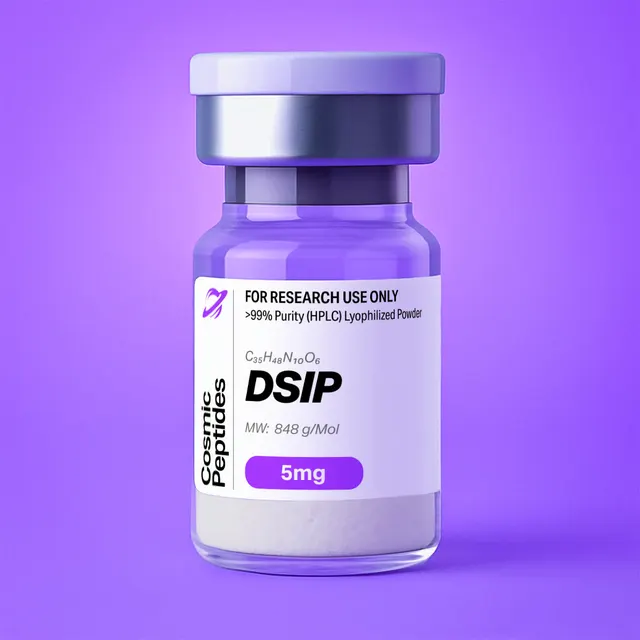
DSIP
Commonly researched with these items
Patent-pending lot tracking: Complete transparency from production to vial.
DSIP (Delta Sleep-Inducing Peptide): An Acidic, Tryptophan-Initiated Nonapeptide, H-Trp-Ala-Gly-Gly-Asp-Ala-Ser-Gly-Glu-OH
Disclaimer
Products described here are supplied for research use only and are not intended for diagnostic, therapeutic, or clinical application. All statements regarding biological activity reflect preclinical and in vitro findings exclusively and have not been evaluated by the U.S. Food and Drug Administration. These materials and related content are provided for educational and investigational purposes only and are intended solely for qualified researchers in laboratory and academic settings.
Key Facts
- Compound class
- Linear, unmodified nonapeptide; acidic (two side-chain carboxylic acids plus the C-terminal acid, no basic side chains)
- Sequence
H-Trp-Ala-Gly-Gly-Asp-Ala-Ser-Gly-Glu-OH(one-letter code: WAGGDASGE); the six chiral residues of the native sequence are L-configured, the three glycines are achiral- Molecular formula
- C35H48N10O15
- Molecular weight
- 848.8 g/mol (average; 848.82 g/mol to two decimals); monoisotopic mass 848.3301 Da
- CAS number
- 62568-57-4
- PubChem CID
- 68816 (L-configured structure); International Nonproprietary Name emideltide
- Physical form as supplied
- Lyophilized powder
- Lot traceability
- Every unit is tied to a tracked lot; the lot number, recorded purity and verified quantity of the lot on sale are shown live on this page, with its certificate of analysis.
Molecular Identity and Classification
Delta Sleep-Inducing Peptide, abbreviated DSIP, is the nonapeptide tryptophyl-alanyl-glycyl-glycyl-aspartyl-alanyl-seryl-glycyl-glutamic acid, written H-Trp-Ala-Gly-Gly-Asp-Ala-Ser-Gly-Glu-OH or, in one-letter code, WAGGDASGE. Its molecular formula is C35H48N10O15, its average molecular weight 848.8 g/mol and its monoisotopic mass 848.3301 Da. It is registered under CAS number 62568-57-4, carries the International Nonproprietary Name emideltide, and appears in PubChem as compound 68816, whose IUPAC name specifies six (2S) stereocentres: one each for Trp1, Ala2, Asp5, Ala6, Ser7 and Glu9. A second PubChem record, CID 3623358, describes the same connectivity with the stereocentres left undefined; it is a database convention for an unspecified stereoisomer mixture and not a description of the native peptide.
The name is historical and is kept as the conventional identifier; it describes neither a molecular target nor a mechanism. Schoenenberger and Monnier in Basel reported the isolation and characterization of the peptide in 1977. The sequence was established by amino-acid analysis and sequencing of the isolated material, and the nonapeptide was then synthesized so that natural and synthetic material could be compared. That combination, isolation followed by total synthesis of the assigned structure, is the classical proof of a peptide's identity, and every synthetic DSIP sample in use today traces back to it.
Classification is unusual for a peptide of this size. Most bioactive peptides are defined by their precursor protein and the cleavage sites that release them; for DSIP, no gene encoding the sequence and no precursor protein from which it is cut has been identified, a point emphasised in the 2006 review by Kovalzon and Strekalova, who described the molecule as a still unresolved riddle. DSIP is therefore classified chemically, as a short, acidic, glycine-rich linear peptide with a single aromatic residue, and pharmacologically only provisionally: no receptor has been characterised for it. For a research laboratory this makes the molecular identity of the material especially important, because there is no receptor-binding or gene-expression assay that can serve as an independent identity check.
Structure and Chemical Features
Reading the sequence from the N-terminus, DSIP divides naturally into three chemical regions. The N-terminal tryptophan carries the free α-amine, the only basic group in the molecule, together with the indole side chain, which is the only aromatic and the only strongly UV-absorbing and fluorescent group. The central Gly-Gly-Asp-Ala segment (residues 3 to 6) is flexible, because glycine lacks a side chain and can adopt backbone dihedral angles that are forbidden to other residues; the aspartic acid in this segment introduces the first negative charge. The C-terminal Ser-Gly-Glu tail contains the single hydroxyl group (serine), the glutamic acid side chain and the C-terminal carboxylate, so two of the molecule's three negative charges sit within the last three residues.
Three of the nine residues are glycine and two are alanine, so more than half of the chain carries either no side chain or a methyl group. This composition gives DSIP a high conformational freedom: it has 25 rotatable bonds by PubChem's computation and no proline, disulfide or cyclic constraint. In aqueous solution a peptide of this composition is expected to populate an ensemble of extended and turn-like conformations rather than a single folded structure, which is why DSIP is treated in the laboratory as a flexible, highly solvated chain.
The charge pattern is the other defining feature. At pH 7 the N-terminal amine is protonated (+1), while the Asp5 β-carboxyl, the Glu9 γ-carboxyl and the C-terminal α-carboxyl are deprotonated (−1 each), giving a net charge of about −2. The indole N–H of tryptophan does not ionise in the aqueous pH range. Using standard side-chain pKa values (Asp about 3.9, Glu about 4.1), the calculated isoelectric point falls between about 2.9 and 3.6 depending on the pKa set chosen for the termini. DSIP is therefore an acidic peptide, the mirror image of basic peptides such as KPV (calculated pI around 10), and this governs its chromatographic retention, its ionisation in mass spectrometry and its interaction with surfaces.
Chemically Reactive Sites
Four positions in the sequence deserve attention because they define how DSIP can change chemically during synthesis, storage or an experiment. Trp1: the electron-rich indole is the most oxidisable group in the molecule; oxidation gives hydroxytryptophan or oxindolylalanine (+15.995 Da), N-formylkynurenine (+31.990 Da) and kynurenine (+3.995 Da). Asp5-Ala6: the aspartyl side-chain carboxyl can attack the following backbone amide to form a five-membered succinimide (aspartimide, −18.011 Da), which reopens to a mixture of the native α-linked and the isomeric β-linked (isoaspartyl) peptide and can also racemise the aspartate. Ser7: the only hydroxyl group and therefore the only possible site of phosphorylation; a phosphoserine analogue of DSIP is well known in the literature. The N-terminal amine: the free α-amine is the attachment point for aminopeptidases and the reactive group in labelling, acylation or reductive amination reactions.
Synthesis and Manufacturing Chemistry
Contemporary DSIP is made by Fmoc-based solid-phase peptide synthesis (SPPS), the solid-phase approach whose Fmoc variant is reviewed in depth by Fields and Noble. For the C-terminal free acid, synthesis begins with Fmoc-Glu(OtBu) attached to an acid-labile support such as Wang or 2-chlorotrityl resin, and the chain is extended towards the N-terminus in eight further coupling cycles: Gly, Ser(tBu), Ala, Asp(OtBu), Gly, Gly, Ala and finally Trp, usually as Fmoc-Trp(Boc)-OH. Each cycle removes the Fmoc group with a secondary amine (typically 20% piperidine in dimethylformamide) and couples the next activated amino acid with reagents such as DIC/Oxyma or HATU.
The sequence is short and lacks the hydrophobic stretches that cause on-resin aggregation, so assembly is generally straightforward, but two side reactions are specific to it. First, the Asp-Ala junction is exposed to repeated exposures to piperidine after Asp5 is incorporated, and base-catalysed aspartimide formation can occur. Mergler and colleagues systematically compared Asp side-chain protecting groups, backbone protection and Fmoc-removal conditions and showed how strongly these choices determine the level of aspartimide-related by-products; for DSIP, standard OtBu protection with short deprotection times, or the addition of an acidic additive to the piperidine solution, keeps this by-product low. Because the reopened isoaspartyl peptide has exactly the same mass as DSIP, it must be controlled chromatographically rather than by mass spectrometry.
Second, the tryptophan indole is vulnerable during the final acidolytic cleavage. Trifluoroacetic acid (TFA) releases tert-butyl cations from the Asp, Glu and Ser protecting groups, and these electrophiles can alkylate the indole ring. Fields and Fields examined how to minimise this tryptophan alkylation after Fmoc SPPS; side-chain Boc protection of the indole and a scavenger-rich cleavage cocktail are the standard countermeasures. DSIP has one advantage here: it contains no arginine, so the sulfonyl-based Arg protecting groups (Pmc, Pbf), a major source of indole modification in many Trp-containing sequences, are absent from the synthesis. A typical cleavage for DSIP therefore uses TFA with water and triisopropylsilane (and, where needed, a thiol scavenger), followed by precipitation in cold ether, preparative reversed-phase HPLC and lyophilization.
Solution-phase routes are also documented. Wang and Xu described a one-pot liquid-phase synthesis of DSIP and its 5-Phe analogue using Fmoc-protected amino acid pentafluorophenyl esters, an approach that avoids a resin altogether and illustrates how active-ester chemistry handles a sequence of this length. Whatever the route, the purified peptide is obtained as a salt of the counterion present in the final purification buffer. Because DSIP has only one basic group, the N-terminal amine, at most about one equivalent of trifluoroacetate is associated with each peptide molecule, a far smaller counterion burden than for basic peptides with several amines.
Analytical Characterization
Reversed-phase HPLC. DSIP is very polar (computed XLogP3 −7.1) and elutes early on C18 columns. Analysts typically start a gradient at low acetonitrile content (for example 0–5% acetonitrile in water with 0.1% TFA) and run a shallow ramp; under acidic mobile-phase conditions the three carboxyl groups are largely protonated, which increases retention. Detection at 214–220 nm captures the peptide bonds, and detection at 280 nm is selective for the tryptophan indole. Comparing the two channels is a useful diagnostic: a peak that absorbs at 214 nm but not at 280 nm cannot contain an intact tryptophan, which flags truncated sequences lacking Trp1 or by-products from oxidised indole. TFA sharpens peaks; formic acid gives better MS sensitivity with slightly broader peaks.
Mass spectrometry. In positive-mode electrospray ionization DSIP gives the protonated molecule [M+H]+ at m/z 849.337 and the doubly protonated ion [M+2H]2+ at m/z 425.172; a sodium adduct [M+Na]+ at m/z 871.319 is common because the acidic side chains bind sodium. Because DSIP is acidic, negative-mode ionization works well and gives [M−H]− at m/z 847.323 and [M−2H]2− at m/z 423.158. With 35 carbon atoms, the first isotope peak (M+1) is roughly 42% of the monoisotopic peak, a pattern that confirms the carbon count in high-resolution spectra.
Tandem MS sequence ladders follow the nomenclature of Roepstorff and Fohlman. Fragmentation of [M+H]+ produces b ions from the N-terminus (b2 m/z 258.124, b3 315.145, b4 372.167, b5 487.194, b6 558.231, b7 645.263, b8 702.284) and y ions from the C-terminus (y1 148.060, y2 205.082, y3 292.114, y4 363.151, y5 478.178, y6 535.199, y7 592.221, y8 663.258). The tryptophan immonium ion at m/z 159.092 is a characteristic low-mass marker. Because DSIP has no arginine, lysine or histidine to sequester the ionising proton, the proton is mobile and fragmentation is spread along the backbone, which usually gives well-populated b and y series and makes full sequence confirmation straightforward.
UV quantitation. Tryptophan's molar absorption coefficient at 280 nm, 5,500 M−1 cm−1 in the widely used values of Pace and colleagues, makes DSIP one of the few short peptides that can be quantified directly by absorbance. A solution containing 1.000 mg/mL of DSIP (as free peptide) is 1.178 mM, and its expected A280 in a 1 cm cell is 1.178 × 10−3 M × 5,500 M−1 cm−1 = 6.48. A 0.100 mg/mL working solution therefore reads about 0.65, within the linear range. Measuring A280 of a weighed solution and comparing it with this value gives a quick estimate of peptide content.
Purity versus peptide content. HPLC purity reports what fraction of the UV-absorbing material is DSIP; peptide content reports what fraction of the weighed powder is peptide at all, as opposed to counterion, water and residual salts. As a worked example, the mono-trifluoroacetate salt of DSIP has a formula weight of 848.82 + 114.02 = 962.85 g/mol, so even a perfectly anhydrous TFA salt is only 88.2% peptide by mass; the mono-acetate salt (848.82 + 60.05 = 908.88 g/mol) is 93.4% peptide. Residual water lowers these values further. Amino-acid analysis after acid hydrolysis (expected ratios Gly 3 : Ala 2 : Asp 1 : Ser 1 : Glu 1, with tryptophan determined separately because it is destroyed by standard 6 M HCl hydrolysis) or quantitative NMR gives an absolute figure when exact molar concentrations matter.
Other techniques. Chiral amino-acid analysis checks that no D-aspartate or other D-isomer was formed; ion chromatography or 19F NMR quantifies trifluoroacetate; Karl Fischer titration measures water. Tryptophan fluorescence (excitation 280–295 nm, emission maximum near 350 nm in water for a solvent-exposed indole) provides a sensitive probe for concentration and local environment.
Physicochemical Properties
| Property | Value | Notes |
|---|---|---|
| Molecular formula | C35H48N10O15 | Free peptide, without counterions |
| Average molecular weight | 848.8 g/mol | PubChem CID 68816; 848.82 by calculation |
| Monoisotopic mass | 848.3301 Da | For high-resolution MS |
| [M+H]+ / [M+2H]2+ | m/z 849.337 / 425.172 | Positive-mode ESI |
| [M−H]− / [M−2H]2− | m/z 847.323 / 423.158 | Negative-mode ESI, favoured by the acidic side chains |
| Heavy atoms | 60 | Computed |
| Stereocentres | 6 (all S in the native peptide) | Three glycines are achiral |
| Net charge at pH 7 | about −2 | One amine; three carboxylates |
| Isoelectric point | about 2.9–3.6 (calculated) | Depends on the pKa set; acidic peptide |
| XLogP3 | −7.1 | Extremely hydrophilic |
| Topological polar surface area | 407 Å2 | Computed |
| Hydrogen-bond donors / acceptors | 14 / 16 | Computed |
| Rotatable bonds | 25 | Computed; no conformational constraints |
| Molar absorption coefficient, 280 nm | 5,500 M−1 cm−1 | One tryptophan; A280 of 1 mg/mL about 6.48 (1 cm) |
| Solubility | Freely soluble in water and near-neutral aqueous buffers | Lowest near the isoelectric point |
The physicochemical profile follows directly from the composition. Three carboxylic acids, one amine and nine amide bonds make DSIP far more polar than its size alone would suggest: its computed partition coefficient of −7.1 and polar surface area of 407 Å2 place it at the hydrophilic extreme even among peptides, so it does not cross lipid membranes by passive diffusion. Solubility is high at neutral and mildly basic pH, where the molecule carries a net negative charge, and lowest near its isoelectric point around pH 3, a fact worth remembering when diluting a stock into strongly acidic HPLC mobile phases at high concentration.
In solution, indole oxidation is accelerated by light, dissolved oxygen and trace metals, and the Asp5 aspartimide pathway by basic pH and heat. DSIP contains no cysteine or methionine, so disulfide scrambling and methionine sulfoxide formation do not arise. Lyophilized DSIP is hygroscopic, as is typical for a peptide rich in carboxyl groups.
Research Context
Lacking a known gene or receptor, in vitro DSIP research has followed three lines: the molecular origin of DSIP-like material, the peptide's stability in biological matrices, and the enzymes it interacts with. The broad literature up to the mid-1980s is collected in the review by Graf and Kastin (1986), and the open questions are summarised by Kovalzon and Strekalova (2006).
DSIP-Like Immunoreactivity and the TSC22D3 Protein
Antisera raised against synthetic DSIP were used from the 1980s to look for the peptide in tissue extracts. Graf and Kastin reported that DSIP-like immunoreactive material exists in large forms that can be dissociated, suggesting that the immunoreactivity is carried, at least in part, by larger molecules rather than by the free nonapeptide. The search for such a carrier led Sillard and colleagues in 1993 to purify a 77-residue peptide from brain extract that was recognised by an anti-DSIP antiserum and contained a leucine-zipper motif. They named it DSIP-immunoreactive peptide (DIP).
The structural biology of DIP was solved by Seidel and colleagues in 1997: their NMR solution structure showed a homologue of the product of the shortsighted gene, a member of the TSC-22 family of leucine-zipper proteins. In the same year, D'Adamio and colleagues identified the orthologous protein as glucocorticoid-induced leucine zipper (GILZ), a transcript induced by glucocorticoid in T lymphocytes. The gene is now catalogued as TSC22D3 (NCBI Gene ID 1831), and its recorded aliases, DIP and DSIPI ("delta sleep-inducing peptide immunoreactor"), preserve this history. The important point for researchers is that DIP/GILZ was found through antibody cross-reactivity: it is not an established precursor of DSIP, and immunoassay signals reported as "DSIP-like" may reflect such cross-reacting proteins. Methods that identify the nonapeptide itself, such as LC-MS with the exact masses listed above, resolve this ambiguity.
Peptidase Susceptibility
As early as 1977, Marks, Stern, Kastin and Coy showed that DSIP and several of its analogues are degraded by brain extract preparations, establishing that the peptide is a substrate for tissue peptidases. Nyberg and colleagues later purified an aminopeptidase from cerebrospinal fluid that degrades DSIP: an enzyme of apparent molecular weight about 80,000, optimally active at neutral pH, sensitive to amastatin, bestatin and EDTA (consistent with a metallo-aminopeptidase), and also able to degrade other neuropeptides such as the enkephalins. Removal of the N-terminal tryptophan by an aminopeptidase yields free tryptophan ([M+H]+ m/z 205.097) and the octapeptide Ala-Gly-Gly-Asp-Ala-Ser-Gly-Glu (neutral monoisotopic mass 662.2508 Da, [M+H]+ m/z 663.258), which also loses the 280 nm chromophore. These two species are the first things to look for when DSIP is incubated in serum-containing culture media, tissue homogenates or other matrices with exopeptidase activity. Typical designs follow the disappearance of the parent ion and the appearance of the des-Trp octapeptide by LC-MS over time, with heat-inactivated matrix or the inhibitors identified by Nyberg (amastatin, bestatin, EDTA) as controls.
Phosphorylation at Ser7
Nakamura and Shiomi demonstrated in 1991 that DSIP is phosphorylated in vitro by casein kinase II (CK2), an acidophilic serine/threonine kinase that favours sites flanked by acidic residues. Their kinetic analysis reported an apparent Km of 20 mM and a Vmax of 90.9 nmol/min/mg protein; both ATP and GTP served as phosphoryl donors, and the reaction was inhibited by heparin and enhanced by spermine, the classical biochemical signature of CK2. The very high apparent Km marks DSIP as a weak substrate, which matters when designing an assay: substrate concentrations in the millimolar range are needed to approach saturation. Because Ser7 is the only hydroxyl-bearing residue, the product is [Ser(PO3H2)7]-DSIP, often abbreviated P-DSIP (C35H49N10O18P, monoisotopic mass 928.2964 Da, [M+H]+ m/z 929.304). Kinase assays with DSIP as a substrate follow either the transfer of radiolabelled phosphate from ATP or, more conveniently today, the +79.966 Da mass shift by LC-MS. One analytical subtlety is worth knowing: the characteristic neutral loss of phosphoric acid (97.977 Da) from P-DSIP in tandem MS gives an ion at m/z 831.327, which has exactly the same elemental composition as the protonated aspartimide of DSIP. The two are distinguished by retention time and by where the 18-Da deficit falls in the fragment ladder (at Ser7, as dehydroalanine, for P-DSIP; at Asp5 for the aspartimide).
Enzyme Systems Studied In Vitro
The table collects the enzyme systems in which DSIP has been examined as a substrate or effector; Khvatova and colleagues added the catalytic properties of brain mitochondrial malate dehydrogenase. In all such kinetic work the counterion and peptide-content corrections described above matter, because a few percent of trifluoroacetate or an inaccurate molar concentration shifts sensitive parameters.
| System | Role of DSIP | Reported findings | Analytical readout |
|---|---|---|---|
| Brain extract peptidases (Marks et al. 1977) | Substrate | DSIP and analogues degraded | Loss of parent peptide over time |
| Cerebrospinal-fluid aminopeptidase (Nyberg et al. 1990) | Substrate | About 80 kDa; neutral pH optimum; inhibited by amastatin, bestatin, EDTA | des-Trp1-DSIP, [M+H]+ m/z 663.258; free Trp |
| Casein kinase II (Nakamura and Shiomi 1991) | Substrate | Apparent Km 20 mM, Vmax 90.9 nmol/min/mg; ATP or GTP; heparin-inhibited | pSer7-DSIP, [M+H]+ m/z 929.304 |
| Mitochondrial malate dehydrogenase (Khvatova et al. 1995) | Effector | Catalytic properties examined | Enzyme kinetics (typically NADH at 340 nm) |
Related Molecular Species: An Exact-Mass Reference Table
Whether a DSIP sample is intact, degraded, phosphorylated or oxidised can be answered with one LC-MS run if the expected masses are known.
| Species | Formula | Monoisotopic mass (Da) | [M+H]+ (m/z) | Origin |
|---|---|---|---|---|
| DSIP | C35H48N10O15 | 848.3301 | 849.337 | Intact nonapeptide |
| P-DSIP (pSer7) | C35H49N10O18P | 928.2964 | 929.304 | Phosphorylation at Ser7 |
| Aspartimide (Asp5) | C35H46N10O14 | 830.3195 | 831.327 | Succinimide at Asp5-Ala6 |
| isoAsp5-DSIP | C35H48N10O15 | 848.3301 | 849.337 | Aspartimide hydrolysis; isobaric, separated by HPLC |
| Trp1 + O | C35H48N10O16 | 864.3250 | 865.332 | Hydroxytryptophan or oxindolylalanine |
| Trp1 + 2O | C35H48N10O17 | 880.3199 | 881.327 | N-formylkynurenine |
| Kyn1-DSIP | C34H48N10O16 | 852.3250 | 853.332 | Kynurenine (deformylated) |
| des-Trp1-DSIP | C24H38N8O14 | 662.2508 | 663.258 | Aminopeptidase cleavage of the Trp-Ala bond |
| L-Tryptophan | C11H12N2O2 | 204.0899 | 205.097 | Released N-terminal residue |
Two practical lessons follow from the table. The isoaspartyl isomer and stereoisomers formed by racemisation have the same mass as DSIP and cannot be seen by MS alone, so a purity statement for DSIP always rests on chromatography. And two different modifications, β-elimination after phosphorylation and aspartimide formation, converge on the same formula, so an ion at m/z 831.327 needs MS/MS before it can be assigned.
DSIP Compared With Other Short Research Peptides
The table uses the catalogue formula weights and approximate net charges calculated from ionisable groups at pH 7 (histidine counted as about 10% protonated).
| Peptide | Sequence | MW (g/mol) | Net charge, pH 7 | A280 chromophore | Main chemical liability |
|---|---|---|---|---|---|
| DSIP | WAGGDASGE | 848.8 | −2 | Trp | Trp oxidation; Asp-Ala aspartimide |
| KPV | KPV | 342.43 | +1 | None | Few labile groups; elutes very early |
| Epitalon | AEDG | 390.35 | −2 | None | Asp-Gly, the most aspartimide-prone motif |
| Pinealon | EDR | 418.4 | −1 | None | N-terminal Glu can cyclise to pyroglutamate |
| Selank | TKPRPGP | 751.9 | +2 | None | Proline cis/trans isomers in NMR and HPLC |
| Semax | MEHFPGP | 813.9 | about −1 | None (Phe absorbs near 257 nm) | Met oxidation (+15.995 Da) |
DSIP is the only one of these with a usable 280 nm chromophore, which makes it the easiest to quantify by absorbance. Paired with KPV it is also a convenient check of a generic peptide LC-MS method: KPV ionises best in positive mode and binds cation-exchange media, while DSIP also ionises well in negative mode and binds anion-exchange media.
Laboratory Handling and Storage
Storage of the solid. Lyophilized DSIP is best kept tightly closed at −20 °C, protected from light and moisture. The N-terminal tryptophan makes light protection important. Let a vial reach room temperature in a desiccator before opening it, so moisture does not condense on the cold, hygroscopic powder.
Preparing solutions. DSIP dissolves readily in high-purity water and in near-neutral buffers such as phosphate-buffered saline or dilute ammonium bicarbonate. If dissolution is slow at high concentration, a small amount of dilute aqueous ammonia or bicarbonate raises the pH away from the isoelectric point and accelerates it; strongly acidic solvents are the least favourable choice for concentrated stocks. Correct the weighed mass for peptide content (see Analytical Characterization), or verify concentration by A280 using 5,500 M−1 cm−1. Stock solutions of 1–10 mM are typical; for cell culture they are passed through a 0.22 µm membrane, divided into single-use aliquots and frozen at −20 °C or −80 °C. Avoid repeated freeze–thaw cycles.
Limiting oxidation and aspartimide formation. Use degassed buffers for long incubations, avoid solutions containing peroxides (for example aged polysorbate or polyethylene glycol stocks), keep solutions cold and dark, and avoid prolonged storage above pH 8, where the aspartimide pathway is fastest. A metal chelator such as EDTA in the buffer reduces metal-catalysed indole oxidation where the experimental design allows.
Matrices with peptidase activity. In media containing serum, tissue homogenates or other biological matrices, DSIP is a substrate for aminopeptidases. Researchers who need the intact nonapeptide throughout an experiment use serum-free media, heat-inactivated matrices or exopeptidase inhibitors, and confirm the result by LC-MS, looking for the des-Trp octapeptide at m/z 663.258. A vehicle control matched for counterion (for example an equivalent concentration of trifluoroacetate) separates effects of the peptide from effects of the salt.
Verification. A short LC-MS check of a stock solution, confirming [M+H]+ at m/z 849.34 and the absence of +16 and −18 Da satellites, establishes identity and integrity after long storage.
Frequently Asked Research Questions
- What is DSIP?
- DSIP, Delta Sleep-Inducing Peptide, is the nonapeptide H-Trp-Ala-Gly-Gly-Asp-Ala-Ser-Gly-Glu-OH (WAGGDASGE), with molecular formula C35H48N10O15 and molecular weight 848.8 g/mol. It was first isolated and characterised by Schoenenberger and Monnier in 1977.
- What is the CAS number of DSIP?
- DSIP is registered under CAS number 62568-57-4. It appears in PubChem as compound 68816, and its International Nonproprietary Name is emideltide.
- What is the exact mass of DSIP, and which ions should I see by mass spectrometry?
- The monoisotopic mass is 848.3301 Da. In positive-mode ESI, expect [M+H]+ at m/z 849.337 and [M+2H]2+ at 425.172, often with [M+Na]+ at 871.319; in negative mode, [M−H]− at 847.323 and [M−2H]2− at 423.158.
- Is DSIP acidic or basic?
- Acidic. It has one amine (the N-terminus) and three carboxylic acids (Asp5, Glu9 and the C-terminus), giving a net charge of about −2 at pH 7 and a calculated isoelectric point of about 2.9–3.6.
- Can DSIP be quantified by UV absorbance?
- Yes. Its single tryptophan gives a molar absorption coefficient of about 5,500 M−1 cm−1 at 280 nm, so a 1.000 mg/mL solution of free peptide has an expected A280 of about 6.48 in a 1 cm cell, and 0.100 mg/mL reads about 0.65.
- Does DSIP have a known gene, precursor or receptor?
- No gene encoding the DSIP sequence, no precursor protein and no characterised receptor have been identified, as reviewed by Kovalzon and Strekalova in 2006. DSIP is therefore defined by its chemical structure.
- What is the relationship between DSIP and TSC22D3 (GILZ)?
- A 77-residue leucine-zipper protein recognised by an anti-DSIP antiserum was isolated in 1993 and named DSIP-immunoreactive peptide (DIP). It belongs to the TSC-22 family and is encoded by TSC22D3, also known as GILZ. The link is immunological cross-reactivity, not a precursor relationship.
- Which residue of DSIP is phosphorylated?
- Ser7, the only hydroxyl-bearing residue. Casein kinase II phosphorylates DSIP in vitro (Nakamura and Shiomi, 1991), giving P-DSIP with a monoisotopic mass of 928.2964 Da.
- Which degradation products should I monitor?
- Oxidation of Trp1 (+16 and +32 Da, and kynurenine at +4 Da), aspartimide formation at Asp5 (−18 Da) and its isobaric isoaspartyl hydrolysis product, and in biological matrices the aminopeptidase product des-Trp1-DSIP at [M+H]+ m/z 663.258.
- What is the difference between purity and peptide content for DSIP?
- HPLC purity is the fraction of UV-absorbing material that is DSIP. Peptide content is the fraction of the weighed powder that is peptide; an anhydrous mono-trifluoroacetate salt is only 88.2% peptide by mass (848.82 of 962.85 g/mol), and water lowers this further.
- Where can I see the purity of the DSIP I am buying?
- This page shows the lot number, recorded purity and verified quantity of the lot currently on sale, taken live from the shop's lot-tracking database, together with that lot's certificate of analysis.
Sources & Further Reading
- National Center for Biotechnology Information. PubChem Compound Summary for CID 68816, Emideltide (Delta Sleep-Inducing Peptide). PubChem CID 68816. See also PubChem CID 3623358 (stereochemistry undefined).
- Schoenenberger GA, Monnier M. Characterization of a delta-electroencephalogram (-sleep)-inducing peptide. Proceedings of the National Academy of Sciences of the USA 1977;74(3):1282–1286. doi:10.1073/pnas.74.3.1282
- Schoenenberger GA, Maier PF, Tobler HJ, Wilson K, Monnier M. The delta EEG (sleep)-inducing peptide (DSIP). XI. Amino-acid analysis, sequence, synthesis and activity of the nonapeptide. Pflügers Archiv: European Journal of Physiology 1978;376(2):119–129. doi:10.1007/BF00581575
- Graf MV, Kastin AJ. Delta-sleep-inducing peptide (DSIP): an update. Peptides 1986;7(6):1165–1187. doi:10.1016/0196-9781(86)90148-8
- Kovalzon VM, Strekalova TV. Delta sleep-inducing peptide (DSIP): a still unresolved riddle. Journal of Neurochemistry 2006;97(2):303–309. doi:10.1111/j.1471-4159.2006.03693.x
- Graf MV, Kastin AJ. Delta sleep-inducing peptide (DSIP)-like material exists in peripheral organs of rats in large dissociable forms. Proceedings of the Society for Experimental Biology and Medicine 1984;177(1):197–204. doi:10.3181/00379727-177-41932
- Sillard R, Schulz-Knappe P, Vogel P, Raida M, Bensch KW, Forssmann WG, Mutt V. A novel 77-residue peptide from porcine brain contains a leucine-zipper motif and is recognized by an antiserum to delta-sleep-inducing peptide. European Journal of Biochemistry 1993;216(2):429–436. doi:10.1111/j.1432-1033.1993.tb18160.x
- Seidel G, Adermann K, Schindler T, Ejchart A, Jaenicke R, Forssmann WG, Rösch P. Solution structure of porcine delta sleep-inducing peptide immunoreactive peptide: a homolog of the shortsighted gene product. Journal of Biological Chemistry 1997;272(49):30918–30927. doi:10.1074/jbc.272.49.30918
- D'Adamio F, Zollo O, Moraca R, et al. A new dexamethasone-induced gene of the leucine zipper family protects T lymphocytes from TCR/CD3-activated cell death. Immunity 1997;7(6):803–812. doi:10.1016/S1074-7613(00)80398-2
- Marks N, Stern F, Kastin AJ, Coy DH. Degradation of delta sleep inducing peptide (DSIP) and its analogs by brain extracts. Brain Research Bulletin 1977;2(6):491–493. doi:10.1016/0361-9230(77)90058-2
- Nyberg F, Thörnwall M, Hetta J. Aminopeptidase in human CSF which degrades delta-sleep inducing peptide (DSIP). Biochemical and Biophysical Research Communications 1990;167(3):1256–1262. doi:10.1016/0006-291X(90)90659-B
- Nakamura A, Shiomi H. Phosphorylation of delta sleep-inducing peptide (DSIP) by casein kinase II in vitro. Peptides 1991;12(6):1375–1377. doi:10.1016/0196-9781(91)90222-B
- Khvatova EM, Gainullin MR, Mikhaleva II. Effect of delta sleep-inducing peptide on catalytic properties of mitochondrial malate dehydrogenase from rat brain. Bulletin of Experimental Biology and Medicine 1995;119(2):132–134. doi:10.1007/BF02445857
- Fields GB, Noble RL. Solid phase peptide synthesis utilizing 9-fluorenylmethoxycarbonyl amino acids. International Journal of Peptide and Protein Research 1990;35(3):161–214. doi:10.1111/j.1399-3011.1990.tb00939.x
- Mergler M, Dick F, Sax B, Weiler P, Vorherr T. The aspartimide problem in Fmoc-based SPPS. Part I. Journal of Peptide Science 2003;9(1):36–46. doi:10.1002/psc.430
- Fields CG, Fields GB. Minimization of tryptophan alkylation following 9-fluorenylmethoxycarbonyl solid-phase peptide synthesis. Tetrahedron Letters 1993;34(42):6661–6664. doi:10.1016/S0040-4039(00)61669-6
- Wang YH, Xu JC. One-pot liquid-phase synthesis of DSIP and 5-Phe-DSIP using fluoren-9-yl-methoxycarbonyl-protected amino acid pentafluorophenyl esters. Synthesis 1990;1990(9):845–846. doi:10.1055/s-1990-27032
- Roepstorff P, Fohlman J. Letter to the editors (proposal for a common nomenclature for sequence ions in mass spectra of peptides). Biomedical Mass Spectrometry 1984;11(11):601. doi:10.1002/bms.1200111109
- Pace CN, Vajdos F, Fee L, Grimsley G, Gray T. How to measure and predict the molar absorption coefficient of a protein. Protein Science 1995;4(11):2411–2423. doi:10.1002/pro.5560041120
Last reviewed:
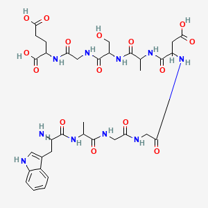
| CAS Number | 62568-57-4 |
|---|---|
| Molecular Formula | C35H48N10O15 |
| Molecular Weight | 848.8 g/Mol |
| Purity | 99.7% |
| Lot Number | 25067 |
| Quantity | 5mg |
| Sequence | H-Trp-Ala-Gly-Gly-Asp-Ala-Ser-Gly-Glu-OH |
Cosmic Peptides Certificate of Analysis
Unable to load certificate. Download COA