FOXO4-DRI
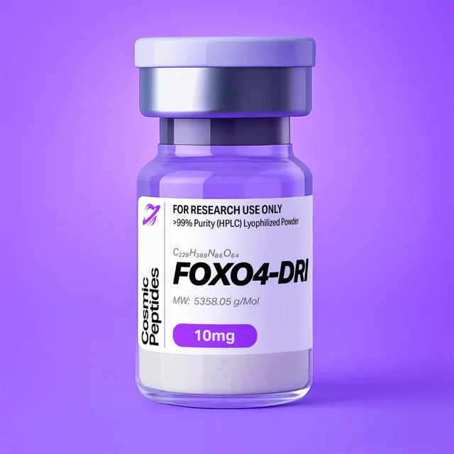
FOXO4-DRI
Commonly researched with these items

Patent-pending lot tracking: Complete transparency from production to vial.
FOXO4-DRI: An All-D Retro-Inverso 46-mer Built From a FOXO4 Forkhead Segment and an Arginine-Rich Carrier Sequence
Disclaimer
Products described here are supplied for research use only and are not intended for diagnostic, therapeutic, or clinical application. All statements regarding biological activity reflect preclinical and in vitro findings exclusively and have not been evaluated by the U.S. Food and Drug Administration. These materials and related content are provided for educational and investigational purposes only and are intended solely for qualified researchers in laboratory and academic settings.
Key Facts
- Compound class
- Synthetic linear 46-residue peptide; all-D retro-inverso (DRI) isomer of a chimera combining a forkhead-domain segment of the transcription factor FOXO4 with an arginine-rich carrier sequence from the HIV-1 Tat basic domain
- Sequence
D-(Leu-Thr-Leu-Arg-Lys-Glu-Pro-Ala-Ser-Glu-Ile-Ala-Gln-Ser-Ile-Leu-Glu-Ala-Tyr-Ser-Gln-Asn-Gly-Trp-Ala-Asn-Arg-Arg-Ser-Gly-Gly-Lys-Arg-Pro-Pro-Pro-Arg-Arg-Arg-Gln-Arg-Arg-Lys-Lys-Arg-Gly), written N-terminus to C-terminus, every chiral residue in the D configuration- Molecular formula
- C228H388N86O64 (free base, no counterions)
- Molecular weight
- 5358.06 g/mol (average); monoisotopic mass 5354.9750 Da
- CAS number
- 2460055-10-9
- PubChem CID
- 167312269 (a second record, CID 168431240, carries the same formula and masses)
- Stereochemistry
- 45 stereocentres: 42 α-carbons (46 residues minus 4 achiral glycines) plus the β-carbons of the single D-threonine and the two D-isoleucines
- Physical form as supplied
- Lyophilized powder
- Lot traceability
- Every unit is tied to a tracked lot; the lot number, recorded purity and verified quantity of the lot on sale are shown live on this page, with its certificate of analysis.
Molecular Identity and Classification
FOXO4-DRI is a 46-residue synthetic peptide in which every chiral amino acid is of the D configuration and the residue order is the reverse of the parent L-peptide. The suffix DRI stands for D-retro-inverso: retro because the sequence is read backwards, inverso because the configuration at each α-carbon is inverted. The molecular formula is C228H388N86O64, the average molecular weight 5358.06 g/mol and the monoisotopic mass 5354.9750 Da. The compound is registered under CAS number 2460055-10-9 and catalogued as PubChem compound 167312269, with a duplicate record at CID 168431240.
Because mirror-image isomers are constitutionally identical, the formula and both masses are exactly those of the all-L parent sequence; no mass measurement distinguishes the two, which is why chiral analysis is central here. The chain draws on fourteen of the twenty proteinogenic amino acids, arginine alone filling ten of the 46 positions (full composition tabulated under Analytical Characterization). Those ten arginines and four lysines dominate its physical behaviour: the calculated net charge at pH 7.0 is +10.8, effectively +11, and the calculated isoelectric point lies near 12.5.
Reading the supplied sequence in reverse recovers the parent L-peptide, GRKKRRQRRRPPPRKGGSRRNAWGNQSYAELISQAIESAPEKRLTL, which resolves into two modules joined by a proline-rich hinge. Its first ten residues, GRKKRRQRRR, are the basic domain of the trans-activator of transcription (Tat) protein of human immunodeficiency virus type 1, residues 48–57 of UniProt P04608, followed by a seven-residue PPPRKGG spacer. The last 29 residues, SRRNAWGNQSYAELISQAIESAPEKRLTL, match residues 96–124 of FOXO4 (UniProt P98177, 505 residues) exactly, spanning the four residues immediately upstream of the forkhead DNA-binding domain (annotated 100–188) and that domain's first helix. The peptide's single tryptophan is therefore FOXO4 Trp101 and its single tyrosine FOXO4 Tyr106.
The design was reported in 2017 by Baar, Brandt, Putavet and colleagues in the laboratory of Peter de Keizer, who sought to interfere competitively with the association between FOXO4 and the tumour-suppressor protein p53 in senescent cells. Building the chimera as its D-retro-inverso isomer was a deliberate chemical choice: a molecule presenting the parent helix's side-chain surface while unreadable by proteases that recognize L-peptide backbones.
Structure and Chemical Features
Chemically this is an ordinary linear peptide: 45 amide bonds, a free α-amine at one end, a free α-carboxylic acid at the other, no disulfides, no cyclization, no non-proteinogenic residues. Its unusual character lies entirely in stereochemistry and in the direction the backbone runs relative to the side chains it carries.
Retro-Inverso Topochemistry
The retro-inverso concept was formalized by Goodman and Chorev in 1979 and reviewed by the same authors in 1993. The observation is geometric: reverse the residue order and invert every α-carbon from L to D, and the side chains can occupy almost the positions they held in the original while the backbone amide bonds point the opposite way. Such a pair is topochemically equivalent — the side-chain pattern a binding partner reads is conserved, while the backbone carrying it is a different molecule to any enzyme that reads main-chain stereochemistry and directionality. Three consequences matter.
Hydrogen-bond polarity is inverted. Each amide unit -C(=O)-NH- becomes -NH-C(=O)- at the equivalent position: carbonyl acceptors and N–H donors swap places. An α-helix accommodates this well, since i to i+4 geometry can still be satisfied; extended and β-type structures generally do not, which is why retro-inverso mimicry of helical epitopes succeeds far more often than of strands. Li and colleagues documented the limits in 2010: a retro-inverso isomer can present its side chains correctly and still lose affinity where backbone hydrogen bonds contribute directly to the interface.
The helix changes hand. An all-D peptide folds into the mirror image of the corresponding L structure, so where the FOXO4 96–124 segment forms a right-handed α-helix, its D-retro-inverso counterpart forms a left-handed α-helix with the same side-chain register — an inversion directly observable by circular dichroism.
The chain termini are formally mismatched. Strict topochemical equivalence also requires replacing the end residues: the new N-terminus becomes a gem-diaminoalkyl unit and the new C-terminus a 2-substituted malonate, so the first and last side chains sit where the parent's did. FOXO4-DRI carries neither; its formula and mass are exactly those of an unmodified free-amine, free-acid 46-mer. It is therefore a partial retro-inverso peptide in the nomenclature reviewed by Fischer in 2003: all-D, fully reversed, ordinary end groups. Over 46 residues that offset is a small perturbation, but the match with the parent helix is not exact at the ends.
The Two Sequence Modules
The arginine-rich module — ten arginines and four lysines, concentrated in the RRRQRRKKRG stretch at the C-terminal end of the DRI isomer — makes the molecule polycationic. Frankel and Pabo reported in 1988 that the Tat protein of HIV-1 is taken up by cultured cells; Vivès, Brodin and Lebleu narrowed that behaviour in 1997 to the basic domain, and Futaki and colleagues showed in 2001 that guanidinium density rather than any specific Tat sequence governs association with cell-surface polyanions. Guanidinium, pKa near 12.5, is the most basic proteinogenic side chain, so these residues stay protonated across the laboratory pH range; being achiral, the group is indifferent to the configuration of the carbon carrying it. Richard and co-workers showed in 2003 that much of what had been read as direct translocation in fixed-cell images was an artefact of fixation and surface adsorption.
The FOXO4-derived module contributes the recognition surface: its AELISQAIESAPEKRLTL segment (FOXO4 107–124) places hydrophobic residues on one face and acidic Glu residues on another, the amphipathic pattern typical of a helix that docks into a hydrophobic groove. All four prolines sit in the RPPPR stretch at the junction between the modules, adjacent to the Tat basic decapeptide, acting as a rigid hinge, since a Pro-Pro-Pro run forms a short polyproline segment with little conformational freedom.
Charge, Stereocentres and Spectroscopic Handles
At pH 7.0 the peptide presents fifteen protonatable basic sites (ten arginine guanidinium groups, four lysine ε-amines, the N-terminal α-amine) against four acidic sites (three glutamate carboxylates, the C-terminal acid). Only two residues absorb above 260 nm. With the residue absorptivities tabulated by Pace and colleagues (5500 M−1cm−1 for Trp, 1490 M−1cm−1 for Tyr), the predicted molar absorption coefficient at 280 nm is 6990 M−1cm−1 — an A280 of 1.305 for a 1 mg/mL solution of the free base; that tryptophan also makes intrinsic fluorescence available as a binding read-out. Three of the 45 stereocentres sit where a wrong diastereomer is easily introduced: the correct building blocks are D-threonine, not D-allo-threonine, and D-isoleucine, (2R,3R), not D-allo-isoleucine, (2R,3S).
Synthesis and Manufacturing Chemistry
FOXO4-DRI is made by solid-phase peptide synthesis, the technique introduced by Merrifield in 1963, using Fmoc/tBu chemistry and D-amino acid building blocks throughout. Assembly runs C-terminus to N-terminus, so the glycine that ends the DRI sequence is anchored first, to an acid-labile linker such as 2-chlorotrityl chloride or a Wang-type resin, and the N-terminal D-leucine is coupled last. Side-chain protection is conventional — Pbf on arginine, Boc on lysine and tryptophan, tBu on glutamate, serine, threonine and tyrosine, trityl on asparagine and glutamine — with Fmoc removal by 20% piperidine in dimethylformamide. Three features make this a far harder target than a short peptide.
Ten arginines and a Pro-Pro-Pro run. Arginine couplings are slow and side-reactive, since activated derivatives cyclize to δ-lactams, consuming the building block and truncating the chain, so double couplings or elevated temperature are usual; at cleavage, ten bulky Pbf groups release a large quantity of sulfonyl cation for scavengers to intercept. Polyproline and arginine-rich stretches also promote on-resin aggregation, countered by low-loading resins, polar solvent mixtures and chaotropic additives.
Tryptophan protection. The single D-Trp is vulnerable to alkylation by carbocations released during acid cleavage, so Fmoc-D-Trp(Boc)-OH and a cation-scavenger cocktail are standard precautions.
Epimerization. Base-mediated epimerization at the α-carbon during activation and Fmoc removal is the main route by which an unwanted L-residue enters an all-D chain, and since an epimerized residue is a diastereomer of identical mass, its control must be verified analytically. Carbodiimide activation with an additive such as ethyl cyano(hydroxyimino)acetate, short activation times and little excess base all reduce it.
Final cleavage uses trifluoroacetic acid with scavengers — for example 92.5% TFA, 2.5% water, 2.5% triisopropylsilane, 2.5% 3,6-dioxa-1,8-octanedithiol — which also removes the Pbf, Boc, tBu and trityl groups. The crude peptide is precipitated in cold diethyl ether and purified by preparative reversed-phase HPLC. Because TFA is used in both steps, the isolated material is normally a trifluoroacetate salt unless ion exchange converts it to acetate or chloride.
Analytical Characterization
Reversed-phase HPLC. A polycationic 46-mer is strongly retained only when ion pairing suppresses its charge. Analysis is typically done on a C18 column with 0.1% trifluoroacetic acid in both mobile phases and an acetonitrile gradient, detecting at 214 nm, where the amide bonds absorb, with a parallel channel at 280 nm for the Trp/Tyr pair; the ratio of the two responses is a useful identity check. Formic acid in place of TFA, as is usual for LC-MS, shortens retention markedly and often broadens the peak, so purity and LC-MS commonly run on different ion-pairing systems. A mirror-image isomer cannot be resolved on an achiral stationary phase: the all-L parent and the DRI isomer co-elute, so HPLC alone cannot establish stereochemical identity.
Mass spectrometry. With fifteen basic sites, positive-mode electrospray gives an extensive charge envelope, usually most intense between 5+ and 9+. From the neutral monoisotopic mass of 5354.9750 Da the expected monoisotopic m/z values are 1072.00 (5+), 893.50 (6+), 766.00 (7+), 670.38 (8+) and 596.00 (9+); the 4+ ion falls at 1339.75. A residue-by-residue calculation gives 5354.9749 Da, agreeing with the monoisotopic mass field of the PubChem record, 5354.9750 Da; the same record carries a separate exact mass field of 5356.9817 Da, 2.007 Da higher, which should not be used to set an extraction window. Where the isotope pattern is resolved, charge state follows from the 1/z spacing of the isotope peaks, and deconvolution should return the neutral mass within a few parts per million. Collision-induced dissociation gives b and y ions confirming residue order but not configuration: an all-D peptide and its all-L enantiomer give identical spectra, and a single-residue epimer differs in mass not at all.
Chiral and Enzymatic Verification
Chiral amino-acid analysis. The peptide is hydrolysed, conventionally in 6 M hydrochloric acid at 110 °C, and the liberated amino acids derivatized with a chiral reagent. Marfey's reagent, FDAA (1-fluoro-2,4-dinitrophenyl-5-L-alanine amide), is the chiral mono-substitution product of the bifunctional reagent 1,5-difluoro-2,4-dinitrobenzene described by Marfey in 1984; its fixed L-alanine amide centre turns D- and L-amino acids into diastereomeric adducts that separate on an ordinary achiral reversed-phase column. Every chiral residue should appear in the D form, in the proportions below. Hydrolysis itself racemizes slightly, so a blank hydrolysis of an authentic standard sets the baseline for the minor enantiomer.
| Residue | Count | Behaviour on acid hydrolysis |
|---|---|---|
| Arg | 10 | Stable; dominant peak, easily overloaded |
| Ala | 4 | Stable; convenient reference ratio |
| Gly | 4 | Achiral, so no D/L pair |
| Lys | 4 | ε-amine also derivatizes |
| Pro | 4 | Secondary amine; different yield |
| Ser | 4 | Partial loss; use a co-hydrolysed standard |
| Glu + Gln | 3 + 3 | Gln deamidates; reported as Glx = 6 |
| Leu | 3 | Stable |
| Ile | 2 | Must resolve as D-Ile, not D-allo-Ile |
| Asn | 2 | Deamidates; reported as Asx = 2 (no Asp present) |
| Thr | 1 | Partial loss; D-Thr, not D-allo-Thr |
| Trp | 1 | Largely destroyed without a protective additive |
| Tyr | 1 | Partial loss; halogenation possible |
| Total | 46 | 42 chiral α-carbons; 4 Gly achiral |
Protease resistance. Trypsin cleaves C-terminal to L-Arg and L-Lys, so with ten arginines and four lysines the all-L parent is cut into many small fragments within minutes, whereas the all-D chain is no substrate at all, these active sites being themselves chiral. Following the intact mass by LC-MS after incubation with trypsin or chymotrypsin is therefore decisive: persistence of the intact 5355 Da species is positive evidence of D configuration. The same property removes enzymatic peptide mapping as an option, so sequence confirmation rests on intact-mass and MS/MS data.
Circular dichroism gives a third, non-destructive check, since an all-D peptide shows the point reflection of its L enantiomer's spectrum: positive bands near 208 and 222 nm and a negative band near 192 nm, where a right-handed α-helical L-peptide shows the opposite signs. A helix-promoting cosolvent such as 2,2,2-trifluoroethanol tests whether the FOXO4 segment's helical propensity survives the inversion.
Purity Versus Peptide Content
HPLC purity is the fraction of UV-absorbing material that is the target peptide; peptide content is the fraction of the weighed powder that is peptide at all, the remainder being counterions, water and residual salts. With eleven net positive charges the counterion mass is worth calculating explicitly. Modelled as the free base plus eleven equivalents of acid, trifluoroacetic acid at 114.02 g/mol adds 1254.2 g/mol to the 5358.06 g/mol free base: a nominal salt mass of 6612.3 g/mol and a theoretical peptide content of 81.0% by weight. Acetic acid, at 60.05 g/mol, totals 6018.6 g/mol for 89.0%. Add the 5–10% water typical of a lyophilized polycation and the shortfall dominates any calculation that ignores it.
A worked example: 10 mg of powder at 81.0% content holds 8.10 mg of peptide, which is 8.10 × 10⁻³ g ÷ 5358.06 g/mol = 1.512 µmol. In 1.00 mL that is a 1.51 mM stock, where the weighed mass alone implies 1.87 mM — an overstatement of 23%. Conversely, 1.00 mL of a 1.00 mM solution needs 6.61 mg of the trifluoroacetate salt. Content is measured by quantitative amino-acid analysis, quantitative NMR against a certified internal standard, or absorbance at 280 nm; the counterion by ion chromatography or 19F NMR, and water by Karl Fischer titration.
Physicochemical Properties
| Property | Value | Notes |
|---|---|---|
| Monoisotopic mass | 5354.9750 Da | Free base; identical to the all-L parent |
| Stereocentres | 45 | 42 α-carbons plus β-carbons of Thr, 2 × Ile |
| Basic / acidic sites | 15 / 4 | 10 Arg, 4 Lys, N-terminus; 3 Glu, C-terminus |
| Net charge at pH 7.0 | +10.8 (effectively +11) | Calculated from standard side-chain pKa values |
| Isoelectric point | approximately 12.5 | Calculated; strongly basic |
| ε280 | 6990 M−1cm−1 | 1 Trp + 1 Tyr, per Pace et al. |
| A280, 1 mg/mL | 1.305 | Free base; correct for peptide content |
| Principal ESI charge states | 5+ to 9+ | m/z 1072.00, 893.50, 766.00, 670.38, 596.00 |
| Aqueous solubility | High in water and dilute acid | Polycationic; no hydrophobic core |
| Peptide content, TFA salt | 81.0% w/w | Theoretical, 11 × 114.02 g/mol |
| Peptide content, acetate salt | 89.0% w/w | Theoretical, 11 × 60.05 g/mol |
| Trypsin or chymotrypsin cleavage | None expected | All-D backbone is not a substrate |
| Oxidation-prone residues | None (no Cys, no Met) | Trp photo-oxidizes slowly in light |
Charge governs practical work here. Eleven net positive charges make the peptide freely soluble in water and dilute acetic acid but also an avid binder of anionic surfaces: polystyrene and untreated glass adsorb it, and heparin, nucleic acids and sulfated glycans bind it strongly. Aggregation follows charge neutralization rather than hydrophobic collapse, so solutions turn cloudy when the pH approaches the isoelectric point or a polyanion is introduced, not when organic content falls.
The solution liabilities are those of any peptide without cysteine or methionine: slow amide hydrolysis at extreme pH, deamidation of the two asparagines and three glutamines, fastest above pH 8 and accelerated by a following glycine (Asn22-Gly23 of the DRI sequence is the classic fast-deamidating context), and slow photo-oxidation of the single tryptophan. Deamidation adds 0.984 Da per event, a +1 Da shoulder on the deconvoluted mass.
Research Context
FOXO4 is one of four mammalian FOXO transcription factors, a family whose DNA-binding module is the winged-helix or forkhead fold. Boura and co-workers solved that domain bound to DNA at 1.9 Å resolution and described how its flanking segments and wing regions contact the duplex; Obsil and Obsilova reviewed the family's structure–function relationships. FOXO4 96–124 begins just upstream of the domain and includes its first helix, so the peptide samples a surface that in the full-length protein takes part in both DNA contact and protein–protein association.
That surface attracted attention because of its interaction with p53. In senescent cells FOXO4 and p53 can be found in the same nuclear foci, and the working model advanced by Baar and colleagues in 2017 is that the association restrains the pro-apoptotic branch of p53 output, so a competitive peptide occupying the FOXO4 interface would release that restraint. The molecular detail was sharpened in 2025 by Bourgeois and co-workers, who used solution NMR to place the binding site for both the FOXO4 forkhead domain and FOXO4-DRI in the intrinsically disordered transactivation domain of p53 rather than in its folded DNA-binding core. That is chemically consequential: the region has no single structure, so interactions with it are usually of modest affinity, involve short motifs that order locally on binding, and are better studied by chemical-shift perturbation and relaxation than by co-crystallization.
Micromolar affinity means titrations must span a wide concentration window, read by a method of matched dynamic range: isothermal titration calorimetry, surface plasmon resonance with attention to the non-specific binding of a +11 analyte, microscale thermophoresis, fluorescence anisotropy of a labelled tracer, or intrinsic tryptophan fluorescence. Because the peptide is polycationic and this p53 region acidic, charge-driven association is a serious confounder: vary buffer ionic strength deliberately alongside the sequence controls listed under Isomer Nomenclature.
In cell-based work the systems are cultured senescent cells and their non-senescent counterparts. Senescence is established in vitro by replicative exhaustion of primary fibroblast strains such as IMR-90 or WI-38, by ionizing radiation, by oxidative stress, or by expression of an activated oncogene, and the state is read as a panel rather than a single marker; Hernandez-Segura, Nehme and Demaria catalogued the panel and its limits, and the senescence-associated β-galactosidase stain standardized by Debacq-Chainiaux and co-workers remains the most widely used single read-out. A selectivity experiment then compares matched senescent and proliferating cultures over one concentration series, reading viability by a metabolic assay, nuclear counting or live/dead staining, confirming apoptosis by caspase-3/7 activity, annexin V binding or PARP cleavage, and reporting a ratio between the two curves rather than a single potency value. Localization of p53 by immunofluorescence or fractionation is the read-out closest to the proposed mechanism, and proximity ligation or co-immunoprecipitation of the FOXO4–p53 pair tests whether the interaction has been displaced. Since the compound carries an arginine-rich carrier module, each experiment needs a vehicle control matched for counterion content, and the fixation artefacts noted above must be excluded by imaging live cells.
Isomer Nomenclature: Four Ways to Rewrite a Peptide
Discussions of this compound routinely confuse the four stereochemical and directional variants of a sequence, and the wrong label makes a literature comparison meaningless.
| Isomer | Residue order | α-carbon configuration | Side-chain topology | Backbone amide direction |
|---|---|---|---|---|
| Parent (all-L) | GRKKRRQRRR…KRLTL | L | Reference | Reference |
| Retro (all-L, reversed) | LTLRK…RRRQRRKKRG | L | Different | Reversed |
| Inverso (all-D, same order) | GRKKRRQRRR…KRLTL | D | Mirror image | Mirrored geometry |
| Retro-inverso (all-D, reversed) — FOXO4-DRI | LTLRK…RRRQRRKKRG | D | Closely superimposable | Reversed |
Only the fourth row restores the parent's side-chain arrangement, which is why retro and inverso isomers taken singly are poor mimics of a native helix while their combination is often a good one. The rows also set an experiment's comparators: the all-L parent chimera, a scrambled all-D peptide of identical composition, and the carrier module alone isolate backbone stereochemistry, sequence specificity and polycationic charge respectively. Guichard and colleagues established the practical validity of the topochemical argument in 1994, when a retro-inverso analogue of a short epitope was still recognized by antibodies raised against the natural L-peptide.
Related Research Peptides
Among compounds whose design depends on D-residues, SS-31 is the closest analytical relative: a short peptide of alternating aromatic and basic residues in which a D-arginine confers resistance to aminopeptidases, so the same chiral analysis and protease-resistance logic applies. At the polycationic end, LL-37 is a 37-residue cationic peptide whose handling problems — adsorption to plastics, polyanion binding, sensitivity to buffer ionic strength — mirror these despite an all-L backbone. The much shorter Epitalon and Pinealon are useful reference materials when validating amino-acid analysis and quantitative NMR, their composition being simple enough that the expected result is unambiguous.
Laboratory Handling and Storage
Storage of the solid. Keep the lyophilized powder tightly closed at −20 °C or below, dark and dry, and let a container equilibrate to room temperature in a desiccator before opening: weighing cold open glassware adds condensed water to the mass.
Dissolving the peptide. FOXO4-DRI dissolves readily in high-purity water and dilute aqueous acid; for a difficult batch, 0.1% acetic acid or water at pH 3–4 sits well away from the isoelectric point. Stocks of 1–5 mM are convenient: dissolve in the minimum volume, verify clarity, then dilute, since a concentrated polycation added straight into phosphate-rich medium invites local precipitation.
Adsorption losses. At low micromolar concentrations a +11 peptide is lost to surfaces faster than most users expect. Use low-binding polypropylene labware, avoid untreated glass, and prefer polyethersulfone or regenerated-cellulose membranes over nylon for filtration. Where the experiment permits, 0.1% bovine serum albumin in dilution buffers substantially reduces losses; otherwise prepare working dilutions immediately before use and quantify at 280 nm.
Aliquoting and re-verification. Divide stocks into single-use aliquots, store at −20 °C for weeks or −80 °C for longer, and avoid freeze–thaw cycles, which concentrate solutes in the unfrozen phase and aggregate charged peptides. Avoid alkaline buffers for long storage. After extended storage an LC-MS check establishes integrity: the expected charge envelope, a deconvoluted monoisotopic mass of 5354.98 Da, no +0.98 Da deamidation shoulder or truncation products. A trypsin-resistance check confirms the material still behaves as an all-D chain.
Frequently Asked Research Questions
- What does the "DRI" in FOXO4-DRI stand for?
- D-retro-inverso: the residue order is the reverse of the parent sequence and every chiral α-carbon is in the D configuration. Together the two inversions place the side chains in approximately the positions they occupy in the parent while reversing the direction of the backbone amide bonds.
- What are the molecular formula, molecular weight and CAS number?
- The free base is C228H388N86O64, average molecular weight 5358.06 g/mol, monoisotopic mass 5354.9750 Da. These values are identical to those of the all-L parent isomer, because enantiomers and sequence-reversed isomers share an atomic composition. The CAS number is 2460055-10-9 and the PubChem identifier CID 167312269, with a duplicate record at CID 168431240.
- Where does the sequence come from?
- Reversing the supplied 46-residue D-peptide gives the parent chimera GRKKRRQRRRPPPRKGGSRRNAWGNQSYAELISQAIESAPEKRLTL. Its first ten residues are the basic domain of the HIV-1 Tat protein (residues 48–57 of UniProt P04608), a PPPRKGG spacer follows, and the last 29 residues match residues 96–124 of FOXO4 (UniProt P98177) exactly, spanning the start of its forkhead DNA-binding domain. The single Trp and Tyr are FOXO4 Trp101 and Tyr106.
- Is FOXO4-DRI a strict retro-inverso peptide?
- No. Full topochemical equivalence requires modified end residues, a gem-diaminoalkyl unit at the new N-terminus and a 2-substituted malonate at the new C-terminus. FOXO4-DRI has ordinary free-amine and free-acid termini, which its formula confirms, so it is a partial retro-inverso isomer: all-D and fully reversed, unmodified at the ends. The gain is a backbone that proteases, whose active sites are chiral, do not recognize; the cost is that backbone hydrogen-bond donors and acceptors swap positions.
- How can a laboratory prove the material is the D isomer and not the L peptide of reversed sequence?
- Mass spectrometry cannot distinguish them; two methods can. Chiral amino-acid analysis after acid hydrolysis, using Marfey's reagent (FDAA) to form separable diastereomeric derivatives, should show every chiral residue in the D form. Independently, incubation with trypsin or chymotrypsin followed by LC-MS should leave the intact 5355 Da species unchanged, whereas the all-L parent, with ten arginines and four lysines, would be cleaved extensively.
- Which masses should appear in the mass spectrum?
- Positive-mode electrospray of a peptide with fifteen basic sites gives a broad charge envelope, usually most intense between 5+ and 9+. Expected monoisotopic m/z values are 1072.00 (5+), 893.50 (6+), 766.00 (7+), 670.38 (8+) and 596.00 (9+), and deconvolution should return a neutral monoisotopic mass of 5354.975 Da. Circular dichroism complements this: an all-D peptide gives positive bands near 208 and 222 nm where an α-helical L-peptide gives negative ones.
- Why do weighed mass and molar concentration disagree, and by how much?
- Because the powder is a salt containing water. With eleven net positive charges the trifluoroacetate form has a theoretical peptide content of 81.0% by weight, and residual water adds a further 5–10%. Ten milligrams of such a powder holds 1.51 µmol of peptide, not the 1.87 µmol the weighed mass implies, and 1.00 mL of a 1.00 mM solution needs about 6.61 mg of the salt. Quantification at 280 nm uses a calculated coefficient of 6990 M−1cm−1.
- What is the molecular target studied in vitro, and how is binding measured?
- The peptide is designed to compete with the FOXO4 forkhead region for binding to p53. Solution-NMR work published in 2025 localizes that interaction to the intrinsically disordered transactivation domain of p53 rather than the folded DNA-binding core. Because such interactions are usually of micromolar affinity, binding is assessed by chemical-shift perturbation, isothermal titration calorimetry, surface plasmon resonance, microscale thermophoresis or tryptophan fluorescence, with ionic strength varied to separate electrostatics from motif-specific binding.
- Which comparator peptides belong in an experiment with FOXO4-DRI?
- Three, each isolating one variable. The all-L parent chimera holds the sequence constant and changes only backbone stereochemistry. A scrambled all-D peptide of identical composition holds net charge and D configuration constant while destroying the sequence, separating motif-specific binding from the electrostatics of a +11 polycation. The carrier module alone reports what the carrier contributes without the FOXO4-derived surface.
- Which in vitro senescence read-outs are used alongside this compound?
- Matched senescent and proliferating cultures of primary fibroblast strains such as IMR-90 or WI-38 are compared after replicative exhaustion, ionizing radiation, oxidative stress or oncogene expression. Senescence is confirmed by a panel: senescence-associated β-galactosidase staining, elevated p21CIP1 and p16INK4a, loss of lamin B1, absence of Ki-67 and EdU incorporation, and secretory markers. Viability and apoptosis are then expressed as a selectivity ratio between the two populations.
- How should the peptide be stored and handled to avoid losses?
- Store the lyophilized solid at −20 °C or below, closed, dry and dark, and warm it in a desiccator before opening. Dissolve in high-purity water or dilute acetic acid, aliquot for single use, and avoid freeze–thaw cycles and alkaline buffers. At low micromolar concentrations use low-binding polypropylene labware, because a +11 peptide adsorbs quickly to glass, nylon and untreated plastics.
- Where is the purity of the material on sale shown?
- This page displays the lot number, recorded purity and verified quantity of the lot currently on sale, taken live from the shop's lot-tracking database, together with that lot's certificate of analysis.
Sources & Further Reading
- National Center for Biotechnology Information. PubChem Compound Summary for CID 167312269, FOXO4-DRI. PubChem CID 167312269 (duplicate record: PubChem CID 168431240).
- UniProt Consortium. UniProtKB P98177, Forkhead box protein O4, 505 residues, forkhead domain 100–188; and UniProtKB P04608, protein Tat of HIV-1 (HXB2), 86 residues, GRKKRRQRRR at 48–57.
- Baar MP, Brandt RMC, Putavet DA, Klein JDD, Derks KWJ, Bourgeois BRM, et al. Targeted apoptosis of senescent cells restores tissue homeostasis in response to chemotoxicity and aging. Cell 2017;169(1):132–147.e16. doi:10.1016/j.cell.2017.02.031
- Bourgeois B, Spreitzer E, Platero-Rochart D, Paar M, Zhou Q, Usluer S, et al. The disordered p53 transactivation domain is the target of FOXO4 and the senolytic compound FOXO4-DRI. Nature Communications 2025;16:5672. doi:10.1038/s41467-025-60844-9
- Goodman M, Chorev M. On the concept of linear modified retro-peptide structures. Accounts of Chemical Research 1979;12(1):1–7. doi:10.1021/ar50133a001
- Chorev M, Goodman M. A dozen years of retro-inverso peptidomimetics. Accounts of Chemical Research 1993;26(5):266–273. doi:10.1021/ar00029a007
- Fischer PM. The design, synthesis and application of stereochemical and directional peptide isomers: a critical review. Current Protein & Peptide Science 2003;4(5):339–356. doi:10.2174/1389203033487054
- Guichard G, Benkirane N, Zeder-Lutz G, van Regenmortel MHV, Briand JP, Muller S. Antigenic mimicry of natural L-peptides with retro-inverso-peptidomimetics. Proceedings of the National Academy of Sciences 1994;91(21):9765–9769. doi:10.1073/pnas.91.21.9765
- Li C, Pazgier M, Li J, Li C, Liu M, Zou G, et al. Limitations of peptide retro-inverso isomerization in molecular mimicry. Journal of Biological Chemistry 2010;285(25):19572–19581. doi:10.1074/jbc.M110.116814
- Frankel AD, Pabo CO. Cellular uptake of the tat protein from human immunodeficiency virus. Cell 1988;55(6):1189–1193. doi:10.1016/0092-8674(88)90263-2
- Vivès E, Brodin P, Lebleu B. A truncated HIV-1 Tat protein basic domain rapidly translocates through the plasma membrane and accumulates in the cell nucleus. Journal of Biological Chemistry 1997;272(25):16010–16017. doi:10.1074/jbc.272.25.16010
- Futaki S, Suzuki T, Ohashi W, Yagami T, Tanaka S, Ueda K, Sugiura Y. Arginine-rich peptides: an abundant source of membrane-permeable peptides having potential as carriers for intracellular protein delivery. Journal of Biological Chemistry 2001;276(8):5836–5840. doi:10.1074/jbc.M007540200
- Richard JP, Melikov K, Vivès E, Ramos C, Verbeure B, Gait MJ, et al. Cell-penetrating peptides: a reevaluation of the mechanism of cellular uptake. Journal of Biological Chemistry 2003;278(1):585–590. doi:10.1074/jbc.M209548200
- Boura E, Rezabkova L, Brynda J, Obsilova V, Obsil T. Structure of the human FOXO4-DBD–DNA complex at 1.9 Å resolution reveals new details of FOXO binding to the DNA. Acta Crystallographica Section D 2010;66(12):1351–1357. doi:10.1107/S0907444910042228
- Obsil T, Obsilova V. Structure/function relationships underlying regulation of FOXO transcription factors. Oncogene 2008;27(16):2263–2275. doi:10.1038/onc.2008.20
- Hernandez-Segura A, Nehme J, Demaria M. Hallmarks of cellular senescence. Trends in Cell Biology 2018;28(6):436–453. doi:10.1016/j.tcb.2018.02.001
- Debacq-Chainiaux F, Erusalimsky JD, Campisi J, Toussaint O. Protocols to detect senescence-associated β-galactosidase (SA-βgal) activity, a biomarker of senescent cells in culture and in vivo. Nature Protocols 2009;4(12):1798–1806. doi:10.1038/nprot.2009.191
- Merrifield RB. Solid phase peptide synthesis. I. The synthesis of a tetrapeptide. Journal of the American Chemical Society 1963;85(14):2149–2154. doi:10.1021/ja00897a025
- Marfey P. Determination of D-amino acids. II. Use of a bifunctional reagent, 1,5-difluoro-2,4-dinitrobenzene. Carlsberg Research Communications 1984;49(6):591–596. doi:10.1007/BF02908688
- Pace CN, Vajdos F, Fee L, Grimsley G, Gray T. How to measure and predict the molar absorption coefficient of a protein. Protein Science 1995;4(11):2411–2423. doi:10.1002/pro.5560041120
Last reviewed:
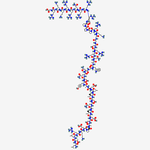
| CAS Number | 2460055-10-9 |
|---|---|
| Molecular Formula | C228H388N86O64 |
| Molecular Weight | 5358.06 g/Mol |
| Sequence | D-(Leu-Thr-Leu-Arg-Lys-Glu-Pro-Ala-Ser-Glu-Ile-Ala-Gln-Ser-Ile-Leu-Glu-Ala-Tyr-Ser-Gln-Asn-Gly-Trp-Ala-Asn-Arg-Arg-Ser-Gly-Gly-Lys-Arg-Pro-Pro-Pro-Arg-Arg-Arg-Gln-Arg-Arg-Lys-Lys-Arg-Gly) |
Cosmic Peptides Certificate of Analysis
Unable to load certificate. Download COA