Ipamorelin
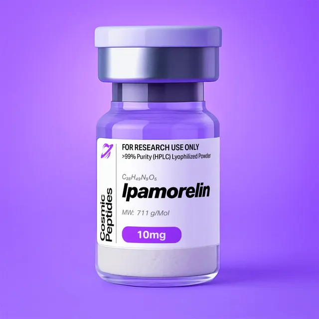
Ipamorelin
Commonly researched with these items
Patent-pending lot tracking: Complete transparency from production to vial.
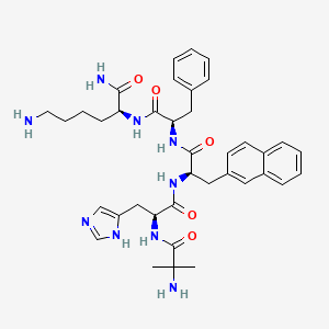
| CAS Number | 170851-70-4 |
|---|---|
| Molecular Formula | C38H49N9O5 |
| Molecular Weight | 711.9 g/Mol |
| Purity | 99.8% |
| Lot Number | 26108 |
| Quantity | 10.13mg |
| Sequence | H-Aib-His-D-2Nal-D-Phe-Lys-NH2 |
Ipamorelin (Aib-His-D-2Nal-D-Phe-Lys-NH2): A Pentapeptide Ligand of the Ghrelin Receptor (GHS-R1a)
Disclaimer
Products described here are supplied for research use only and are not intended for diagnostic, therapeutic, or clinical application. All statements regarding biological activity reflect preclinical and in vitro findings exclusively and have not been evaluated by the U.S. Food and Drug Administration. These materials and related content are provided for educational and investigational purposes only and are intended solely for qualified researchers in laboratory and academic settings.
Key Facts
- Compound class
- Synthetic linear pentapeptide amide containing non-proteinogenic and D-amino acids; growth hormone-releasing peptide (GHRP) family; agonist ligand of the growth hormone secretagogue receptor type 1a (GHS-R1a, the ghrelin receptor)
- Sequence
H-Aib-His-D-2Nal-D-Phe-Lys-NH2(Aib = 2-aminoisobutyric acid; D-2Nal = 3-(2-naphthyl)-D-alanine; C-terminal primary amide)- Molecular formula
- C38H49N9O5
- Molecular weight
- 711.9 g/mol (711.87 g/mol calculated, free base); monoisotopic mass 711.3857 Da
- CAS number
- 170851-70-4
- PubChem CID
- 9831659
- Other identifiers
- Development code NNC 26-0161; InChIKey NEHWBYHLYZGBNO-BVEPWEIPSA-N
- Physical form as supplied
- Lyophilized powder
- Lot traceability
- Every unit is tied to a tracked lot; the lot number, recorded purity and verified quantity of the lot on sale are shown live on this page, with its certificate of analysis.
Molecular Identity and Classification
Ipamorelin is a synthetic pentapeptide, H-Aib-His-D-2Nal-D-Phe-Lys-NH2. Its systematic name, (2S)-6-amino-2-[[(2R)-2-[[(2R)-2-[[(2S)-2-[(2-amino-2-methylpropanoyl)amino]-3-(1H-imidazol-5-yl)propanoyl]amino]-3-naphthalen-2-ylpropanoyl]amino]-3-phenylpropanoyl]amino]hexanamide, reads from the C-terminus inwards and records its four stereocentres: L-lysine (2S), D-phenylalanine (2R), D-3-(2-naphthyl)alanine (2R) and L-histidine (2S). The fifth residue, 2-aminoisobutyric acid (Aib, or 2-methylalanine), is achiral, carrying two methyl groups on its α-carbon. In CAS style the peptide is 2-methylalanyl-L-histidyl-3-(2-naphthalenyl)-D-alanyl-D-phenylalanyl-L-lysinamide.
The free base has the molecular formula C38H49N9O5, an average molecular weight of 711.87 g/mol (listed as 711.9 in PubChem) and a monoisotopic mass of 711.3857 Da. It carries CAS number 170851-70-4 and PubChem CID 9831659, and was disclosed in 1998 by Raun and colleagues at Novo Nordisk in Måløv, Denmark, under the code NNC 26-0161; the name follows the "-relin" stem for peptides acting on the pituitary hormone-release axis.
Ipamorelin belongs to the growth hormone-releasing peptides (GHRPs), small synthetic peptides acting at the growth hormone secretagogue receptor type 1a (GHS-R1a). That receptor was cloned only in 1996, years after the first GHRPs had been made, and its endogenous ligand, the acylated stomach peptide ghrelin, in 1999: this is a ligand class designed by structure–activity chemistry before its target was known. It is distinct from the peptides that act on the growth hormone-releasing hormone receptor (GHRHR), such as Sermorelin, which are longer, all-L sequences derived from the 44-residue hypothalamic hormone and signal through a different G protein-coupled receptor.
Structure and Chemical Features
Each of the five residues was chosen for a structural reason, and each leaves a mark on the chemistry or the analytics:
| Position | Residue | Configuration | Residue formula / mass | Structural role |
|---|---|---|---|---|
| 1 | Aib (2-aminoisobutyric acid) | Achiral (α,α-dimethyl) | C4H7NO, 85.0528 Da | Free N-terminal amine; gem-dimethyl substitution restricts backbone φ/ψ angles and hinders aminopeptidase cleavage |
| 2 | His (L-histidine) | L (2S) | C6H7N3O, 137.0589 Da | Imidazole side chain (pKa near 6), partly protonated near neutral pH; tautomeric |
| 3 | D-2Nal (3-(2-naphthyl)-D-alanine) | D (2R) | C13H11NO, 197.0841 Da | Flat bicyclic aromatic surface; dominant hydrophobic group and UV/fluorescence chromophore |
| 4 | D-Phe (D-phenylalanine) | D (2R) | C9H9NO, 147.0684 Da | Second aromatic ring; D-configuration conserved across the GHRP family |
| 5 | Lys-NH2 (L-lysinamide) | L (2S) | C6H12N2O, 128.0950 Da (+ NH2 terminus) | Cationic ε-amine; the primary amide removes the carboxylate charge |
Summing the five residue masses (694.3591 Da) and adding the terminal groups (together NH3, 17.0265 Da) gives 711.3857 Da, a worked check that matches the PubChem monoisotopic mass exactly.
The N-terminal Aib. 2-Aminoisobutyric acid is the simplest α,α-disubstituted amino acid. Its two Cα methyl groups restrict the backbone dihedral angles to the helical regions of the Ramachandran map (φ near ±57°, ψ near ±47°), and with no α-hydrogen it cannot racemize and is a poor substrate for many peptidases. It replaces the N-terminal alanine of the parent sequence.
Histidine. The imidazole pKa near 6.0 leaves roughly 4% of these side chains protonated at pH 7.4 and the ring fully protonated in an acidic HPLC mobile phase (about pH 2 with 0.1% trifluoroacetic acid). That pH-dependent charge is why retention and charge-state distribution shift with buffer conditions.
The two D-aromatic residues. The central D-2Nal-D-Phe pair is the hydrophobic core. 3-(2-Naphthyl)alanine is a non-proteinogenic residue whose naphthalene ring presents roughly twice the flat aromatic surface of phenylalanine. Adjacent D-aromatic residues recur across the GHRPs: the D-configuration resists proteolysis by enzymes that recognize L-residues, and the conformational-energy calculations of Momany, Bowers and colleagues used exactly this kind of D-residue placement to steer the aromatic side chains into the energetically favoured arrangements their active analogues shared.
The C-terminal lysinamide. The D-Phe-Lys-NH2 motif is shared by essentially every peptidic GHRP. The primary amide removes what would be a negative carboxylate, so the molecule carries no acidic group at all. The lysine ε-amine (pKa near 10.5) and the N-terminal Aib amine (pKa near 8 in a peptide context) are largely protonated at neutral pH, giving a net charge of about +2 at pH 7 and +3 under acidic conditions.
The molecule has 52 heavy atoms, 8 hydrogen-bond donors, 8 acceptors and 19 rotatable bonds, with a computed topological polar surface area of 240 Å2 and an XLogP3 of 1.8. That positive XLogP, unusual for a peptide of this charge, reflects the naphthyl and phenyl rings.
From Enkephalin Analogues to Ipamorelin: A Structural Lineage
The chemistry of ipamorelin is the end point of nearly two decades of structure–activity work. Analogues of the opioid pentapeptide methionine-enkephalin (Tyr-Gly-Gly-Phe-Met) were found to release growth hormone (GH) weakly from pituitary tissue in vitro. Momany, Bowers and co-workers used conformational-energy calculations to design better sequences, reporting in 1981 that Tyr-D-Trp-Ala-Trp-D-Phe-NH2 released GH in vitro at 10–30 ng/mL, about a thousand-fold above the enkephalin starting points. Iteration produced GHRP-6, His-D-Trp-Ala-Trp-D-Phe-Lys-NH2, described in 1984 and the reference compound of the field.
Later GHRPs swapped the indole of D-Trp for the naphthalene of D-2Nal, giving GHRP-1 (Ala-His-D-2Nal-Ala-Trp-D-Phe-Lys-NH2) and GHRP-2. Raun and colleagues describe ipamorelin as emerging from a series of compounds lacking the central Ala-Trp dipeptide of GHRP-1; with the N-terminal alanine also replaced by Aib, the result is the shortest of the classical GHRPs in the table below and the only one without tryptophan, yet still active at low nanomolar concentrations in pituitary cell assays.
| Peptide | Sequence | Length | Notable feature |
|---|---|---|---|
| Met-enkephalin | Tyr-Gly-Gly-Phe-Met | 5 | D-substituted analogues were the first weak in vitro GH releasers |
| Momany 1981 lead | Tyr-D-Trp-Ala-Trp-D-Phe-NH2 | 5 | Introduces the D-aromatic, D-Phe-amide pattern |
| GHRP-6 | His-D-Trp-Ala-Trp-D-Phe-Lys-NH2 | 6 | Adds the C-terminal Lys-NH2 |
| GHRP-1 | Ala-His-D-2Nal-Ala-Trp-D-Phe-Lys-NH2 | 7 | D-2Nal for D-Trp; direct parent of ipamorelin |
| GHRP-2 | D-Ala-D-2Nal-Ala-Trp-D-Phe-Lys-NH2 | 6 | D-Ala N-terminus |
| Ipamorelin | Aib-His-D-2Nal-D-Phe-Lys-NH2 | 5 | GHRP-1 minus Ala-Trp; Aib N-terminus; no tryptophan |
Ipamorelin in turn became a lead structure. Ankersen and colleagues set out to reduce its size and its number of hydrogen-bonding sites, replacing the N-terminal Aib-His dipeptide with the peptidomimetic fragment 3-(aminomethyl)benzoic acid and introducing sequential backbone N-methylations. The tetrapeptide NNC 26-0235, 3-(aminomethyl)benzoyl-D-2Nal-N-Me-D-Phe-Lys-NH2, kept high potency and efficacy in a pituitary cell assay. That result suggests that the D-2Nal-D-Phe-Lys-NH2 C-terminal half carries most of the pharmacophore, while the N-terminal dipeptide can be exchanged for a simpler fragment that keeps a basic amine at a similar distance.
Synthesis and Manufacturing Chemistry
Ipamorelin is made by solid-phase peptide synthesis (SPPS), introduced by Merrifield in 1963, with Fmoc (9-fluorenylmethoxycarbonyl) α-amine protection, introduced by Carpino and Han in 1972 and reviewed for SPPS by Fields and Noble. Because the C-terminus is a primary amide, the chain is built on an amide-generating support, so that acidolytic cleavage releases the peptide directly as the carboxamide.
Assembly runs from the C-terminus, lysine first and Aib last. Side-chain protection is chosen to fall away in the final acid cleavage — tert-butyloxycarbonyl (Boc) on the lysine ε-amine and trityl (Trt) on the histidine imidazole, as surveyed by Isidro-Llobet, Álvarez and Albericio — and each cycle removes the Fmoc group under mild base before the next residue is coupled.
Three points are specific to this sequence and explain its characteristic impurities:
- Histidine racemization. Histidine is the residue most prone to epimerization during activation, because the imidazole nitrogen can abstract the α-proton of the activated ester. Trityl protection, low-racemization coupling conditions and short activation times limit formation of the D-His diastereomer,
Aib-D-His-D-2Nal-D-Phe-Lys-NH2. - Hindered Aib coupling. The α,α-dimethyl substitution makes the Aib carboxyl sterically hindered, so the final coupling onto histidine is slow, and an incomplete step leaves the des-Aib tetrapeptide
His-D-2Nal-D-Phe-Lys-NH2. Because Aib has no α-hydrogen it cannot epimerize, an advantage over the alanine it replaced. - Enantiopure building blocks. The D-2Nal and D-Phe units must be of high enantiomeric purity; L-contaminants give diastereomers of identical mass, separable only by chromatography.
Acidolytic cleavage with trifluoroacetic acid (TFA) and scavengers removes the Boc and Trt groups and releases the peptide amide; the crude product is then purified by preparative reversed-phase HPLC and lyophilized. Because TFA is present throughout purification, the peptide is isolated as a trifluoroacetate salt unless exchanged into another counterion; ipamorelin is also commonly handled as the acetate salt, and the PubChem record lists an acetate-form entry among its synonyms. The certificate of analysis for each lot states the results that apply to that material.
Analytical Characterization
Reversed-phase HPLC. Unlike very polar short peptides, ipamorelin is well retained on C18 columns, because its naphthyl and phenyl rings provide a substantial hydrophobic surface. A typical method pairs water and acetonitrile, both with 0.1% TFA, in a linear gradient over the low-to-mid organic range (say 10–50% acetonitrile), with detection at 214–220 nm. Because the naphthalene chromophore also absorbs between about 260 and 290 nm, a second wavelength near 280 nm confirms that a peak is the aromatic peptide rather than a non-aromatic impurity. Mant, Hodges and colleagues give a thorough account of how charge, hydrophobicity and ion-pairing reagent govern peptide retention.
Diastereomers are the critical separation. The D-His epimer and any L-2Nal or L-Phe variants have the same mass, so mass spectrometry alone cannot distinguish them; they differ in shape, and therefore in hydrophobic contact with the stationary phase, so a shallow gradient (0.25–0.5% acetonitrile per minute across the elution window) usually resolves them. Chiral amino-acid analysis after hydrolysis, using a reagent such as Marfey's reagent, confirms the configuration of each residue independently.
Expected Ions in Mass Spectrometry
In positive electrospray ionization the peptide protonates readily on its three nitrogen bases. From the monoisotopic mass of 711.3857 Da and a proton mass of 1.00728 Da:
| Species | Calculated m/z | Notes |
|---|---|---|
| [M+H]+ | 712.3929 | Gains intensity at higher mobile-phase pH |
| [M+2H]2+ | 356.7001 | Usually the base peak in acidic mobile phases |
| [M+3H]3+ | 238.1358 | Observed when the imidazole is also protonated |
| [M+Na]+ / [M+K]+ | 734.3749 / 750.3488 | Alkali adducts |
| [M+H]+, first 13C isotope | 713.3963 | About 45% of the monoisotopic peak |
| C-terminal free acid (deamidated), [M+H]+ | 713.3770 | +0.9840 Da; overlaps the 13C peak at low resolution |
| des-Aib, [M+H]+ | 627.3402 | Incomplete final coupling |
| des-His, [M+H]+ | 575.3340 | Deletion impurity |
| des-D-Phe, [M+H]+ | 565.3245 | Deletion impurity |
| des-D-2Nal, [M+H]+ | 515.3089 | Deletion impurity |
Note that the hydrolysed free acid (m/z 713.3770) and the natural 13C isotope peak of the intact peptide (m/z 713.3963) differ by only 0.019 m/z units, which requires a resolving power near 37,000 to separate. On lower-resolution instruments this impurity is better found chromatographically, or by an isotope pattern that deviates from the theoretical ratio.
In tandem MS, dissociation of the peptide bonds yields b and y ions: calculated singly charged values are b2 223.1190 (Aib-His), b3 420.2030, b4 567.2714, y1 146.1288 (Lys-NH2), y2 293.1972, y3 490.2813 and y4 627.3402. The b2 ion is typically prominent, because cleavage C-terminal to histidine is favoured, and the D-2Nal immonium ion at m/z 170.0964 marks the naphthylalanine residue. Note that y4 and the des-Aib impurity share an m/z: one is a fragment formed in the collision cell, the other a separate molecule with its own retention time.
Purity versus peptide content. HPLC purity is the fraction of UV-absorbing material that is ipamorelin; peptide content is the fraction of the weighed powder that is peptide at all, rather than counterions, water and residual salts. With up to three basic sites, a lyophilized preparation carries a proportionally large counterion mass, worked through below for the two counterions met in practice.
| Salt form | Formula weight of the salt | Peptide as a fraction of mass |
|---|---|---|
| Free base | 711.87 g/mol | 100% |
| Mono-acetate | 771.92 g/mol | 92.2% |
| Di-acetate | 831.97 g/mol | 85.6% |
| Mono-trifluoroacetate | 825.89 g/mol | 86.2% |
| Di-trifluoroacetate | 939.91 g/mol | 75.7% |
Residual water, which a lyophilized peptide readily holds, lowers the peptide fraction further. A weighed portion of di-trifluoroacetate salt is only about three quarters peptide, so a concentration calculated from mass alone can be high by 25% or more. Quantitative amino-acid analysis, quantitative 1H NMR against a certified internal standard, or UV against a calibrated reference solution give the true molar concentration; ion chromatography or 19F NMR quantifies the trifluoroacetate, and Karl Fischer titration the water.
UV quantification. Unlike peptides whose only chromophore is the amide bond, this one can be quantified in the near-UV. The naphthalene ring absorbs with maxima near 275 and 283 nm and a molar absorptivity of the order of 5,000–6,000 M-1cm-1, more than twenty times phenylalanine's weak 257 nm band (about 200 M-1cm-1). Calibrate this absorptivity against a gravimetric or amino-acid-analysis standard rather than relying on a literature figure; a 1 mM solution in a 1 cm cuvette is far above the useful range, so dilute into 5–50 µM for a spectrophotometric check. The same chromophore makes the peptide intrinsically fluorescent, with excitation near 280 nm and emission near 335 nm.
Orthogonal identity tests. A complete package combines HPLC purity at two wavelengths, high-resolution ESI-MS, MS/MS sequence confirmation, amino-acid analysis after hydrolysis (expected His:2Nal:Phe:Lys of 1:1:1:1; Aib survives hydrolysis but is not reported by standard methods, and the C-terminal amide is lost as ammonia), chiral analysis, counterion and water determination, and 1H NMR.
Physicochemical Properties
| Property | Value | Notes |
|---|---|---|
| Molecular formula | C38H49N9O5 | Free base |
| Average molecular weight | 711.87 g/mol (711.9) | PubChem CID 9831659 |
| Monoisotopic mass | 711.3857 Da | For high-resolution MS |
| Elemental composition | C 64.1%, H 6.9%, N 17.7%, O 11.2% | Calculated by mass |
| [M+H]+ / [M+2H]2+ | m/z 712.3929 / 356.7001 | Positive-mode ESI, calculated |
| Ionizable groups | Three bases, no acid | Aib α-amine; His imidazole (about 6); Lys ε-amine (about 10.5) |
| Net charge at pH 7 | approximately +2 | Rises towards +3 below pH 4 |
| Isoelectric point | Basic; no acidic group present | No carboxylate balances the amines |
| XLogP3 | 1.8 | Unusually lipophilic for a charged peptide |
| Topological polar surface area | 240 Å2 | Computed |
| Hydrogen-bond donors / acceptors | 8 / 8 | Computed |
| Rotatable bonds | 19 | Computed |
| Heavy atoms | 52 | Computed |
| UV absorbance | Amide bonds near 214 nm; naphthalene bands near 275 and 283 nm | Detection at two wavelengths |
| Intrinsic fluorescence | Excitation about 280 nm, emission about 335 nm | From the 2-naphthyl group |
| Solubility | Water, aqueous buffers, dimethyl sulfoxide | Amphipathic: cationic groups with an aromatic core |
This is the profile of an amphipathic peptide rather than a simple polar one: two or three positive charges keep it water-soluble, while the fused aromatic surface of D-2Nal gives it real lipophilic character, reflected in the positive computed logP. Hence two practical consequences — dilute solutions lose material to hydrophobic surfaces and glass, and the peptide is well retained on reversed-phase media, which makes HPLC analysis and solid-phase extraction straightforward.
The sequence is comparatively robust: no cysteine (no disulfide chemistry or thiol oxidation), no methionine (no sulfoxide formation) and no tryptophan (no indole oxidation or alkylation, an advantage over the earlier peptides of this family). The liabilities are the C-terminal primary amide, which hydrolyses slowly to the free acid (+0.984 Da) under acidic or strongly basic conditions; slow backbone hydrolysis at extremes of pH; and photochemistry of the naphthalene chromophore, which argues for protection from light. The Aib and D-residues make the chain resistant to the exo- and endopeptidases present in biological matrices used in vitro.
Research Context
Ipamorelin is studied as a chemical probe of one receptor: the growth hormone secretagogue receptor type 1a (GHS-R1a, gene GHSR), a class A G protein-coupled receptor. The history is inverted relative to most pharmacology: peptides of this family were optimized through the 1980s against a functional read-out, growth hormone release from pituitary cell cultures in vitro. Howard and colleagues cloned the receptor only in 1996, from pituitary and hypothalamic tissue, and Kojima and colleagues identified its endogenous ligand in 1999 — ghrelin, a 28-residue peptide bearing an octanoyl ester on Ser3, installed by ghrelin O-acyltransferase (Gutierrez and colleagues, 2008) and required for receptor activation. Ipamorelin is thus a non-acylated synthetic ligand of a receptor evolved to read a lipid-modified peptide.
Nomenclature matters when searching the literature. The IUPHAR nomenclature committee (Davenport and colleagues, 2005) recommended the name ghrelin receptor for the protein long called GHS-R1a. The same gene also gives a truncated splice variant, GHS-R1b, with only five transmembrane domains, which does not respond to ghrelin or to synthetic secretagogues; assays of ipamorelin therefore use the full-length 1a form.
The defining in vitro result is the 1998 report by Raun and colleagues: in primary pituitary cell cultures ipamorelin released growth hormone with an EC50 of 1.3 ± 0.4 nmol/L and a maximal effect of 85 ± 5% of the reference hexapeptide GHRP-6 (EC50 2.2 ± 0.3 nmol/L). Profiling in the same work with GHRP-type and growth hormone-releasing hormone (GHRH) receptor antagonists showed that the response runs through the GHRP-like receptor and not the GHRH receptor. The title of that paper, "Ipamorelin, the first selective growth hormone secretagogue," is the origin of the descriptor still attached to the molecule; at the level of cells and receptors, the antagonist profiling is the evidence that places it in the GHRP-receptor class.
Receptor Assays Used With This Ligand
In cell-based work the receptor is expressed heterologously in COS-7, HEK293 or BHK cells, and ligands are characterized through orthogonal read-outs:
- Competition binding. Displacement of a labelled reference secretagogue (for example 35S-labelled ibutamoren, MK-677) from membranes expressing the human receptor gives an inhibition constant (Ki). Because affinity at a recombinant receptor and potency in a functional read-out reflect different equilibria, the two need not rank a ligand series identically, so both are reported.
- Inositol phosphate turnover. GHS-R1a couples principally to Gq/11 and phospholipase C, so inositol phosphate accumulation is the canonical proximal read-out.
- Calcium mobilization. Downstream of phospholipase C, fluorescent indicators report the cytosolic calcium rise in real time.
- Reporter genes. Read-outs driven by cAMP-response elements or the serum response element integrate signalling over hours.
- β-Arrestin recruitment. Evron and colleagues showed in 2014 that GHS-R1a and β-arrestin form metastable complexes at the plasma membrane after agonist exposure, and that single substitutions in the second intracellular loop (Pro-148, Leu-149) bias the receptor towards G protein or β-arrestin signalling, so ligands are profiled for bias as well as potency.
- Constitutive activity. Holst and colleagues showed in 2003, by inositol phosphate turnover and a cAMP-response-element reporter in transfected COS-7 and HEK293 cells, that GHS-R1a signals strongly with no ligand present. Agonists are therefore measured against a high basal level, and inverse agonists drive signalling below it: the substance P analogue [D-Arg1,D-Phe5,D-Trp7,9,Leu11]-substance P, long used as a low-potency antagonist, proved a full inverse agonist with an EC50 of 5.2 nM. Untransfected cells, and the homologous motilin receptor, which shows no constitutive activity, are the natural controls.
Structural Pharmacology of the Receptor
Structures of GHS-R1a published since 2020 let this chemistry be read against the binding site. Shiimura and colleagues solved a crystal structure of the antagonist-bound receptor, finding a pocket bifurcated by a salt bridge between Glu124 and Arg283 and a wide hydrophobic crevasse between the transmembrane 6 and 7 bundles, lined by a cluster of phenylalanine residues. In 2021 two groups reported cryo-electron microscopy structures of the activated receptor. Wang and colleagues solved the Gq-coupled receptor bound to ghrelin and to the synthetic hexapeptide GHRP-6, describing a distinct pocket for the octanoyl group that positions the hormone for activation. Liu and colleagues solved the Gi-coupled receptor bound to ghrelin and to the non-peptide agonist ibutamoren, and identified a salt bridge and an aromatic cluster near the agonist pocket as important motifs in receptor activation. The GHRP-6 complex is the closest experimental template for ipamorelin: the two peptides share the D-aromatic-D-Phe-Lys-NH2 C-terminal motif, so the GHRP-6 pose is the natural starting point for docking ipamorelin.
Those features map onto ipamorelin's design directly. The adjacent D-aromatic residues present a large, rigidly oriented hydrophobic surface of the kind the phenylalanine-rich crevasse accommodates, oriented differently than an all-L sequence would be, while the lysine and histidine supply cations able to engage acidic residues near the pocket entrance. Docking of GHRP-family ligands into these structures, with mutagenesis of the pocket residues, is the current way of testing such interpretations — a structure-based rationalization of a decades-old structure–activity series.
Related Research Compounds
Ipamorelin is most often compared with peptides acting through a different receptor on the same axis. Sermorelin is the 29-residue N-terminal fragment of growth hormone-releasing hormone, an all-L sequence that binds the GHRH receptor; CJC-1295 (no DAC) is a modified analogue of the same family, and Tesamorelin a further one, distinguished by an N-terminal trans-3-hexenoyl group. Testing a GHRP against a GHRH-receptor ligand in one cell system is the standard way to separate the two signalling routes, the comparison Raun and colleagues made with receptor antagonists in 1998.
Among peptides handled by similar analytical methods, AOD-9604 is a fragment of growth hormone itself rather than a receptor ligand, and Kisspeptin-10 another short agonist of a class A G protein-coupled receptor, both quantified by reversed-phase HPLC with aromatic detection.
Laboratory Handling and Storage
The lyophilized solid. Keep the closed container at −20 °C, dark and dry. The powder is hygroscopic, so let a container from the freezer equilibrate to room temperature in a desiccator before opening it; otherwise water condenses on the cold solid, inflating the weighed mass and seeding slow hydrolysis. Light protection matters more here than for an aliphatic sequence, because the naphthalene chromophore absorbs in the near-UV. Each lot's certificate of analysis records the analytical results for that material.
Preparing solutions. Ipamorelin dissolves in high-purity water, common aqueous buffers and dimethyl sulfoxide. The molar calculation is easy to get wrong because of the counterion. For the free base, 10 mg ÷ 711.87 g/mol = 14.05 µmol, so 10 mg in 1.00 mL of water is a 14.0 mM solution. If that same 10 mg is the di-trifluoroacetate salt (939.91 g/mol, 75.7% peptide by mass), it holds 10.64 µmol and the true concentration is 10.6 mM — 25% lower. Multiplying by the peptide-content figure on the certificate of analysis, or measuring concentration directly, removes the error. A 1–10 mM stock suits most in vitro work, since assays on this receptor span the picomolar-to-micromolar range.
Solutions in storage. Divide stocks into single-use portions, freeze at −20 °C or −80 °C, and avoid repeated freeze–thaw cycles, which concentrate solutes at the ice interface and promote adsorption losses. Aqueous solutions kept at 4 °C are best regarded as short-lived; the chemistry to expect is slow hydrolysis of the C-terminal amide to the free acid, seen as a +0.98 Da species and a new, slightly earlier-eluting HPLC peak. For solutions added to cell cultures, a 0.22 µm filter removes particulates and microbial contamination at the cost of some adsorptive loss, which a low-protein-binding membrane (polyethersulfone or PVDF) limits.
Adsorption. Cationic charge plus a large aromatic surface makes this peptide more adsorptive than a simple polar one. At low micromolar and especially nanomolar concentrations, losses to tubes, tips, vials and filter membranes can shift an apparent EC50 several-fold. Low-binding polypropylene labware, carrier protein in the assay buffer where the design permits, and fresh dilutions all help; comparing dilution series prepared in two labware types is a cheap control.
Buffer compatibility. As a polycation across the usable pH range, the peptide binds cation-exchange media and can interact with polyanions such as heparin or nucleic acids. Phosphate-buffered saline and HEPES-buffered media are suitable; buffers above pH 9 accelerate amide hydrolysis. As a general-purpose laboratory diluent, bacteriostatic water is compatible, provided its benzyl alcohol content is accounted for in assays sensitive to organic co-solvents.
Re-verification. After long storage, a short LC-MS run is the quickest check: [M+2H]2+ at m/z 356.70, [M+H]+ at m/z 712.39, one main HPLC peak at both 214 nm and 280 nm, and no growing +0.98 Da peak.
Frequently Asked Research Questions
- What is ipamorelin, chemically?
- A synthetic pentapeptide amide, H-Aib-His-D-2Nal-D-Phe-Lys-NH2, where Aib is 2-aminoisobutyric acid and D-2Nal is 3-(2-naphthyl)-D-alanine. Molecular formula C38H49N9O5, molecular weight 711.87 g/mol (listed as 711.9), and in in vitro assays an agonist ligand of the growth hormone secretagogue receptor type 1a (GHS-R1a, the ghrelin receptor).
- What are the CAS number and PubChem CID for ipamorelin?
- CAS number 170851-70-4 and PubChem CID 9831659. Its development code at Novo Nordisk was NNC 26-0161, and its InChIKey is NEHWBYHLYZGBNO-BVEPWEIPSA-N.
- What masses should ipamorelin show by mass spectrometry?
- The monoisotopic mass of the neutral peptide is 711.3857 Da. In positive electrospray ionization the calculated ions are [M+H]+ at m/z 712.3929, [M+2H]2+ at m/z 356.7001 and [M+3H]3+ at m/z 238.1358. In acidic mobile phases the doubly charged ion is usually the base peak.
- How is ipamorelin related to GHRP-1, GHRP-2 and GHRP-6, and why the unusual residues?
- It is the shortest member of that series: formally GHRP-1 (Ala-His-D-2Nal-Ala-Trp-D-Phe-Lys-NH2) with the central Ala-Trp dipeptide deleted and the N-terminal alanine replaced by Aib. All four share the C-terminal D-Phe-Lys-NH2 motif, and ipamorelin alone has no tryptophan. The D-configuration of the two aromatic residues resists proteases that recognize L-residues and orients the side chains as the structure–activity work required; Aib is α,α-dimethylated, so it restricts backbone conformation, cannot racemize and resists aminopeptidase cleavage.
- Which receptor does ipamorelin act on in vitro, and how was that established?
- GHS-R1a, cloned by Howard and colleagues in 1996, whose endogenous ligand is the acylated peptide ghrelin (Kojima and colleagues, 1999). Raun and colleagues showed in 1998 that growth hormone release from primary pituitary cell cultures was blocked by antagonists of the GHRP-like receptor and not by a GHRH receptor antagonist.
- What in vitro potency is reported for ipamorelin?
- In primary pituitary cell cultures, Raun and colleagues reported an EC50 of 1.3 ± 0.4 nmol/L with a maximal effect of 85 ± 5% relative to GHRP-6, whose EC50 in the same system was 2.2 ± 0.3 nmol/L.
- Why does the constitutive activity of GHS-R1a matter when designing experiments?
- Holst and colleagues showed in 2003 that this receptor signals strongly with no ligand present, so unstimulated transfected cells already show a large inositol phosphate signal relative to untransfected cells. Concentration–response work therefore needs a well-defined basal control, and the system supports inverse agonists that fall below it.
- Can mass spectrometry alone confirm that a sample is ipamorelin?
- No. The diastereomeric impurities that matter most — a D-histidine epimer, or an L-2Nal or L-Phe contaminant — have the same elemental composition and mass. Confirmation needs chromatographic resolution of the diastereomers on a shallow reversed-phase gradient, ideally with chiral amino-acid analysis after hydrolysis.
- Can ipamorelin be quantified spectrophotometrically?
- Yes, unusually for a short peptide: the 2-naphthylalanine residue absorbs near 275 and 283 nm with a molar absorptivity of the order of 5,000–6,000 M-1cm-1, and fluoresces with excitation near 280 nm and emission near 335 nm. Calibrate the absorptivity against a gravimetric or amino-acid-analysis standard before relying on it.
- How do I calculate a molar concentration correctly from a weighed amount?
- Divide the mass by 711.87 g/mol for the free base, then correct for peptide content. 10 mg of free base is 14.05 µmol, giving 14.0 mM in 1.00 mL. The same mass of the di-trifluoroacetate salt (939.91 g/mol, 75.7% peptide) contains only 10.64 µmol, giving 10.6 mM. Ignoring the counterion overstates the concentration by about 25% in that case.
- How should ipamorelin be stored, and what can degrade?
- Keep the lyophilized solid tightly closed at −20 °C, dark and dry, and warm it in a desiccator before opening; keep solutions as single-use frozen portions and avoid buffers above pH 9. With no cysteine, methionine or tryptophan, disulfide chemistry and side-chain oxidation are not concerns. The liabilities are slow hydrolysis of the C-terminal amide to the free acid (+0.984 Da), backbone hydrolysis at extreme pH, moisture uptake, naphthalene photochemistry and adsorption of dilute solutions to labware.
- Where do I find the purity of the material I am buying?
- This page shows the lot number, recorded purity and verified quantity of the lot currently on sale, taken live from the shop's lot-tracking database, together with that lot's certificate of analysis.
Sources & Further Reading
- National Center for Biotechnology Information. PubChem Compound Summary for CID 9831659, Ipamorelin. PubChem CID 9831659.
- Raun K, Hansen BS, Johansen NL, et al. Ipamorelin, the first selective growth hormone secretagogue. European Journal of Endocrinology 1998;139(5):552–561. doi:10.1530/eje.0.1390552
- Ankersen M, Johansen NL, Madsen K, et al. A new series of highly potent growth hormone-releasing peptides derived from ipamorelin. Journal of Medicinal Chemistry 1998;41(19):3699–3704. doi:10.1021/jm9801962
- Momany FA, Bowers CY, Reynolds GA, et al. Design, synthesis, and biological activity of peptides which release growth hormone in vitro. Endocrinology 1981;108(1):31–39. doi:10.1210/endo-108-1-31
- Bowers CY, Momany FA, Reynolds GA, Hong A. On the in vitro and in vivo activity of a new synthetic hexapeptide that acts on the pituitary to specifically release growth hormone. Endocrinology 1984;114(5):1537–1545. doi:10.1210/endo-114-5-1537
- Howard AD, Feighner SD, Cully DF, et al. A receptor in pituitary and hypothalamus that functions in growth hormone release. Science 1996;273(5277):974–977. doi:10.1126/science.273.5277.974
- Kojima M, Hosoda H, Date Y, Nakazato M, Matsuo H, Kangawa K. Ghrelin is a growth-hormone-releasing acylated peptide from stomach. Nature 1999;402(6762):656–660. doi:10.1038/45230
- Gutierrez JA, Solenberg PJ, Perkins DR, et al. Ghrelin octanoylation mediated by an orphan lipid transferase. Proceedings of the National Academy of Sciences 2008;105(17):6320–6325. doi:10.1073/pnas.0800708105
- Holst B, Cygankiewicz A, Jensen TH, Ankersen M, Schwartz TW. High constitutive signaling of the ghrelin receptor — identification of a potent inverse agonist. Molecular Endocrinology 2003;17(11):2201–2210. doi:10.1210/me.2003-0069
- Evron T, Peterson SM, Urs NM, et al. G protein and β-arrestin signaling bias at the ghrelin receptor. Journal of Biological Chemistry 2014;289(48):33442–33455. doi:10.1074/jbc.M114.581397
- Shiimura Y, Horita S, Hamamoto A, et al. Structure of an antagonist-bound ghrelin receptor reveals possible ghrelin recognition mode. Nature Communications 2020;11:4160. doi:10.1038/s41467-020-17554-1
- Wang Y, Guo S, Zhuang Y, et al. Molecular recognition of an acyl-peptide hormone and activation of ghrelin receptor. Nature Communications 2021;12:5064. doi:10.1038/s41467-021-25364-2
- Liu H, Sun D, Myasnikov A, et al. Structural basis of human ghrelin receptor signaling by ghrelin and the synthetic agonist ibutamoren. Nature Communications 2021;12:6410. doi:10.1038/s41467-021-26735-5
- Davenport AP, Bonner TI, Foord SM, et al. International Union of Pharmacology. LVI. Ghrelin receptor nomenclature, distribution, and function. Pharmacological Reviews 2005;57(4):541–546. doi:10.1124/pr.57.4.1
- Merrifield RB. Solid phase peptide synthesis. I. The synthesis of a tetrapeptide. Journal of the American Chemical Society 1963;85(14):2149–2154. doi:10.1021/ja00897a025
- Carpino LA, Han GY. 9-Fluorenylmethoxycarbonyl amino-protecting group. The Journal of Organic Chemistry 1972;37(22):3404–3409. doi:10.1021/jo00795a005
- Fields GB, Noble RL. Solid phase peptide synthesis utilizing 9-fluorenylmethoxycarbonyl amino acids. International Journal of Peptide and Protein Research 1990;35(3):161–214. doi:10.1111/j.1399-3011.1990.tb00939.x
- Isidro-Llobet A, Álvarez M, Albericio F. Amino acid-protecting groups. Chemical Reviews 2009;109(6):2455–2504. doi:10.1021/cr800323s
- Mant CT, Chen Y, Yan Z, et al. HPLC analysis and purification of peptides. Methods in Molecular Biology 2007;386:3–55. doi:10.1007/978-1-59745-430-8_1
Last reviewed:
In the research library
Everything the library holds on Ipamorelin, and the methods behind the numbers on its certificate.
- Ipamorelin reference entry Identity, calculated properties, the mass spectrometry ions to expect and analytical notes, from the structure.
- Check a certificate against this sequence Paste the masses from a spectrum or a certificate; the mass-match tool compares them with what this sequence should give.
- Peptide calculator, with this sequence Formula, average and monoisotopic mass, charge across pH, isoelectric point and absorbance, recalculated as you edit.
- Growth hormone secretagogues The family side by side: sequences aligned, each member's relation to the reference peptide, and how analysis tells them apart.
- Ipamorelin and Tesamorelin compared Different classes. Ipamorelin is a five-residue synthetic amide built from non-proteinogenic and D-amino acids (Aib, D-2-naphthylalanine, D-phenylalanine); tesamorelin is the full 44-residue amide of growth hormone–releasing hormone with a trans-3-hexenoyl group on its N-terminus.
- How to read a certificate of analysis What a peptide certificate of analysis should contain, how to check that it belongs to the material in hand, how to read its identity and purity results, and the signs that it deserves a second look.
- HPLC purity analysis of peptides How reversed-phase HPLC measures the chromatographic purity of a peptide, what the figure includes, and what it leaves out.
- Mass spectrometry for peptide identity How mass spectrometry confirms that a peptide is the intended sequence: electrospray and MALDI, working out the expected mass, reading charge states and adducts, sequence confirmation, and the limits of the method.
- Net peptide content and purity: two different numbers Why a peptide that is 99% pure by HPLC can be only 75–85% peptide by weight: what the powder contains, how each part is measured, and how the two figures combine.
- Preparing peptide solutions in the laboratory From a freeze-dried vial to a solution of known concentration: the arithmetic, the solvent, the charge chemistry and the handling, with the errors each step can introduce and the article that covers it.
- Peptide stability and storage How peptides degrade, chemically and physically, why the dry solid lasts far longer than a solution, which sequences are most at risk, and the storage and handling that slow it down.