Kisspeptin-10
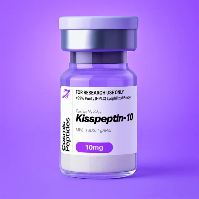
Kisspeptin-10
Commonly researched with these items
Patent-pending lot tracking: Complete transparency from production to vial.
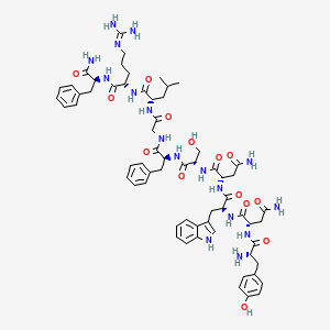
| CAS Number | 374675-21-5 |
|---|---|
| Molecular Formula | C63H83N17O14 |
| Molecular Weight | 1302.4 g/Mol |
| Purity | 99.7% |
| Lot Number | 26112 |
| Quantity | 5.76mg |
| Sequence | H-Tyr-Asn-Trp-Asn-Ser-Phe-Gly-Leu-Arg-Phe-NH2 |
Kisspeptin-10 (KP-10): The Minimal C-Terminal RF-Amide Decapeptide of the KISS1 Precursor
Disclaimer
Products described here are supplied for research use only and are not intended for diagnostic, therapeutic, or clinical application. All statements regarding biological activity reflect preclinical and in vitro findings exclusively and have not been evaluated by the U.S. Food and Drug Administration. These materials and related content are provided for educational and investigational purposes only and are intended solely for qualified researchers in laboratory and academic settings.
Key Facts
- Compound class
- Linear C-terminally amidated decapeptide; RF-amide peptide family; C-terminal fragment of the KISS1 gene product
- Sequence
H-Tyr-Asn-Trp-Asn-Ser-Phe-Gly-Leu-Arg-Phe-NH2(one-letter code: YNWNSFGLRF-NH2), all chiral residues in the L-configuration- Molecular formula
- C63H83N17O14
- Molecular weight
- 1302.4 g/mol (average, free base); monoisotopic mass 1301.6305 Da
- CAS number
- 374675-21-5
- PubChem CID
- 25240297
- Common synonyms
- KP-10, Kisspeptin-10 (human sequence), metastin (45–54), kisspeptin-13 (4–13)
- Molecular target studied in vitro
- Kisspeptin receptor KISS1R (formerly GPR54, AXOR12, hOT7T175); UniProt Q969F8, 398 residues
- Physical form as supplied
- Lyophilized powder
- Lot traceability
- Every unit is tied to a tracked lot; the lot number, recorded purity and verified quantity of the lot on sale are shown live on this page, with its certificate of analysis.
Molecular Identity and Classification
Kisspeptin-10 is the decapeptide H-Tyr-Asn-Trp-Asn-Ser-Phe-Gly-Leu-Arg-Phe-NH2, the shortest fully active C-terminal fragment of the precursor protein encoded by the KISS1 gene. In the mature precursor (UniProt Q15726, 138 residues, of which residues 1–19 form a signal sequence), the decapeptide occupies positions 112–121 and terminates in a phenylalanine amide, a post-translational modification recorded in the same database entry. Because the 54-residue fragment of this precursor was originally named metastin and numbered 1–54 beginning at residue 68, the same decapeptide appears in the older literature as metastin (45–54); PubChem retains both names as synonyms of CID 25240297.
The molecular formula is C63H83N17O14, with an average molecular weight of 1302.4 g/mol and a monoisotopic mass of 1301.6305 Da for the neutral free base. Summing the ten residue masses with a water-equivalent terminus and the C-terminal amide reproduces both values exactly; relative to the corresponding free acid, the amide terminus trades an oxygen for a nitrogen and a hydrogen, lowering the mass by 0.984 Da. The compound is registered under CAS number 374675-21-5 and carries the UNII code FS1N52VS3S.
Chemically, Kisspeptin-10 belongs to the RF-amide family, a group of peptides defined by the Arg-Phe-NH2 C-terminus. That terminal dipeptide amide is the family's signature recognition element. The receptor itself, first cloned as the orphan GPR54, was renamed the kisspeptin receptor (KISS1R) by the IUPHAR nomenclature committee once the ligand pairing had been established, following the convention of naming a receptor after its endogenous ligand. Longer natural fragments of the same precursor — the 54-, 14- and 13-residue forms, all sharing the identical C-terminus — are catalogued alongside the decapeptide in UniProt, and the decapeptide is described in the receptor-nomenclature literature as the smallest fragment retaining full activity at the receptor.
Note that "kisspeptin-13 (4–13)" is a synonym for the identical decapeptide, not a different molecule, because it counts the same ten residues from the N-terminus of the 13-residue fragment. Sequences also differ at position 10 between orthologues: the decapeptide annotated in UniProt entries Q7TSB7 and Q6Y4S4 is YNWNSFGLRY-NH2, ending in Tyr-NH2 rather than Phe-NH2. That single extra oxygen gives C63H83N17O15, a monoisotopic mass of 1317.6254 Da (15.9949 Da above the Phe10 form), a y1 ion at m/z 181.097 instead of 165.102 and a second tyrosine chromophore at 280 nm. A reported affinity or structure is therefore not automatically transferable between the two, and the material described here is the sequence ending in Phe-NH2.
Structure and Chemical Features
The decapeptide divides functionally into a polar, aromatic N-terminal half and a compact C-terminal recognition motif. Tyr1 contributes a phenolic hydroxyl with a side-chain pKa near 10.1 and, with Trp3, accounts for essentially all of the molecule's absorbance near 280 nm. Asn2 and Asn4 contribute neutral carboxamide side chains that are the principal chemical liability of the sequence, because asparagine undergoes non-enzymatic deamidation through a cyclic succinimide intermediate, converting Asn to Asp (or isoAsp) with a mass increase of 0.984 Da. The rate depends strongly on the following residue: Asn4 is followed by Ser5, one of the faster-deamidating contexts, while Asn2 is followed by the bulky Trp3 and is slower. Ser5 adds a small hydroxyl; Phe6 and Phe10 provide two of the three aromatic rings; Gly7 is a flexible hinge with no side chain; Leu8 is the most hydrophobic aliphatic residue; and Arg9 carries a guanidinium group with a pKa of roughly 12.5, which remains protonated across the entire practical pH range for aqueous work.
At pH 7 the molecule carries an N-terminal α-amino group (pKa near 8.6) and the Arg9 guanidinium, both protonated, with no ionisable acid at all: the C-terminus is an amide rather than a carboxylate, and the sequence contains no Asp or Glu. The net charge is therefore approximately +2, and the calculated isoelectric point lies above pH 11, far more basic than most decapeptides of comparable size. This has two immediate practical consequences. On reversed-phase columns it behaves as a strongly basic analyte and benefits from an acidic ion-pairing additive such as 0.1% trifluoroacetic acid; on cation-exchange media it binds tightly at neutral pH, which is sometimes used as an orthogonal purification step.
The absence of proline, cysteine and methionine simplifies the chemistry: no cis/trans amide isomers to double NMR resonances, no disulfide chemistry to control, no thioether to oxidise. The one oxidation-sensitive residue is Trp3, which can add a single oxygen (+15.995 Da) or form kynurenine-type products under light or peroxide exposure. Together with the two asparagines it defines the degradation profile expected on a stressed sample: a +0.98 Da family from deamidation and a +15.99 Da family from indole oxidation.
Solution Conformation and the RF-Amide Motif
Short peptides are usually described as disordered in water, but this sequence is an interesting exception in structuring solvents and membrane-mimetic media. NMR work with restrained molecular modelling reported by Gutiérrez-Pascual and colleagues in 2009, carried out on the Tyr10 orthologue of the decapeptide, found a helicoidal segment spanning Asn4 to Tyr10, with mixed α-helical and 310-helical character; alanine substitution at position 10 abolished that helix in the C-terminal region, while substitution at position 6 perturbed it only locally. Because the two sequences differ only at the final residue, the result is a reasonable structural model for the Phe10 form, but it was not measured on it. An independent NMR structure of the 13-residue fragment in sodium dodecyl sulfate micelles, reported by Orsini and colleagues in 2007, likewise described a relatively stable helix across the C-terminal seven residues and identified three residues — corresponding to Phe6, Arg9 and Phe10 of the decapeptide numbering — that lie on a single face of that helix and define the pharmacophore for receptor recognition.
In 2024 two independent cryo-electron microscopy studies resolved the receptor-bound conformation directly. Shen and colleagues determined KISS1R in complex with heterotrimeric Gq and with this decapeptide bound, at 3.06 Å resolution (Protein Data Bank entry 8ZJD), and described a markedly different intracellular transmembrane-helix-6 geometry — a roughly 40° angular deviation relative to other Gq-coupled receptors of the same class. Wu and colleagues, working with a synthetic agonist and with the 54-residue form rather than the decapeptide itself, solved complexes with the receptor coupled to both Gq and Gi, showed functionally that the receptor engages Gi/o as well as Gq/11, and attributed the coupling preference to conformational differences in the extracellular and intracellular loops and in the Gα "wavy hook". These structures turn an inference from alanine scanning into an observed binding mode.
Synthesis and Manufacturing Chemistry
The decapeptide is assembled by solid-phase peptide synthesis, the method introduced by Merrifield in 1963, almost always in the Fmoc mode that follows from the base-labile 9-fluorenylmethoxycarbonyl protecting group described by Carpino and Han in 1972. Because the C-terminus is a primary amide rather than a free acid, the synthesis starts not on a Wang or chlorotrityl resin but on a Rink amide or similar amide-generating support: the first residue, Phe10, is coupled to the resin's protected amine, and final acidolytic cleavage liberates the C-terminal carboxamide directly. The wrong resin yields the free-acid impurity, which differs from the target by only +0.984 Da and co-elutes closely in reversed-phase systems.
Chain extension runs from Phe10 towards Tyr1. Side-chain protection follows standard practice: tert-butyl for the Tyr1 phenol and the Ser5 hydroxyl, trityl for the two asparagine carboxamides, 2,2,4,6,7-pentamethyldihydrobenzofuran-5-sulfonyl (Pbf) for the Arg9 guanidinium, and tert-butyloxycarbonyl for the Trp3 indole nitrogen. The Boc group on tryptophan suppresses alkylation of the electron-rich indole ring by tert-butyl and Pbf cations released during final cleavage.
Unprotected asparagine side chains can dehydrate to the nitrile (−18.011 Da) under carbodiimide activation, hence trityl protection; arginine couplings are slow and prone to δ-lactam formation, so the Arg9 step is often double-coupled.
Final cleavage uses trifluoroacetic acid with a scavenger cocktail designed for tryptophan- and arginine-containing sequences — typically water with 1,2-ethanedithiol or triisopropylsilane and thioanisole — followed by precipitation in cold ether, preparative reversed-phase purification and lyophilization. Because trifluoroacetic acid is present through cleavage and purification, the isolated material is normally a trifluoroacetate salt unless it is deliberately exchanged into acetate or chloride. With two basic sites, the peptide can carry up to two trifluoroacetate counterions, adding about 228 g/mol to the 1302 g/mol free base; the salt form therefore has a direct and quantitatively significant effect on peptide content, discussed below.
Analytical Characterization
A complete package rests on four orthogonal measurements: reversed-phase HPLC (purity), high-resolution MS (identity), UV or amino-acid analysis (content) and ion chromatography (counterion).
Reversed-phase HPLC. A C18 column with a water/acetonitrile gradient containing 0.1% trifluoroacetic acid is the standard system. Record two wavelengths at once: 210–220 nm reports the amide backbone and therefore every peptidic species, while 280 nm reports only the Trp3 and Tyr1 chromophores. Comparing the traces is diagnostic — an impurity visible at 214 nm but absent at 280 nm cannot contain the aromatic N-terminal residues and is most likely a C-terminal truncation product.
Mass spectrometry. In positive electrospray ionisation the peptide gives a singly protonated ion [M+H]+ at m/z 1302.638, a doubly protonated ion [M+2H]2+ at m/z 651.823 (usually the base peak, since the molecule has two basic sites) and a measurable triply protonated ion at m/z 434.884. Tandem mass spectrometry of the 2+ precursor produces the b/y series tabulated below. The tryptophan immonium ion at m/z 159.092 is a convenient confirmation that Trp3 is present and unmodified. Note that y3 (434.287) and the 3+ precursor (434.884) sit only 0.597 units apart; the isotope spacing is the cleaner discriminator, 1.003 for the singly charged y3 against 0.334 for the triply charged precursor.
| n | bn (residues from Tyr1) | m/z bn | yn (residues from Phe10) | m/z yn |
|---|---|---|---|---|
| 1 | Y | 164.071 | F-NH2 | 165.102 |
| 2 | YN | 278.114 | RF | 321.203 |
| 3 | YNW | 464.193 | LRF | 434.287 |
| 4 | YNWN | 578.236 | GLRF | 491.309 |
| 5 | YNWNS | 665.268 | FGLRF | 638.377 |
| 6 | YNWNSF | 812.336 | SFGLRF | 725.409 |
| 7 | YNWNSFG | 869.358 | NSFGLRF | 839.452 |
| 8 | YNWNSFGL | 982.442 | WNSFGLRF | 1025.532 |
| 9 | YNWNSFGLR | 1138.543 | NWNSFGLRF | 1139.574 |
Two features of the ladder are diagnostic. The b4–b5 and y5–y6 gaps of 87.032 locate Ser5, and a deamidated Asn appears as a +0.984 shift beginning at b2 (Asn2) or b4 (Asn4), so the ladder localises which asparagine has converted. Because Arg9 sits next to the C-terminus, the singly charged y series tends to dominate low-energy collision spectra of the 2+ precursor.
Quantitation by UV. With one tryptophan and one tyrosine, the molar extinction coefficient at 280 nm follows from residue contributions by the approach of Pace and colleagues: ε280 ≈ 5500 + 1490 = 6990 M−1 cm−1 for the free base in water. A 1.00 mg/mL free-base-equivalent solution (768 µM) therefore reads about 5.37 absorbance units in a 1 cm cell, so a 10- to 20-fold dilution lands in the reliable 0.2–1.0 range — a route to concentration that does not depend on weighing a hygroscopic powder.
Purity versus peptide content. These are different quantities and are routinely conflated. HPLC purity is the fraction of UV-absorbing material that is the target peptide. Peptide content is the fraction of the weighed mass that is peptide at all, the remainder being counterions, bound water and residual salts. For this sequence the arithmetic is substantial: a di-trifluoroacetate salt has a nominal mass near 1530.5 g/mol, so even a chromatographically pure powder is only about 85% peptide by mass (1302.4 / 1530.5), before any water of hydration is counted. Worked example: 5.00 mg of such a salt contains roughly 4.25 mg of peptide, which is 3.27 µmol; dissolved in 2.00 mL of water this yields 1.63 mM rather than the 1.92 mM that a naïve calculation from the weighed mass would predict — a 15% error that propagates directly into every reported concentration. Amino-acid analysis after acid hydrolysis (expected molar ratios Asn/Asp 2, Phe 2, Tyr 1, Ser 1, Gly 1, Leu 1, Arg 1, with tryptophan destroyed by standard hydrolysis and requiring a separate determination) or quantitative NMR settles the question definitively.
Physicochemical Properties
| Property | Value | Notes |
|---|---|---|
| Molecular formula | C63H83N17O14 | Free base, no counterions |
| Average molecular weight | 1302.4 g/mol | PubChem CID 25240297 |
| Monoisotopic mass | 1301.6305 Da | For high-resolution MS |
| [M+H]+ / [M+2H]2+ / [M+3H]3+ | m/z 1302.638 / 651.823 / 434.884 | Positive-mode ESI; 2+ usually dominant |
| Nominal mass of di-trifluoroacetate salt | ≈1530.5 g/mol | Free base + 2 × 114.02; affects peptide content |
| Net charge at pH 7 | approximately +2 | N-terminal amine and Arg9; no acidic side chain |
| Isoelectric point | above pH 11 (calculated) | Strongly basic; C-terminus is an amide |
| ε280 (water) | ≈6990 M−1 cm−1 | One Trp + one Tyr (Pace method) |
| A280 of a 1 mg/mL solution | ≈5.37 (1 cm path) | Dilute before reading |
| 1 mg/mL in molar terms | 768 µM | Free-base-equivalent basis |
| XLogP3 | −1.9 | Computed; hydrophilic overall |
| Topological polar surface area | 538 Å2 | Computed |
| Hydrogen-bond donors / acceptors | 18 / 16 | Computed |
| Rotatable bonds | 38 | Computed; flexible in free solution |
| Aromatic residues | Tyr1, Trp3, Phe6, Phe10 | Source of 280 nm absorbance and of UV sensitivity |
| Chemically labile positions | Asn2, Asn4 (deamidation, +0.984 Da); Trp3 (oxidation, +15.995 Da) | Expected degradation products on stressed samples |
| Solubility | Readily soluble in water and in aqueous buffers; also soluble in dimethyl sulfoxide | Basic, polar, no disulfide |
The descriptors describe a flexible, strongly hydrogen-bonding, net-cationic peptide: 38 rotatable bonds and 538 Å2 of polar surface fit a weak free-solution helical preference that membrane-mimetic or receptor-bound environments stabilise, and argue strongly against passive diffusion across lipid bilayers.
In the solid state the lyophilized powder is hygroscopic; in solution the dominant risks are deamidation at the two asparagines (base- and temperature-accelerated), indole oxidation at Trp3 (light- and peroxide-accelerated) and adsorption losses at sub-micromolar concentration. With no cysteine there is no disulfide scrambling or dimerisation pathway, which makes this sequence more forgiving than many of similar length.
Research Context
The molecule's history illustrates how a gene product can be characterised before its chemistry is understood. In 1996 Lee and colleagues identified KiSS-1 by subtractive hybridisation as a complementary DNA expressed in non-metastatic but not metastatic melanoma cell lines. Separately, in 1999, Lee and colleagues cloned the orphan G-protein-coupled receptor GPR54, noting 44–45% transmembrane identity with galanin receptors but no galanin binding. The threads met in 2001: Ohtaki and colleagues isolated a 54-residue C-terminally amidated peptide as the ligand of the orphan receptor hOT7T175, Kotani and colleagues isolated the 54-, 14- and 13-residue forms, named them kisspeptins and showed low-nanomolar binding at the receptor expressed in Chinese hamster ovary K1 cells. The nomenclature review by Kirby and colleagues in 2010 consolidated the naming and identified the decapeptide as the smallest active fragment.
Receptor Binding and Signalling In Vitro
In receptor-expressing cell lines, binding of the decapeptide to KISS1R couples predominantly to Gq/11 and the phospholipase C pathway: hydrolysis of phosphatidylinositol 4,5-bisphosphate, accumulation of inositol 1,4,5-trisphosphate, mobilisation of intracellular Ca2+, activation of protein kinase C, release of arachidonic acid, and phosphorylation of ERK1/2 and p38 mitogen-activated protein kinases — the set of read-outs Kotani and colleagues reported in transfected CHO-K1 cells. The signalling review by Castaño and colleagues adds the phosphatidylinositol-3-kinase/Akt and nuclear-factor-κB branches and cross-talk to the chemokine receptor CXCR4, while the 2024 structural work shows that the receptor also couples to Gi/o, so a Ca2+-only read-out captures one arm of the response.
Quantitatively, alanine-scanning work by Gutiérrez-Pascual and colleagues in CHO cells transfected with the Tyr10 orthologue's receptor placed half-maximal [Ca2+]i responses for the unmodified decapeptides in the 10−8 M range (1.54 × 10⁻⁸ M for the Tyr10 form and 2.6 × 10⁻⁸ M for the Phe10 form), while alanine substitution of the Tyr10 form at position 6 or position 10 raised the value above 6.46 × 10⁻⁶ M, reduced the proportion of responsive cells and lengthened the time to maximal response — a shift of more than two orders of magnitude from two single-residue changes, and the clearest available demonstration that the C-terminal face of the helix carries the recognition determinants. Immortalised neuronal cell lines provide a second in vitro system: Novaira and colleagues documented receptor expression in the GT1-7 and GN11 lines and reported that kisspeptin produced concentration- and time-dependent increases in gonadotropin-releasing hormone transcript levels and release, with the effect attenuated by the ERK1/2 pathway inhibitor PD98059 and the phosphatidylinositol-3-kinase inhibitor LY294002, and with increased phosphorylation of MAPK and Akt substrates by immunoblot.
Selectivity and Proteolytic Processing
Two chemical findings shape experimental design with this sequence. First, selectivity is not absolute within the RF-amide family: Oishi and colleagues reported that the decapeptide binds and activates the neuropeptide FF receptors NPFFR1 and NPFFR2, whereas shortened agonist analogues bound those receptors much more weakly and were correspondingly more selective for KISS1R. Experiments attributing an in vitro effect to KISS1R therefore benefit from a selective control compound or a receptor-knockdown comparison. Second, the peptide is a defined substrate for matrix metalloproteinases: Takino and colleagues showed in 2003 that MMP-2, MMP-9, MT1-MMP, MT3-MMP and MT5-MMP all cleave the Gly–Leu bond of the decapeptide — the Gly7–Leu8 bond in decapeptide numbering, Gly118–Leu119 in precursor numbering — and that cleavage abolishes ligand activity. The cut yields products matching b7 (869.358) and y3 (434.287), so proteolysis in assay medium is easy to follow by LC-MS and ligand half-life in the well is best measured rather than assumed.
Structure–Activity and Analogue Chemistry
Because the pharmacophore sits in the C-terminal half, analogue chemistry on this scaffold has focused on truncation and backbone modification. Niida and colleagues ran alanine and D-amino-acid scans and then synthesised shortened derivatives in a yeast reporter system expressing the human receptor: derivatives of only a few residues were strong agonists provided they kept an N-terminal basic group and a C-terminal aromatic-amide motif. Orsini and colleagues used the NMR-derived pharmacophore of the 13-residue fragment to screen small-molecule databases and reported non-peptidic full agonists with submicromolar affinity.
The Kisspeptin Fragment Family at a Glance
| Fragment | Residues in precursor | Length | Formula / average mass | Notes |
|---|---|---|---|---|
| Kisspeptin-54 (metastin) | 68–121 | 54 | — | Originally isolated form; "metastin" in UniProt |
| Kisspeptin-14 | 108–121 | 14 | — | Annotated cleavage product |
| Kisspeptin-13 | 109–121 | 13 | C78H107N21O18, 1626.8 g/mol (CID 101134421) | Subject of the SDS-micelle NMR structure |
| Kisspeptin-10 | 112–121 | 10 | C63H83N17O14, 1302.4 g/mol (CID 25240297) | Smallest fragment with full in vitro activity; the material here |
All share the C-terminus and so give identical y1–y3 ions; the b series and intact mass (1626.8 against 1302.4 g/mol for the 13- and 10-residue forms) discriminate between them.
Related Research Peptides
The chemically closest analytical neighbours are other C-terminally amidated sequences, which share both the amide-terminus mass arithmetic and the free-acid impurity problem described above. Oxytocin is a useful method-development reference: likewise amidated, likewise carrying a tyrosine chromophore, and likewise demanding that an analyst separate the amide from its free-acid counterpart. For counterion correction and peptide-content determination, the small, highly basic tripeptide KPV shows the same arithmetic in its most extreme form, since the counterion mass fraction grows as peptide mass falls.
Laboratory Handling and Storage
Storing the solid. Keep the lyophilized powder tightly closed at −20 °C or below, protected from light and moisture; the powder is hygroscopic and the indole of Trp3 is photosensitive, so amber or foil-wrapped containers and a desiccated environment are worth the trouble. Let a container equilibrate to room temperature inside a desiccator before opening, so atmospheric water does not condense on cold powder and inflate the weighed mass.
Preparing solutions. The peptide dissolves readily in high-purity water; for stock solutions, mildly acidic water (for example water with 0.1% acetic acid) is a common choice because the two basic sites are fully protonated there and deamidation is slowest at acidic pH. Dimethyl sulfoxide is an alternative for concentrated stocks where the downstream assay tolerates it. Prepare working dilutions in the assay buffer immediately before use, and correct the weighed mass for peptide content rather than assuming the powder is 100% peptide; alternatively, verify the concentration spectrophotometrically using the ε280 value of about 6990 M−1 cm−1.
Aliquoting and freeze–thaw. Divide stocks into single-use aliquots at 1–10 mM in low-binding polypropylene tubes and store them at −80 °C; repeated freeze–thaw cycles are the most avoidable cause of apparent loss of activity. At sub-micromolar concentrations, adsorption of this net-cationic peptide to plastic and glass becomes measurable, so low-binding labware and, where the design permits, a carrier protein in the buffer reduce those losses.
Counterion controls. For trifluoroacetate salts, include a vehicle control matched for trifluoroacetate concentration: Cornish and colleagues showed that trifluoroacetate alone influences the proliferation of cultured cells, so an unmatched control can misattribute a counterion effect to the peptide.
Re-verification. After long storage, an LC-MS check of the stock shows the 2+ ion at m/z 651.82, with deamidation 0.49 and Trp oxidation 8.00 m/z units above it. Each lot's certificate of analysis, shown live on this page, records the analytical results for that lot.
Frequently Asked Research Questions
- What is Kisspeptin-10?
- Kisspeptin-10 is the C-terminally amidated decapeptide H-Tyr-Asn-Trp-Asn-Ser-Phe-Gly-Leu-Arg-Phe-NH2, corresponding to residues 112–121 of the KISS1 precursor protein. It has the molecular formula C63H83N17O14, an average molecular weight of 1302.4 g/mol and CAS number 374675-21-5, and it is the shortest natural fragment of that precursor described in the receptor-nomenclature literature as retaining full activity at KISS1R in vitro.
- Why is it also called metastin (45–54) or kisspeptin-13 (4–13)?
- All three names describe the same molecule counted from different reference points. "Metastin" named the 54-residue fragment, numbered 1–54 from precursor residue 68, so this decapeptide is its residues 45–54; "kisspeptin-13 (4–13)" counts the same ten residues from the N-terminus of the 13-residue fragment. PubChem lists all of them as synonyms of CID 25240297.
- What masses should the peptide show by mass spectrometry?
- Monoisotopic mass 1301.6305 Da; in positive ESI, [M+H]+ m/z 1302.638, [M+2H]2+ 651.823 (usually the base peak) and [M+3H]3+ 434.884. Characteristic fragments include y1 165.102, y2 321.203, y3 434.287, b2 278.114, b7 869.358 and the tryptophan immonium ion at 159.092.
- What is the net charge and isoelectric point?
- At pH 7 the net charge is approximately +2, from the N-terminal α-amino group and the Arg9 guanidinium; the sequence contains no acidic side chain and the C-terminus is an amide, not a carboxylate, so the calculated isoelectric point lies above pH 11.
- How can the concentration be verified without relying on weighing?
- The sequence contains one tryptophan and one tyrosine, giving a predicted molar extinction coefficient at 280 nm of about 6990 M−1 cm−1. A 1 mg/mL free-base-equivalent solution (768 µM) therefore reads about 5.37 absorbance units in a 1 cm cell, so a 10- to 20-fold dilution gives a reading in the accurate range.
- Why does the trifluoroacetate counterion matter so much here?
- With two basic sites, a di-trifluoroacetate salt weighs roughly 1530.5 g/mol against a free base of 1302.4 g/mol, so only about 85% of the weighed mass is peptide; ignoring the correction overstates molar concentration by about 17% (the true value is about 15% below the naive one) before any bound water is counted.
- Which degradation products should a stability check look for?
- Deamidation of Asn2 or Asn4 (+0.984 Da, 0.49 m/z above the 2+ ion) and oxidation of the Trp3 indole (+15.995 Da, 8.00 above it). With no cysteine, disulfide scrambling is not a route.
- Which residues are critical for receptor recognition in vitro?
- Alanine scanning of the Tyr10 orthologue in receptor-transfected CHO cells showed that substitution at position 6 or position 10 raises the half-maximal Ca2+-response concentration from the 10−8 M range to above 6.46 × 10⁻⁶ M. NMR-based pharmacophore analysis of the 13-residue fragment in sodium dodecyl sulfate micelles places Phe6, Arg9 and Phe10 on one face of a C-terminal helix; in free aqueous solution the decapeptide is largely flexible, and 2024 cryo-electron microscopy structures (PDB 8ZJD) show its receptor-bound conformation directly.
- Which signalling read-outs are used in cell-based assays?
- Intracellular Ca2+ mobilisation, inositol phosphate accumulation, and phospho-specific assays for ERK1/2, p38, Akt and protein kinase C substrates. Because the receptor couples to Gi/o as well as Gq/11, a Ca2+-only read-out samples one branch.
- Is the peptide selective within the RF-amide family?
- Not entirely. The decapeptide has been reported to bind and activate the neuropeptide FF receptors NPFFR1 and NPFFR2, while shortened agonist analogues show greater selectivity for KISS1R. Assays attributing an effect to KISS1R are stronger with a selective comparator or a receptor-knockdown control.
- How stable is the peptide in assay medium?
- It is a defined substrate for matrix metalloproteinases, which cleave the Gly7–Leu8 bond (Gly118–Leu119 in precursor numbering) and abolish ligand activity, so in media with metalloproteinase activity the ligand half-life should be measured rather than assumed. The cleavage is easy to follow by LC-MS as loss of intact mass with appearance of the 869.36 and 434.29 fragments.
- How should the lyophilized powder be stored?
- Closed, at −20 °C or below, protected from light and moisture, and warmed to room temperature in a desiccator before opening; solutions as single-use aliquots at −80 °C.
Sources & Further Reading
- National Center for Biotechnology Information. PubChem Compound Summary for CID 25240297, Kisspeptin-10. PubChem CID 25240297.
- UniProt Consortium. Metastasis-suppressor KiSS-1 (Homo sapiens). UniProtKB entry Q15726 (KISS1_HUMAN). KiSS-1 receptor, UniProtKB entry Q969F8 (KISS1R_HUMAN).
- Lee JH, Miele ME, Hicks DJ, Phillips KK, Trent JM, Weissman BE, Welch DR. KiSS-1, a novel human malignant melanoma metastasis-suppressor gene. JNCI Journal of the National Cancer Institute 1996;88(23):1731–1737. doi:10.1093/jnci/88.23.1731
- Lee DK, Nguyen T, O'Neill GP, Cheng R, Liu Y, Howard AD, et al. Discovery of a receptor related to the galanin receptors. FEBS Letters 1999;446(1):103–107. doi:10.1016/S0014-5793(99)00009-5
- Ohtaki T, Shintani Y, Honda S, Matsumoto H, Hori A, Kanehashi K, et al. Metastasis suppressor gene KiSS-1 encodes peptide ligand of a G-protein-coupled receptor. Nature 2001;411(6837):613–617. doi:10.1038/35079135
- Kotani M, Detheux M, Vandenbogaerde A, Communi D, Vanderwinden JM, Le Poul E, et al. The metastasis suppressor gene KiSS-1 encodes kisspeptins, the natural ligands of the orphan G protein-coupled receptor GPR54. Journal of Biological Chemistry 2001;276(37):34631–34636. doi:10.1074/jbc.M104847200
- Kirby HR, Maguire JJ, Colledge WH, Davenport AP. International Union of Basic and Clinical Pharmacology. LXXVII. Kisspeptin receptor nomenclature, distribution, and function. Pharmacological Reviews 2010;62(4):565–578. doi:10.1124/pr.110.002774
- Takino T, Koshikawa N, Miyamori H, Tanaka M, Sasaki T, Okada Y, et al. Cleavage of metastasis suppressor gene product KiSS-1 protein/metastin by matrix metalloproteinases. Oncogene 2003;22(30):4617–4626. doi:10.1038/sj.onc.1206542
- Orsini MJ, Klein MA, Beavers MP, Connolly PJ, Middleton SA, Mayo KH. Metastin (KiSS-1) mimetics identified from peptide structure-activity relationship-derived pharmacophores and directed small molecule database screening. Journal of Medicinal Chemistry 2007;50(3):462–471. doi:10.1021/jm0609824
- Niida A, Wang Z, Tomita K, Oishi S, Tamamura H, Otaka A, et al. Design and synthesis of downsized metastin (45–54) analogs with maintenance of high GPR54 agonistic activity. Bioorganic & Medicinal Chemistry Letters 2006;16(1):134–137. doi:10.1016/j.bmcl.2005.09.054
- Gutiérrez-Pascual E, Leprince J, Martínez-Fuentes AJ, Ségalas-Milazzo I, Pineda R, Roa J, et al. In vivo and in vitro structure-activity relationships and structural conformation of kisspeptin-10-related peptides. Molecular Pharmacology 2009;76(1):58–67. doi:10.1124/mol.108.053751
- Castaño JP, Martínez-Fuentes AJ, Gutiérrez-Pascual E, Vaudry H, Tena-Sempere M, Malagón MM. Intracellular signaling pathways activated by kisspeptins through GPR54: do multiple signals underlie function diversity? Peptides 2009;30(1):10–15. doi:10.1016/j.peptides.2008.07.025
- Novaira HJ, Ng Y, Wolfe A, Radovick S. Kisspeptin increases GnRH mRNA expression and secretion in GnRH secreting neuronal cell lines. Molecular and Cellular Endocrinology 2009;311(1–2):126–134. doi:10.1016/j.mce.2009.06.011
- Oishi S, Misu R, Tomita K, Setsuda S, Masuda R, Ohno H, et al. Activation of neuropeptide FF receptors by kisspeptin receptor ligands. ACS Medicinal Chemistry Letters 2011;2(1):53–57. doi:10.1021/ml1002053
- Shen S, Wang D, Liu H, He X, Cao Y, Chen J, et al. Structural basis for hormone recognition and distinctive Gq protein coupling by the kisspeptin receptor. Cell Reports 2024;43(7):114389. doi:10.1016/j.celrep.2024.114389 (Protein Data Bank entry 8ZJD, 3.06 Å)
- Wu Z, Chen G, Qiu C, Yan X, Xu L, Jiang S, et al. Structural basis for the ligand recognition and G protein subtype selectivity of kisspeptin receptor. Science Advances 2024;10(33):eadn7771. doi:10.1126/sciadv.adn7771
- Merrifield RB. Solid phase peptide synthesis. I. The synthesis of a tetrapeptide. Journal of the American Chemical Society 1963;85(14):2149–2154. doi:10.1021/ja00897a025
- Carpino LA, Han GY. 9-Fluorenylmethoxycarbonyl amino-protecting group. The Journal of Organic Chemistry 1972;37(22):3404–3409. doi:10.1021/jo00795a005
- Pace CN, Vajdos F, Fee L, Grimsley G, Gray T. How to measure and predict the molar absorption coefficient of a protein. Protein Science 1995;4(11):2411–2423. doi:10.1002/pro.5560041120
- Cornish J, Callon KE, Lin CQX, Xiao CL, Mulvey TB, Cooper GJS, Reid IR. Trifluoroacetate, a contaminant in purified proteins, inhibits proliferation of osteoblasts and chondrocytes. American Journal of Physiology-Endocrinology and Metabolism 1999;277(5):E779–E783. doi:10.1152/ajpendo.1999.277.5.E779
Last reviewed:
In the research library
Everything the library holds on Kisspeptin-10, and the methods behind the numbers on its certificate.
- Kisspeptin-10 reference entry Identity, calculated properties, the mass spectrometry ions to expect and analytical notes, from the structure.
- Check a certificate against this sequence Paste the masses from a spectrum or a certificate; the mass-match tool compares them with what this sequence should give.
- Peptide calculator, with this sequence Formula, average and monoisotopic mass, charge across pH, isoelectric point and absorbance, recalculated as you edit.
- How to read a certificate of analysis What a peptide certificate of analysis should contain, how to check that it belongs to the material in hand, how to read its identity and purity results, and the signs that it deserves a second look.
- HPLC purity analysis of peptides How reversed-phase HPLC measures the chromatographic purity of a peptide, what the figure includes, and what it leaves out.
- Mass spectrometry for peptide identity How mass spectrometry confirms that a peptide is the intended sequence: electrospray and MALDI, working out the expected mass, reading charge states and adducts, sequence confirmation, and the limits of the method.
- Net peptide content and purity: two different numbers Why a peptide that is 99% pure by HPLC can be only 75–85% peptide by weight: what the powder contains, how each part is measured, and how the two figures combine.
- Preparing peptide solutions in the laboratory From a freeze-dried vial to a solution of known concentration: the arithmetic, the solvent, the charge chemistry and the handling, with the errors each step can introduce and the article that covers it.
- Peptide stability and storage How peptides degrade, chemically and physically, why the dry solid lasts far longer than a solution, which sequences are most at risk, and the storage and handling that slow it down.