LL-37
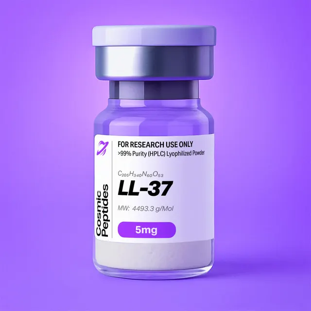
LL-37
Commonly researched with these items
Patent-pending lot tracking: Complete transparency from production to vial.
LL-37: The C-Terminal Antimicrobial Domain of Human Cathelicidin hCAP-18
Disclaimer
Products described here are supplied for research use only and are not intended for diagnostic, therapeutic, or clinical application. All statements regarding biological activity reflect preclinical and in vitro findings exclusively and have not been evaluated by the U.S. Food and Drug Administration. These materials and related content are provided for educational and investigational purposes only and are intended solely for qualified researchers in laboratory and academic settings.
Key Facts
- Compound class
- Cationic, amphipathic α-helical antimicrobial peptide; 37-residue C-terminal domain of the human cathelicidin precursor hCAP-18 (gene CAMP)
- Sequence
H-Leu-Leu-Gly-Asp-Phe-Phe-Arg-Lys-Ser-Lys-Glu-Lys-Ile-Gly-Lys-Glu-Phe-Lys-Arg-Ile-Val-Gln-Arg-Ile-Lys-Asp-Phe-Leu-Arg-Asn-Leu-Val-Pro-Arg-Thr-Glu-Ser-OH(one-letter codeLLGDFFRKSKEKIGKEFKRIVQRIKDFLRNLVPRTES), all residues in the L-configuration, free N-terminal amine and free C-terminal acid- Molecular formula
- C205H340N60O53
- Molecular weight
- 4493.3 g/mol (average, free base); monoisotopic mass 4490.58 Da
- CAS number
- 597562-32-8 (also registered as 154947-66-7)
- PubChem CID
- 16198951
- Net charge at pH 7
- approximately +6 (twelve basic sites, six acidic sites)
- Physical form as supplied
- Lyophilized powder
- Lot traceability
- Every unit is tied to a tracked lot; the lot number, recorded purity and verified quantity of the lot on sale are shown live on this page, with its certificate of analysis.
Molecular Identity and Classification
LL-37 is a 37-residue linear peptide whose name comes from its own chemistry: the chain begins with two leucine residues (LL) and is 37 amino acids long. It is the mature antimicrobial domain released from the C-terminus of hCAP-18, the cationic antimicrobial protein of 18 kDa and the only cathelicidin known in this species. Its gene, CAMP, lies on chromosome 3 at 3p21.3 and encodes a preproprotein consisting of a signal sequence, a conserved cathelin-like domain of roughly 100 residues, and the variable C-terminal antimicrobial segment that becomes LL-37 after proteolytic release. Sørensen and colleagues showed in 2001 that extracellular cleavage by proteinase 3 converts stored hCAP-18 into LL-37, so the peptide is generated by proteolysis of a stored precursor rather than expressed directly as the mature chain.
Chemically, LL-37 belongs to the cationic amphipathic α-helical class of antimicrobial peptides, the structural family that also contains magainins, cecropins and melittin. The defining features of that class are all present: a high content of basic residues (six lysines and five arginines), a modest number of acidic residues (two aspartates and three glutamates), no cysteine and therefore no disulfide constraint, and a residue order in which hydrophobic and cationic side chains segregate onto opposite faces once the backbone is helical. The complete amino-acid composition is Lys6, Arg5, Phe4, Leu4, Glu3, Ile3, Asp2, Gly2, Ser2, Val2, Asn1, Gln1, Pro1 and Thr1.
Three absences shape both the chemistry and the analytics of this sequence. No cysteine, so the peptide forms no disulfides and needs no oxidative folding step after synthesis. No methionine, removing the most common site of oxidative modification in stored peptide solutions. And no tryptophan, tyrosine or histidine, so the molecule has essentially no absorbance at 280 nm and no intrinsic fluorescence; the four phenylalanines absorb only weakly near 257 nm. Every quantitative and spectroscopic method applied to LL-37 has to work around that spectroscopic silence, which is why peptide-bond absorbance, amino-acid analysis and mass spectrometry carry the analytical load.
Residue numbering in the literature counts from Leu1 of LL-37, not from the start of the hCAP-18 preproprotein, and fragment names follow the same convention as the parent: first and last residue in one-letter code plus chain length, as in LL-23, GF-17 and KR-12.
Structure and Chemical Features
LL-37 is conformationally plastic: in dilute aqueous buffer of low ionic strength it is largely disordered, and it folds into an α-helix when the ionic strength rises, when the pH moves away from neutrality, or when it meets an anionic surface such as a lipid bilayer or a detergent micelle. Johansson and colleagues demonstrated this directly in 1998 by circular dichroism, showing that helical content increases with salt and with anion concentration and that antibacterial activity tracks the folded state, which is why the peptide is described as conformation-dependent rather than intrinsically structured.
Solution NMR in membrane-mimetic micelles gives the clearest picture of the folded state. Wang determined structures of LL-37 and of its minimal fragment KR-12 in lipid micelles in 2008 (Protein Data Bank entry 2K6O), and Porcelli and colleagues determined the structure in dodecylphosphocholine micelles in the same year. Both describe a long amphipathic helix occupying most of the chain, a bend near Gly14, and a short, flexible C-terminal segment after Pro33. The single proline is the structural full stop of the molecule: its nitrogen carries no hydrogen, so it cannot donate the backbone hydrogen bond an α-helix requires at that position, the helix terminates there, and the Arg34-Thr35-Glu36-Ser37 tail stays mobile.
Crystallography added a second layer of detail. Sancho-Vaello and colleagues reported in 2017 an atomic-resolution structure of a monomeric form at 0.95 Å (entry 5NMN) together with forms crystallized in the presence of detergents at 1.80, 1.90 and 2.21 Å (entries 5NNK, 5NNM and 5NNT), and proposed that on membranes the peptide remodels into extended, fibril-like assemblies. These structures bear on an observation reported much earlier by Oren and colleagues in 1999: at physiological ionic strength LL-37 self-associates rather than staying monomeric, and that oligomeric state is central both to its resistance to proteolysis and to its low selectivity between anionic and zwitterionic bilayers.
Amphipathic Organization
Mapping the sequence onto a helical wheel with 100° per residue shows why this peptide is a textbook amphipath. The hydrophobic face is built from Phe5, Phe6, Ile13, Phe17, Ile20, Ile24, Phe27 and Leu28 together with Val21 and Leu31, while the opposite face carries the cationic cluster Arg7, Lys8, Lys10, Lys12, Lys15, Lys18, Arg19, Arg23, Lys25 and Arg29. Four of the five acidic residues (Asp4, Glu11, Glu16, Asp26) sit near the boundary between the faces, where they can form intramolecular salt bridges with neighbouring lysines and arginines; that partial charge neutralisation is one reason the peptide tolerates so high a charge density without precipitating. The mean hydrophobicity on the Kyte–Doolittle scale is only −0.72, yet the hydrophobic moment is large, because the hydrophobic residues are segregated onto one face rather than spread evenly.
Residues 1–6 (Leu-Leu-Gly-Asp-Phe-Phe) form the most hydrophobic stretch in the molecule and anchor the helix into the acyl-chain region of a bilayer; truncating or substituting this segment changes insertion depth without altering net charge, which is why it is a frequent target of sequence-modification studies.
Discovery as Chemistry
LL-37 entered the literature in 1995 from two independent directions, each using a different chemical strategy, and the convergence of those routes is a good illustration of how a peptide gets identified. Agerberth and colleagues, working from bone-marrow cDNA, described a putative cysteine-free peptide antibiotic they named FALL-39 after the first four residues and the assumed length of 39 amino acids. Cowland and colleagues, working from protein, isolated a cathelin-like species from the specific granules of neutrophils by chromatography and named it hCAP-18 for its apparent 18 kDa mass. When Gudmundsson and colleagues sequenced the processed peptide released from granulocyte hCAP-18 in 1996, the mature chain proved to begin two residues later than the cDNA-based prediction, at Leu-Leu rather than Phe-Ala, and the 37-residue designation LL-37 replaced FALL-39.
The two names remain useful chemically, because FALL-39 and LL-37 are genuinely different molecules: FALL-39 has the formula C217H354N62O55 and an average mass of 4711.6 g/mol, 218.3 Da heavier than LL-37 by exactly one phenylalanine plus one alanine residue. A mass spectrum that shows 4711 rather than 4493 is reporting the longer construct, and comparisons of activity data between early and later papers need to establish which chain was used. This is the single most common identity ambiguity in the LL-37 literature, and it is resolved entirely by intact mass.
Synthesis and Manufacturing Chemistry
LL-37 is prepared by solid-phase peptide synthesis, in which the chain is assembled on an insoluble support one residue at a time from the C-terminus towards the N-terminus. Because the product carries a free C-terminal acid, assembly starts from a serine residue attached to an acid-labile resin; chains of this length are typically built with Fmoc chemistry, in which a base-labile 9-fluorenylmethoxycarbonyl group protects each incoming α-amine and acid-labile groups protect the side chains.
At 37 residues with eleven protected basic side chains, this sequence sits at the demanding end of routine synthesis, and three features dominate the chemistry. First, cumulative yield: even at 99.5% efficiency per coupling, 36 couplings leave only about 84% of chains intact, so stepwise efficiency rather than any single step sets the crude purity. Second, protecting-group load: the arginines require bulky sulfonyl protection, and the final acidolytic cleavage has to remove the whole protecting-group complement at once. Third, aggregation on the resin: long amphipathic sequences form β-sheet-like associations during assembly that slow deprotection and coupling, which is why such syntheses use elevated temperature, chaotropic additives, pseudoproline or backbone-amide protection, or ligation of two shorter segments.
Known chain-level by-products that purification must resolve include single-residue deletions, incompletely deprotected species that still carry one sulfonyl or tert-butyl group, and aspartimide-related products arising at the Asp4 and Asp26 positions during repeated base exposure. Each of these differs from the target by a defined mass increment, so high-resolution mass spectrometry of the crude mixture is diagnostic before any chromatography is attempted. Preparative reversed-phase HPLC with trifluoroacetic acid as the ion-pairing agent is the standard purification, after which the material is lyophilized; because trifluoroacetate pairs with every basic site, the isolated solid is normally a trifluoroacetate salt unless it is deliberately exchanged into acetate or chloride.
Analytical Characterization
Four methods together establish the identity and quality of a 4.5 kDa cationic peptide, and each answers a different question.
Reversed-phase HPLC answers how much of the UV-absorbing material is the target. Detection is at 210–220 nm, where the amide bonds absorb; the absence of tryptophan and tyrosine means a 280 nm trace is effectively blank and cannot be used. A C18 column with a water/acetonitrile gradient containing 0.1% trifluoroacetic acid is conventional. Two behaviours are characteristic: the peptide is strongly retained for its polarity, because the hydrophobic face binds the stationary phase as a folded helix, and peaks sharpen at elevated column temperature, where monomer–oligomer exchange becomes fast on the chromatographic timescale. Tailing in an LL-37 chromatogram usually reflects that exchange, or interaction with residual silanols, rather than impurity.
Mass spectrometry answers whether the molecule is the right one. In positive-mode electrospray ionization a peptide with twelve basic sites gives a broad charge-state envelope; the ions usually dominant for a chain of this length and charge density are [M+4H]4+ at m/z 1124.3, [M+5H]5+ at m/z 899.7, [M+6H]6+ at m/z 749.9 and [M+7H]7+ at m/z 642.9, all calculated from the average mass, which is the appropriate basis on instruments that do not resolve isotopes. On a high-resolution instrument the isotope pattern is resolved and the monoisotopic peak is no longer the tallest: for C205H340N60O53 the computed envelope peaks at the second 13C isotopologue, with the monoisotopic signal at roughly 31% of the apex (computed relative intensities M+0 31%, M+1 78%, M+2 100%, M+3 88%, M+4 60%). This is also why a database field reporting the mass of the most abundant isotopologue, such as the value 4492.58 Da listed for PubChem CID 16198951, sits about 2 Da above the monoisotopic 4490.58 Da; the two numbers describe different peaks of the same envelope. Comparing a measured mass against the wrong reference produces an apparent 2–3 Da discrepancy that is purely arithmetic.
Sequence confirmation by tandem MS is complicated by the same basicity that makes the peptide easy to ionize: trypsin cleaves after lysine and arginine, and with eleven such residues a tryptic digest consists largely of fragments of one to four residues that carry little sequence information. Chymotrypsin, which cuts after the four phenylalanines, gives informative mid-sized fragments, and top-down electron-transfer dissociation of the intact multiply charged ion handles such highly charged precursors better than collision-induced dissociation.
Amino-acid analysis and peptide content answer how much peptide is actually in the weighed powder. After acid hydrolysis the expected molar ratios are Lys:Arg:Phe:Leu of 6:5:4:4, with Asn and Gln appearing as Asp and Glu, so hydrolysate values for those two pairs are reported combined (Asx 3, Glx 4). The distinction between HPLC purity and peptide content is unusually consequential here. Purity is the fraction of UV-absorbing material that is LL-37; peptide content is the fraction of the weighed solid that is peptide rather than counterion, water or salt. With up to twelve trifluoroacetate ions (114.02 g/mol each) available to pair with the basic sites, a fully paired preparation would be only about 77% peptide by mass and a partially exchanged one about 87%, so a nominal 1 mM stock prepared by weight alone can be 13–23% low in true molarity. Ion chromatography quantifies the counterion, and Karl Fischer titration or thermogravimetry the residual water.
Spectroscopy answers what conformation the molecule is in. With no usable aromatic chromophore, concentration is best determined from absorbance at 205 nm using the sequence-based extinction coefficients described by Anthis and Clore, or from quantitative amino-acid analysis. Far-UV circular dichroism is the conformational read-out: a disordered sample shows a single minimum near 198 nm, while the helical form shows the double minimum at 208 and 222 nm, and helical content is estimated from the mean residue ellipticity at 222 nm. A titration of LL-37 with sodium chloride, with trifluoroethanol, or with anionic vesicles, followed by circular dichroism, is the simplest way for a laboratory to confirm that a given lot folds as expected.
Physicochemical Properties
| Property | Value | Notes |
|---|---|---|
| Molecular formula | C205H340N60O53 | Free base, without counterions |
| Average molecular weight | 4493.3 g/mol | Calculated from the formula; PubChem CID 16198951 reports 4493 |
| Monoisotopic mass | 4490.58 Da | For high-resolution MS; envelope apex is the M+2 isotopologue |
| Chain length | 37 residues | Linear; no cysteine, so no disulfide bond and no oxidative folding step |
| Basic / acidic sites | 12 / 6 | 6 Lys, 5 Arg, N-terminal amine; 2 Asp, 3 Glu, C-terminal acid |
| Net charge | +5.9 at pH 7.0, +5.8 at pH 7.4 (calculated) | Changes by less than one unit between pH 6 and pH 8 (+6.0 to +5.5) |
| Isoelectric point | about 11.1 (calculated) | Strongly basic; arginine-dominated |
| Mean hydrophobicity (GRAVY) | −0.72 | Kyte–Doolittle average over 37 residues |
| XLogP3 | −20.7 | Computed, PubChem; no passive membrane permeation |
| Topological polar surface area | about 1900 Å2 | Computed, PubChem |
| Hydrogen-bond donors / acceptors | 68 / 65 | Computed, PubChem |
| Rotatable bonds / heavy atoms | 166 / 318 | Computed, PubChem |
| UV absorbance | Peptide bond (205–220 nm); weak Phe band near 257 nm | No Trp, Tyr, His or Cys; 280 nm trace is blank |
| Secondary structure | Disordered in dilute buffer; α-helical with salt, anionic lipids or micelles | Circular dichroism minima at 208 and 222 nm when folded |
| Oligomeric state | Self-associating at physiological ionic strength | Monomeric and dimeric crystal forms both reported |
| Solubility | Readily soluble in water and dilute aqueous acid; reduced solubility near its isoelectric point | High charge density at neutral pH aids dissolution |
| Mass-to-molarity factor | 1 µM = 4.49 µg/mL | Free base; correct for peptide content before use |
| Isotope envelope | Apex at M+2; monoisotopic peak 31% of apex | Computed for C205H340N60O53 from natural isotope abundances |
The property table explains two behaviours that recur in practical work. The extreme calculated logP and the enormous polar surface area confirm that this molecule does not cross lipid membranes by passive diffusion; whatever it does at a bilayer, it does at the interface, by binding, inserting its hydrophobic face and reorganizing the lipid, not by partitioning through. And the combination of high positive charge with a large hydrophobic face makes the peptide adhesive: it binds anionic surfaces of every kind, including glass, untreated polystyrene, anionic polymers, phosphate-rich buffers and polyanions such as nucleic acids and glycosaminoglycans. Apparent potency differences between laboratories often trace back to how much peptide survived contact with labware and buffer components rather than to the peptide itself.
Ionic strength is the single most informative variable in LL-37 physical chemistry. Raising salt promotes folding and self-association, which stabilizes the peptide against proteases, while screening the electrostatic attraction to anionic surfaces that initiates binding. The two effects pull in opposite directions, so results obtained in low-salt buffer and in a physiological-strength medium can differ widely; Turner and colleagues documented this salt sensitivity in 1998 across a panel of bacterial strains in vitro.
Research Context
LL-37 is studied in vitro along three lines: as a membrane-active molecule whose mechanism can be resolved biophysically, as an antimicrobial agent measured in defined laboratory assays, and as a receptor ligand in cultured cells. A comprehensive review of this literature, covering structure, membrane interaction and cell-based work together, was given by Xhindoli and colleagues in 2016.
Membrane Biophysics
The mechanistic question is how a water-soluble, highly charged helix reorganizes a lipid bilayer. Solid-state NMR of LL-37 in oriented bilayers led Henzler Wildman and colleagues to the toroidal-pore description in 2003: the peptide lies predominantly in the plane of the membrane interface rather than spanning it, and induces positive curvature strain that bends the bilayer so that lipid headgroups line a transient aqueous channel. Lee and colleagues later used X-ray lamellar diffraction and leakage measurements on model membranes to characterize transmembrane pores and the threshold peptide-to-lipid ratio at which they appear, establishing pore formation as a cooperative transition rather than a gradual one. Oren and colleagues had earlier argued from fluorescence and functional data for a carpet-like surface mechanism with limited bilayer selectivity; the crystallographic oligomers of Sancho-Vaello and colleagues offer a structural route to reconciling these pictures.
The standard in vitro toolkit for this work is worth stating precisely, because it is largely transferable between laboratories: large or small unilamellar vesicles of defined composition, commonly phosphatidylcholine for a zwitterionic reference and phosphatidylglycerol or cardiolipin-containing mixtures to model an anionic bacterial surface; dye-leakage assays using self-quenched carboxyfluorescein or a calcein/cobalt pair; isothermal titration calorimetry or surface plasmon resonance for binding thermodynamics and kinetics; 31P and 2H solid-state NMR for headgroup and acyl-chain order; small-angle X-ray scattering for curvature and phase behaviour; and circular dichroism or oriented-sample linear dichroism for folding and helix tilt. All of these read-outs depend sensitively on lipid-to-peptide ratio, ionic strength and divalent cation content, so those three parameters belong in every methods description.
Antimicrobial Assays In Vitro
Broth microdilution according to the method described by Wiegand, Hilpert and Hancock is the reference format for determining a minimum inhibitory concentration. For cationic peptides the method needs adaptations that are routinely missed: polypropylene or low-binding plates rather than untreated polystyrene, because the peptide adsorbs; a defined medium with controlled cation content, because divalent cations compete for anionic binding sites; and avoidance of albumin-rich or anionic-polymer-containing media unless the sequestration they cause is part of the question. Overhage and colleagues reported in 2008 that concentrations well below the inhibitory concentration already interfere with biofilm formation by Pseudomonas aeruginosa in vitro, which is why biofilm work uses sub-inhibitory concentration series. Checkerboard assays with conventional antibacterial agents, and time-kill curves by colony counting or by flow cytometry with membrane-potential and permeability dyes, complete the repertoire.
Receptor and Cell-Based Assays
Beyond the membrane, LL-37 is a ligand for cell-surface receptors in cultured cells. Yang and colleagues identified formyl peptide receptor-like 1 (FPRL1, now FPR2), a G protein-coupled receptor, as a receptor mediating chemotactic responses of cultured leukocytes to LL-37 in 2000; this is measured in Boyden-chamber or transwell migration assays, by calcium flux in receptor-transfected cell lines, and by competition with receptor-selective ligands. Downstream signalling is followed by immunoblotting for phosphorylated ERK, p38 and JNK and by reporter-gene assays of transcription-factor activation. The peptide also binds polyanions avidly, and its complexes with nucleic acids are studied by electrophoretic mobility shift, isothermal titration calorimetry and small-angle scattering as a model of how a cationic peptide condenses a polyanion.
Fragments and Sequence Variants Used as Comparators
Much of the mechanistic literature is comparative: a defined fragment is tested beside the full-length chain to localize a property to a region of the sequence. Li and colleagues established the fragment approach in 2006 with solution NMR structures of LL-37 segments and the identification of a minimal membrane-targeting region, and Wang's 2008 work defined KR-12 as the shortest fragment retaining measurable antibacterial activity in vitro. The table below gives compositions for the comparators most often encountered, calculated from their sequences.
| Peptide | Residues of LL-37 | Sequence | Formula | Average mass (g/mol) | Net charge at pH 7 |
|---|---|---|---|---|---|
| FALL-39 | Precursor numbering, 2 residues longer | FALLGDFFRKSKEKIGKEFKRIVQRIKDFLRNLVPRTES | C217H354N62O55 | 4711.6 | +6 |
| LL-37 | 1–37 | LLGDFFRKSKEKIGKEFKRIVQRIKDFLRNLVPRTES | C205H340N60O53 | 4493.3 | +6 |
| LL-23 | 1–23 | LLGDFFRKSKEKIGKEFKRIVQR | C130H216N38O32 | 2823.4 | +5 |
| GF-17 | Gly plus 17–32 | GFKRIVQRIKDFLRNLV | C97H164N30O22 | 2102.6 | +4 |
| FK-13 | 17–29 | FKRIVQRIKDFLR | C80H135N25O17 | 1719.1 | +4 |
| KR-12 | 18–29 | KRIVQRIKDFLR | C71H126N24O16 | 1571.9 | +4 |
Two patterns in this table are themselves findings. The central segment around residues 17–32 carries most of the cationic and hydrophobic character per residue, which is why the short fragments stay amphipathic after losing more than half the chain. And charge falls more slowly than mass on shortening, so the fragments have a higher charge-to-mass ratio than the parent, a difference visible directly in their electrospray charge-state envelopes.
Related Research Peptides
Within the catalogue, LL-37 sits alongside other peptides used in innate-immunity and cell-signalling research in vitro. The tripeptide KPV, the C-terminal fragment of α-melanocyte-stimulating hormone, is a useful minimal-sequence counterpoint: short, unstructured and transporter-dependent where LL-37 is long, foldable and membrane-active. Thymosin alpha-1 and Thymalin are studied with comparable cell-based and biochemical read-outs, and GHK-Cu offers a contrast in coordination chemistry, its properties depending on a bound copper ion rather than on an amphipathic fold.
Laboratory Handling and Storage
Storage of the solid. Lyophilized LL-37 is kept tightly closed at −20 °C or below, protected from light and moisture. The powder is hygroscopic; a vial taken from the freezer should be brought to room temperature in a desiccator before opening, so that atmospheric water does not condense onto cold material and add to the weighed mass.
Dissolving the peptide. The peptide dissolves readily in high-purity water and in dilute aqueous acid such as 0.01–0.1% acetic acid, where its net charge is maximal; water or dilute acid is the preferred first solvent, with buffer added afterwards. Dissolving directly into a phosphate-rich or high-salt buffer invites local charge screening and cloudiness, and because the calculated isoelectric point is about 11, solubility is lowest in strongly alkaline buffers.
Concentration and aliquoting. Determine concentration at 205 nm or by amino-acid analysis rather than by weight alone, and correct for peptide content; with up to twelve counterion-pairing sites the peptide mass fraction can be as low as about 77%. Stocks are commonly prepared at 0.5–2 mM, divided into single-use aliquots in low-binding polypropylene and frozen, since repeated freeze–thaw cycles promote aggregation and loss.
Worked molarity calculation. The arithmetic is worth setting out once, because two corrections are easy to omit. For a 1 mM solution of the free base, 1 mM × 4493.3 g/mol = 4.4933 mg/mL, so 1.00 mg of pure free base dissolves to 0.2226 mL of a 1 mM solution. If the solid is instead a trifluoroacetate salt of 80% peptide content, 1.00 mg of powder contains 0.80 mg of peptide and gives only 0.178 mL at 1 mM; adding the full 0.2226 mL to it yields 0.80 mM, 20% below the nominal value. At the dilute end the same factor applies in reverse: a 1 µM working solution is 4.49 µg/mL of peptide, or 5.6 µg/mL of an 80%-content powder. Peptide content, not label mass, is the number to divide by, and each lot's certificate of analysis is the place to find it. If 0.22 µm filtration is needed for cell-culture work, expect measurable loss on the membrane, prefer low-binding filter materials, and verify the concentration afterwards.
Adsorption and sequestration. This is the dominant practical hazard with a +6 peptide. Losses to glass, untreated polystyrene and standard pipette tips are large at low micromolar and submicromolar concentrations; low-binding labware, pre-rinsing tips with the working solution, and inclusion of a carrier protein where the experimental design allows it all reduce the loss. Anionic components of the assay itself also sequester the peptide: heparin and other glycosaminoglycans, nucleic acids released from lysed cells, anionic polymers and some medium additives bind it tightly. A counterion-matched control is worth running alongside the vehicle control, since trifluoroacetate alone can influence cells in culture, as Cornish and colleagues showed.
Stability and re-verification. There is no cysteine or methionine, so oxidative degradation is a minor concern. The main chemical liabilities are slow deamidation at Asn30 and Gln22, which adds 0.98 Da per event and is detectable by high-resolution MS, and hydrolysis near the Asp residues at extreme pH. The main physical liability is self-association, and in protease-containing media the peptide is cleaved at its many basic residues, so long experiments in such media should include a stability time course. After prolonged storage, an LC-MS check of the intact mass plus a circular dichroism scan in the presence of salt or anionic vesicles confirms both chemical integrity and the ability to fold. Each lot's certificate of analysis, shown on this page, records the analytical results for that lot.
Frequently Asked Research Questions
- What is LL-37?
- LL-37 is a 37-residue cationic, amphipathic α-helical antimicrobial peptide, the C-terminal domain released by proteolysis from the human cathelicidin precursor hCAP-18 encoded by the CAMP gene. Its sequence is
LLGDFFRKSKEKIGKEFKRIVQRIKDFLRNLVPRTES, its molecular formula C205H340N60O53 and its average molecular weight 4493.3 g/mol. - What are the CAS number and PubChem CID of LL-37?
- It is registered under CAS number 597562-32-8, and also under 154947-66-7; both registry numbers are listed for PubChem compound 16198951, which reports the formula C205H340N60O53 and a molecular weight of 4493.
- Why is the peptide called LL-37, and how does it differ from FALL-39?
- The name encodes the chemistry: two N-terminal leucines and 37 residues. FALL-39 was the original cDNA-derived designation assuming a chain starting two residues earlier, at Phe-Ala. FALL-39 is a genuinely different molecule, C217H354N62O55, average mass 4711.6 g/mol, 218.3 Da heavier, so intact mass distinguishes the two unambiguously.
- What is the net charge and isoelectric point?
- Twelve basic sites (six lysines, five arginines, the N-terminal amine) and six acidic sites (two aspartates, three glutamates, the C-terminal acid) give a calculated net charge of about +5.8 at pH 7.4, varying by less than one charge unit between pH 6 and pH 8, and a calculated isoelectric point near 11.1.
- What masses should I expect in electrospray mass spectrometry?
- The average mass is 4493.3 Da and the monoisotopic mass 4490.58 Da. Calculated from the average mass, the commonly observed ions are [M+4H]4+ at m/z 1124.3, [M+5H]5+ at 899.7, [M+6H]6+ at 749.9 and [M+7H]7+ at 642.9. On a high-resolution instrument the isotope envelope peaks two mass units above the monoisotopic signal, which is only about 31% of the apex intensity.
- Why is there no useful absorbance at 280 nm?
- The sequence contains no tryptophan, tyrosine, histidine or cysteine; its only aromatic residues are four phenylalanines, which absorb weakly near 257 nm. Quantification therefore uses absorbance at 205 nm or amino-acid analysis, and HPLC detection the peptide-bond region at 210–220 nm.
- Is LL-37 α-helical in solution?
- Not necessarily: in dilute low-ionic-strength buffer it is largely disordered. Helix formation is induced by increased ionic strength, non-neutral pH, trifluoroethanol or anionic surfaces such as lipid vesicles and detergent micelles, appearing in circular dichroism as minima at 208 and 222 nm. Johansson and colleagues characterized this conformational dependence in 1998. Even in the folded state the helix does not run the whole length of the chain: Pro33 cannot donate a backbone amide hydrogen bond, so the helix terminates there and the C-terminal Arg34-Thr35-Glu36-Ser37 segment stays flexible, as the micelle NMR structures show.
- Which structures of LL-37 are available?
- Solution NMR structures in lipid micelles were determined by Wang in 2008 (Protein Data Bank entry 2K6O), and fragment structures by Li and colleagues in 2006 (entry 2FCG); a structure of the LL-23 fragment in membrane-mimetic micelles is deposited separately as entry 2LMF. Crystal structures from Sancho-Vaello and colleagues in 2017 include a monomeric form at 0.95 Å (5NMN) and detergent-bound forms at 1.80, 1.90 and 2.21 Å (5NNK, 5NNM, 5NNT).
- Why do reported in vitro potencies vary so much between papers?
- Three variables dominate: ionic strength and divalent cation content, which modulate folding and electrostatic binding to anionic surfaces; adsorption to labware, which removes peptide from solution at low concentrations; and sequestration by anionic medium components such as glycosaminoglycans and nucleic acids. Salt sensitivity in defined bacterial assays was documented by Turner and colleagues in 1998.
- What is the difference between HPLC purity and peptide content for this peptide?
- HPLC purity is the fraction of UV-absorbing material that is LL-37. Peptide content is the fraction of the weighed solid that is peptide rather than counterion, bound water or salt. With up to twelve trifluoroacetate ions able to pair with the basic sites, a fully paired preparation is only about 77% peptide by mass, so molar concentrations calculated from weight alone can be substantially overstated.
- Which fragment is the shortest one still active in vitro?
- KR-12, corresponding to residues 18–29 (
KRIVQRIKDFLR, C71H126N24O16, 1571.9 g/mol), was identified by Wang in 2008 as the shortest fragment retaining measurable antibacterial activity in laboratory assays; the contribution of its individual basic residues has since been mapped by residue-substitution series. - How should the lyophilized powder be stored and dissolved?
- Store tightly closed at −20 °C or below, protected from light and moisture, and warm to room temperature in a desiccator before opening because the solid is hygroscopic. Dissolve first in high-purity water or dilute acetic acid, then dilute into buffer; aliquot into low-binding polypropylene and avoid repeated freeze–thaw cycles.
Sources & Further Reading
- National Center for Biotechnology Information. PubChem Compound Summary for CID 16198951, LL-37. PubChem CID 16198951.
- Agerberth B, Gunne H, Odeberg J, Kogner P, Boman HG, Gudmundsson GH. FALL-39, a putative human peptide antibiotic, is cysteine-free and expressed in bone marrow and testis. Proceedings of the National Academy of Sciences 1995;92(1):195–199. doi:10.1073/pnas.92.1.195
- Cowland JB, Johnsen AH, Borregaard N. hCAP-18, a cathelin/pro-bactenecin-like protein of human neutrophil specific granules. FEBS Letters 1995;368(1):173–176. doi:10.1016/0014-5793(95)00634-L
- Gudmundsson GH, Agerberth B, Odeberg J, Bergman T, Olsson B, Salcedo R. The human gene FALL39 and processing of the cathelin precursor to the antibacterial peptide LL-37 in granulocytes. European Journal of Biochemistry 1996;238(2):325–332. doi:10.1111/j.1432-1033.1996.0325z.x
- Sørensen OE, Follin P, Johnsen AH, Calafat J, Tjabringa GS, Hiemstra PS, Borregaard N. Human cathelicidin, hCAP-18, is processed to the antimicrobial peptide LL-37 by extracellular cleavage with proteinase 3. Blood 2001;97(12):3951–3959. doi:10.1182/blood.V97.12.3951
- Johansson J, Gudmundsson GH, Rottenberg ME, Berndt KD, Agerberth B. Conformation-dependent antibacterial activity of the naturally occurring human peptide LL-37. Journal of Biological Chemistry 1998;273(6):3718–3724. doi:10.1074/jbc.273.6.3718
- Oren Z, Lerman JC, Gudmundsson GH, Agerberth B, Shai Y. Structure and organization of the human antimicrobial peptide LL-37 in phospholipid membranes: relevance to the molecular basis for its non-cell-selective activity. Biochemical Journal 1999;341(3):501–513. doi:10.1042/bj3410501
- Henzler Wildman KA, Lee DK, Ramamoorthy A. Mechanism of lipid bilayer disruption by the human antimicrobial peptide, LL-37. Biochemistry 2003;42(21):6545–6558. doi:10.1021/bi0273563
- Li X, Li Y, Han H, Miller DW, Wang G. Solution structures of human LL-37 fragments and NMR-based identification of a minimal membrane-targeting antimicrobial and anticancer region. Journal of the American Chemical Society 2006;128(17):5776–5785. doi:10.1021/ja0584875
- Wang G. Structures of human host defense cathelicidin LL-37 and its smallest antimicrobial peptide KR-12 in lipid micelles. Journal of Biological Chemistry 2008;283(47):32637–32643. doi:10.1074/jbc.M805533200
- Porcelli F, Verardi R, Shi L, Henzler-Wildman KA, Ramamoorthy A, Veglia G. NMR structure of the cathelicidin-derived human antimicrobial peptide LL-37 in dodecylphosphocholine micelles. Biochemistry 2008;47(20):5565–5572. doi:10.1021/bi702036s
- Lee CC, Sun Y, Qian S, Huang HW. Transmembrane pores formed by human antimicrobial peptide LL-37. Biophysical Journal 2011;100(7):1688–1696. doi:10.1016/j.bpj.2011.02.018
- Sancho-Vaello E, François P, Bonetti EJ, Lilie H, Finger S, Gil-Ortiz F, Gil-Carton D, Zeth K. Structural remodeling and oligomerization of human cathelicidin on membranes suggest fibril-like structures as active species. Scientific Reports 2017;7:15371. doi:10.1038/s41598-017-14206-1
- Turner J, Cho Y, Dinh NN, Waring AJ, Lehrer RI. Activities of LL-37, a cathelin-associated antimicrobial peptide of human neutrophils. Antimicrobial Agents and Chemotherapy 1998;42(9):2206–2214. doi:10.1128/AAC.42.9.2206
- Overhage J, Campisano A, Bains M, Torfs ECW, Rehm BHA, Hancock REW. Human host defense peptide LL-37 prevents bacterial biofilm formation. Infection and Immunity 2008;76(9):4176–4182. doi:10.1128/IAI.00318-08
- Wiegand I, Hilpert K, Hancock REW. Agar and broth dilution methods to determine the minimal inhibitory concentration (MIC) of antimicrobial substances. Nature Protocols 2008;3(2):163–175. doi:10.1038/nprot.2007.521
- Yang D, Chen Q, Schmidt AP, Anderson GM, Wang JM, Wooters J, Oppenheim JJ, Chertov O. LL-37, the neutrophil granule- and epithelial cell-derived cathelicidin, utilizes formyl peptide receptor-like 1 (FPRL1) as a receptor to chemoattract human peripheral blood neutrophils, monocytes, and T cells. Journal of Experimental Medicine 2000;192(7):1069–1074. doi:10.1084/jem.192.7.1069
- Xhindoli D, Pacor S, Benincasa M, Scocchi M, Gennaro R, Tossi A. The human cathelicidin LL-37 — a pore-forming antibacterial peptide and host-cell modulator. Biochimica et Biophysica Acta (Biomembranes) 2016;1858(3):546–566. doi:10.1016/j.bbamem.2015.11.003
- Anthis NJ, Clore GM. Sequence-specific determination of protein and peptide concentrations by absorbance at 205 nm. Protein Science 2013;22(6):851–858. doi:10.1002/pro.2253
- Cornish J, Callon KE, Lin CQX, Xiao CL, Mulvey TB, Cooper GJS, Reid IR. Trifluoroacetate, a contaminant in purified proteins, inhibits proliferation of osteoblasts and chondrocytes. American Journal of Physiology-Endocrinology and Metabolism 1999;277(5):E779–E783. doi:10.1152/ajpendo.1999.277.5.E779
Last reviewed:
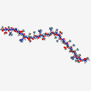
| CAS Number | 597562-32-8 |
|---|---|
| Molecular Formula | C205H340N60O53 |
| Molecular Weight | 4493.0 g/Mol |
| Purity | 99.5% |
| Lot Number | 25028 |
| Quantity | 5.21mg |
| Sequence | H-Leu-Leu-Gly-Asp-Phe-Phe-Arg-Lys-Ser-Lys-Glu-Lys-Ile-Gly-Lys-Glu-Phe-Lys-Arg-Ile-Val-Gln-Arg-Ile-Lys-Asp-Phe-Leu-Arg-Asn-Leu-Val-Pro-Arg-Thr-Glu-Ser-OH |
Cosmic Peptides Certificate of Analysis
Unable to load certificate. Download COA