Oxytocin
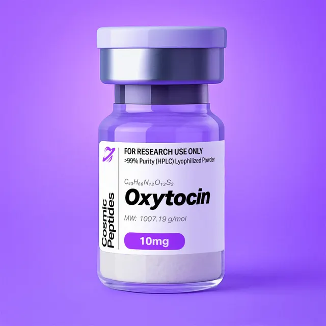
Oxytocin
Commonly researched with these items
Patent-pending lot tracking: Complete transparency from production to vial.
Oxytocin: A Disulfide-Bridged Cyclic Nonapeptide Amide and the Reference Agonist of the Oxytocin Receptor
Disclaimer
Products described here are supplied for research use only and are not intended for diagnostic, therapeutic, or clinical application. All statements regarding biological activity reflect preclinical and in vitro findings exclusively and have not been evaluated by the U.S. Food and Drug Administration. These materials and related content are provided for educational and investigational purposes only and are intended solely for qualified researchers in laboratory and academic settings.
Key Facts
- Compound class
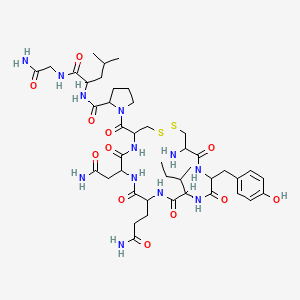
- Cyclic nonapeptide amide of the neurohypophysial peptide family; six-residue disulfide ring (Cys1–Cys6) with a three-residue C-terminal tail
- Sequence / structure
H-Cys-Tyr-Ile-Gln-Asn-Cys-Pro-Leu-Gly-NH2(disulfide bond Cys1–Cys6; one-letter code CYIQNCPLG-NH2), all residues in the L-configuration- Molecular formula
- C43H66N12O12S2
- Molecular weight
- 1007.2 g/mol (average, 1007.19 by calculation); monoisotopic mass 1006.4365 Da
- CAS number
- 50-56-6
- PubChem CID
- 439302
- Molecular target
- Oxytocin receptor (OXTR), a class A G protein-coupled receptor of 389 amino acids
- Physical form as supplied
- Lyophilized powder
- Lot traceability
- Every unit is tied to a tracked lot; the lot number, recorded purity and verified quantity of the lot on sale are shown live on this page, with its certificate of analysis.
Molecular Identity and Classification
Oxytocin is a nonapeptide with a C-terminal carboxamide, written H-Cys-Tyr-Ile-Gln-Asn-Cys-Pro-Leu-Gly-NH2, in which the thiol groups of the two cysteine residues, at positions 1 and 6, are joined in a single intramolecular disulfide bond. The disulfide closes a six-residue ring (Cys1 to Cys6); Pro7-Leu8-Gly9-NH2 forms a short tail ending in a primary amide rather than a free acid. Older literature counts the cystine as one amino acid and so calls oxytocin an "octapeptide amide", the wording of the 1953 synthesis paper; modern usage counts the two half-cystines separately, giving nine residues.
The molecular formula is C43H66N12O12S2, the average molecular weight 1007.2 g/mol and the monoisotopic mass 1006.4365 Da (PubChem CID 439302). Oxytocin is registered under CAS number 50-56-6. Its systematic IUPAC name, (2S)-1-[(4R,7S,10S,13S,16S,19R)-19-amino-7-(2-amino-2-oxoethyl)-10-(3-amino-3-oxopropyl)-13-[(2S)-butan-2-yl]-16-[(4-hydroxyphenyl)methyl]-6,9,12,15,18-pentaoxo-1,2-dithia-5,8,11,14,17-pentazacycloicosane-4-carbonyl]-N-[(2S)-1-[(2-amino-2-oxoethyl)amino]-4-methyl-1-oxopentan-2-yl]pyrrolidine-2-carboxamide, is informative: the disulfide ring appears in it as a twenty-membered macrocycle ("cycloicosane") with two sulfur and five nitrogen atoms, and all nine stereocentres are specified.
Those nine stereocentres are the eight α-carbons of the chiral residues (glycine has none) plus the β-carbon of isoleucine. Both cysteine α-carbons carry the descriptor R and the rest S, yet all residues are L: the R label follows from the Cahn–Ingold–Prelog rules, in which the sulfur-bearing side chain outranks the carboxyl carbon. Isoleucine is the (2S,3S) diastereomer; its (2S,3R) epimer, L-allo-isoleucine, is a possible synthesis-related impurity.
Oxytocin belongs to the neurohypophysial peptide family, whose members all share the Cys1–Cys6 ring, Pro7 and an amidated Gly9. Its closest relative, arginine vasopressin (H-Cys-Tyr-Phe-Gln-Asn-Cys-Pro-Arg-Gly-NH2, C46H65N15O12S2, 1084.2 g/mol), differs only at positions 3 (Phe for Ile) and 8 (Arg for Leu) — enough to shift receptor selectivity, and a classic example in peptide chemistry of how small side-chain changes redirect recognition by related G protein-coupled receptors.
Structure and Chemical Features
The defining structural element is the disulfide-bridged ring. Its twenty atoms are the β-carbon, α-carbon and carbonyl carbon of Cys1, the backbone nitrogen, α-carbon and carbonyl carbon of each of Tyr2, Ile3, Gln4 and Asn5, the nitrogen, α-carbon and β-carbon of Cys6, and the two sulfurs. The N-terminal α-amine lies outside the ring, as does the Cys6 carbonyl that carries the Pro-Leu-Gly-NH2 tail.
Residue-by-Residue Walk-Through
Cys1 carries the only free amine in the molecule. Because the sulfur atom on its β-carbon pulls electron density away from the nitrogen, this α-amine is less basic than a typical N-terminal amine, so the protonation state of oxytocin changes within the near-neutral pH range used in most assays. Tyr2 contributes the phenol responsible for UV absorbance at 275–280 nm and for fluorescence, and is the only residue that electrophilic iodination can target. Ile3 is the branched hydrophobic residue that, against Phe3 in vasopressin, favours OXTR. Gln4 and Asn5 are neutral amides: polar, and the main sites of deamidation. Cys6 closes the disulfide. Pro7 introduces a tertiary amide bond (Cys6–Pro7) that lacks an NH donor and can adopt cis and trans geometries. Leu8 is the second hydrophobic side chain, and Gly9-NH2 ends the chain in a primary amide.
A striking feature of oxytocin is that it contains no acidic groups at all. The C-terminus is an amide, and the side chains of Gln and Asn are amides, not acids; the only ionizable groups are the N-terminal amine and the tyrosine phenol (the cysteine thiols are consumed in the disulfide). Oxytocin therefore carries +1 at acidic pH, is largely uncharged once the N-terminal amine deprotonates, and turns negative only at strongly basic pH when the phenol (pKa near 10) ionizes — a profile that explains its reversed-phase behaviour and its single counterion in the usual salt forms.
Conformation in the Solid State and in Solution
The first high-resolution crystal structures in this family were those of deamino-oxytocin, the analogue in which the N-terminal amine is replaced by hydrogen (Mpa1-oxytocin). Wood and colleagues (1986) solved two crystal forms at better than 1.1 Å resolution; each contained two closely related conformers whose disulfide bridges had opposite chirality, a difference they discussed in terms of conformational flexibility and receptor recognition. In solution, NMR describes a dynamic ensemble: the ring is relatively ordered, with turn-like conformations around residues 2 to 5, while the Pro-Leu-Gly-NH2 tail is mobile. The Cys6–Pro7 bond is mainly trans, with a minor cis population that can double nearby signals in high-field spectra.
The bridge adds its own stereochemistry: the C–S–S–C dihedral angle in peptide disulfides lies close to ±90°, giving left- or right-handed forms whose handedness, in so small a ring, is coupled to the backbone conformation. For anyone modelling oxytocin in the binding pocket, the point is that the receptor-bound conformation is one member of a solution ensemble rather than the only form present.
Synthesis and Manufacturing Chemistry
Oxytocin has a special place in the history of chemistry. In 1953 Vincent du Vigneaud and co-workers reported the synthesis of an "octapeptide amide with the hormonal activity of oxytocin"; the same year, du Vigneaud, Ressler and Trippett published the sequence and proposed the cyclic disulfide structure, and Hans Tuppy independently determined the sequence by partial hydrolysis and end-group analysis. Du Vigneaud received the 1955 Nobel Prize in Chemistry for this work on biochemically important sulfur compounds. The original route was solution-phase: a linear S-benzyl-protected precursor was assembled from fragments, the S-benzyl groups were removed with sodium in liquid ammonia, and the free dithiol was closed to the disulfide by air oxidation.
Today oxytocin is made by Fmoc solid-phase peptide synthesis, built from Gly9 towards Cys1 on a resin that releases a C-terminal amide on cleavage, such as Rink amide. Standard protection is trityl (Trt) for both cysteines and for the Gln and Asn side-chain amides, with tert-butyl for the tyrosine phenol; trityl on Gln and Asn also suppresses dehydration of the side-chain amide to a nitrile during activation.
Two steps are specific to this molecule:
- Cysteine coupling without racemization. Activated cysteine derivatives are prone to epimerization at the α-carbon, particularly with uronium activators in the presence of tertiary amine bases. Carbodiimide/additive activation (for example DIC with Oxyma or HOBt), or a weaker, hindered base such as 2,4,6-collidine, limits formation of the D-Cys diastereomers, which are difficult to separate from the target peptide.
- Disulfide formation. After cleavage with trifluoroacetic acid (TFA) and scavengers (water, triisopropylsilane and a thiol such as 1,2-ethanedithiol to capture trityl cations), the linear dithiol is oxidized to the cyclic monomer: air or dimethyl sulfoxide oxidation at mildly basic pH, potassium ferricyanide, or iodine oxidation applied directly to S-trityl- or S-acetamidomethyl-protected precursors, as described by Kamber and colleagues (1980). On-resin cyclization is an alternative. Whatever the method, high dilution (well below 1 mg/mL) favours the intramolecular reaction over intermolecular coupling to disulfide-linked dimers.
The crude cyclic peptide is purified by preparative reversed-phase HPLC and lyophilized. TFA-containing buffers give the trifluoroacetate salt; the acetate is also common after ion exchange. With a single basic group the salt is ideally 1:1, but counterion and water content are lot-specific, which is why each lot's certificate of analysis reports the results for that material.
Analytical Characterization
Reversed-phase HPLC. Oxytocin is analysed on C18 columns with water/acetonitrile gradients containing 0.1% TFA, or with phosphate buffers. The Tyr, Ile, Leu and Pro side chains give it enough hydrophobicity to elute mid-way through a typical 5–60% acetonitrile gradient. Detection at 214–220 nm (peptide bonds) gives the most sensitive purity profile, reported as percentage of total peak area; 275–280 nm, where only Tyr2 and the disulfide absorb, confirms that a peak is peptide-derived.
Mass spectrometry. In positive electrospray ionization the most abundant ions are the protonated molecule [M+H]+ at m/z 1007.444 and the doubly protonated ion [M+2H]2+ at m/z 504.226. Sodium adducts ([M+Na]+, m/z 1029.426) are common. The isotope pattern is distinctive: with 43 carbons and two sulfurs, the calculated M, M+1, M+2 and M+3 intensities are about 100 : 54 : 26 : 8.5, the M+2 peak enlarged by 34S (4.25% natural abundance per sulfur) — a quick check that both sulfurs are present.
Tandem MS. The disulfide ring resists fragmentation under collision-induced dissociation, so fragments arise mainly from the linear tail. The characteristic product ions are the ring acylium ion b6 at m/z 723.259 (the cyclic Cys1–Cys6 unit after loss of Pro-Leu-Gly-NH2) and the C-terminal y-ions y3 (Pro-Leu-Gly-NH2, m/z 285.192), y2 (m/z 188.139) and y1 (m/z 75.055). Cleavage N-terminal to proline is favoured, so the Cys6–Pro7 cleavage giving b6 and y3 dominates, making the transition m/z 1007.4 → 723.3 a natural choice for targeted LC-MS/MS.
Worked Mass Calculation and Diagnostic Mass Shifts
The monoisotopic mass follows from the residue masses: Cys 103.00919 (×2), Tyr 163.06333, Ile 113.08406, Gln 128.05858, Asn 114.04293, Pro 97.05276, Leu 113.08406 and Gly 57.02146 Da sum to 991.42556 Da. Adding the N-terminal hydrogen and C-terminal NH2 (17.02655 Da) gives 1008.45211 Da for the reduced dithiol; closing the disulfide removes two hydrogens (2.01565 Da), giving 1006.43646 Da, in agreement with PubChem. The same arithmetic predicts the informative variants:
| Species | Monoisotopic mass (Da) | Shift vs oxytocin | Origin |
|---|---|---|---|
| Oxytocin (cyclic disulfide) | 1006.4365 | 0 | Target |
| Reduced, linear dithiol | 1008.4521 | +2.0157 | Incomplete oxidation, or reduction with DTT or TCEP |
| Deamidated (Gln4, Asn5 or Gly9-NH2) | 1007.4205 | +0.9840 | Hydrolysis of an amide to an acid |
| Trisulfide | 1038.4085 | +31.9721 | Sulfur insertion into the disulfide |
| Sulfoxide / single oxidation | 1022.4314 | +15.9949 | Oxidation, for example of the disulfide |
| Covalent dimer (two disulfides) | 2012.8729 | ×2 | Intermolecular disulfide exchange; its [M+2H]2+ falls at the same m/z as the monomer [M+H]+ |
| Des-Gly9-NH2 fragment ion (b8) | — | m/z 933.396 | Tail fragmentation in MS/MS |
The dimer row deserves emphasis: a doubly protonated disulfide-linked dimer appears at exactly the m/z of the monomer's 1+ ion (1007.444), and is told apart by isotope spacing (0.5 versus 1.0 unit) and by its later reversed-phase retention.
Disulfide and thiol tests. Ellman's reagent, 5,5′-dithiobis(2-nitrobenzoic acid), releases the yellow 2-nitro-5-thiobenzoate anion from free thiols, measured at 412 nm: a correctly cyclized sample gives no signal, while reduction with dithiothreitol or tris(2-carboxyethyl)phosphine should reveal two thiols per molecule alongside a +2 Da mass shift and a retention change.
Purity versus peptide content. HPLC purity is the fraction of UV-absorbing material that is oxytocin; peptide content is the fraction of the weighed powder that is peptide rather than counterion, water and residual salt. For the 1:1 acetate salt (C45H70N12O14S2, 1067.25 g/mol) the theoretical peptide content is 94.4%; for a 1:1 trifluoroacetate salt (1121.22 g/mol), 89.8%, before any water. Amino-acid analysis after acid hydrolysis (expected Asp : Glu : Gly : Ile : Leu : Pro : Tyr : half-Cys of 1:1:1:1:1:1:1:2, since Asn and Gln hydrolyse to Asp and Glu) or quantitative NMR gives peptide content when exact molarity is needed.
Physicochemical Properties
| Property | Value | Notes |
|---|---|---|
| Molecular formula | C43H66N12O12S2 | Free base, without counterion |
| Average molecular weight | 1007.2 g/mol | PubChem CID 439302 |
| Monoisotopic mass | 1006.4365 Da | For high-resolution MS |
| [M+H]+ / [M+2H]2+ | m/z 1007.444 / 504.226 | Positive-mode ESI |
| Heavy atoms / defined stereocentres | 69 / 9 | Eight α-carbons plus the Ile β-carbon |
| Ionizable groups | Two | N-terminal α-amine of Cys1; Tyr2 phenol (pKa near 10). No acidic side chain |
| Net charge near pH 7 | Between 0 and +1 | The α-amine of a cysteinyl N-terminus is weakly basic, pKa roughly 6–7 |
| XLogP3 | −2.6 | Computed; hydrophilic overall despite Tyr, Ile and Leu |
| Topological polar surface area | 450 Å2 | Computed |
| Hydrogen-bond donors / acceptors | 12 / 15 | Computed |
| Rotatable bonds | 17 | Computed; fewer than a linear nonapeptide, as the ring is fixed |
| Molar extinction coefficient at 280 nm | approximately 1615 M−1 cm−1 | One Tyr (1490) plus one cystine (125), after Pace and colleagues |
| UV maxima | about 214 nm (amide) and 275 nm (Tyr) | No Trp; weak 280 nm signal |
| Solubility | Freely soluble in water and aqueous buffers; soluble in dimethyl sulfoxide and dilute acetic acid | Typical of a small, amidated cyclic peptide |
| Chemical liabilities | Disulfide exchange, deamidation of Gln4/Asn5, dimerization, trisulfide formation | See Laboratory Handling and Storage |
The charge profile explains much of oxytocin's laboratory behaviour. With no carboxylate, the molecule never carries a net negative charge under ordinary assay conditions, binds weakly to cation-exchange media near neutral pH, and is retained on reversed-phase columns mainly by its Tyr, Ile, Leu and Pro side chains rather than by ion pairing. The computed polar surface area of 450 Å2, twelve hydrogen-bond donors and negative partition coefficient (XLogP3 −2.6) together describe a molecule that hydrates well and does not partition into lipid phases.
A worked example shows how the extinction coefficient is used. Oxytocin contains one tyrosine and one cystine and no tryptophan, so by the method of Pace and colleagues (1995) its molar extinction coefficient at 280 nm is approximately 1490 + 125 = 1615 M−1 cm−1. For a 1.00 mg/mL solution of the free base, the molar concentration is 1.00 g/L ÷ 1007.2 g/mol = 0.993 mM, giving a predicted absorbance in a 1 cm cell of A280 = 1615 × 0.000993 ≈ 1.60. Absorbance well below this value, after correcting for peptide content and light scattering, indicates more counterion and water in the powder than assumed. Tyrosine absorbance is pH-sensitive above pH 9, so such measurements belong in neutral or mildly acidic buffer.
Research Context
Oxytocin is the reference agonist of the oxytocin receptor (OXTR), a 389-residue class A (rhodopsin-like) G protein-coupled receptor first cloned in 1992 by Kimura and colleagues. OXTR sits in the same branch as the three vasopressin receptors (V1a, V1b and V2), and the four share enough of their binding pocket that cross-reactivity is the central methodological problem of the field: oxytocin activates V1a receptors at higher concentrations, and vasopressin activates OXTR. The 2001 review by Gimpl and Fahrenholz remains the standard entry point to the biochemistry.
In vitro, OXTR couples principally to Gq/11. Agonist binding triggers the exchange of GDP for GTP on the Gαq subunit, activation of phospholipase C-β, and hydrolysis of phosphatidylinositol 4,5-bisphosphate into diacylglycerol and inositol 1,4,5-trisphosphate; the latter releases Ca2+ from intracellular stores, and the rise in cytosolic Ca2+ together with diacylglycerol activates protein kinase C isoforms. Recombinant-cell work also shows coupling to Gi/o. Busnelli and colleagues (2012) used oxytocin-derived analogues in bioluminescence resonance energy transfer assays to show that individual ligands discriminate between G protein family subtypes at the same receptor; oxytocin itself serves as the balanced reference against which such biased analogues are measured.
Structural Pharmacology of the Receptor Complex
Three structures transformed the picture of OXTR recognition. Waltenspühl and colleagues (2020) solved the crystal structure of the inactive receptor bound to a small-molecule antagonist, defining the orthosteric pocket between the extracellular ends of the seven transmembrane helices and showing sites for allosteric modulation by cholesterol and by divalent ions. In 2022 two cryo-electron microscopy structures of the active receptor bound to oxytocin followed. Waltenspühl and colleagues described the oxytocin binding mode and the basis of subtype specificity within the oxytocin/vasopressin receptor family. Meyerowitz and colleagues, working with an engineered miniGq/i protein, found that activation involves a Mg2+ coordination complex formed between oxytocin and the receptor together with a disruption of transmembrane helix 7, and that the identity of a single cation-coordinating residue decides whether a receptor of the vasopressin family is cation-dependent. This is the structural basis of the long-known Mg2+ dependence of high-affinity agonist binding at OXTR, and the reason binding-assay buffers are specified for divalent-cation content as carefully as for pH.
A second modulator is cholesterol. Gimpl and Fahrenholz (2002) described cholesterol as a stabilizer of the receptor: in membrane and solubilized preparations it holds OXTR in a high-affinity state for agonists and antagonists, acting through a specific interaction rather than as a bulk membrane property, so cholesterol depletion lowers high-affinity oxytocin binding. The cholesterol site later seen in the 2020 crystal structure gave this biochemistry a structural anchor, and OXTR is accordingly a model system for lipid–receptor interaction studies.
Common In Vitro Read-Outs
Work with oxytocin as a reference agonist rests on a small set of assays: competition binding against a radiolabelled or fluorescent ligand on membranes from OXTR-expressing cell lines; inositol monophosphate (IP-1) accumulation as a stable proxy for phospholipase C activity; calcium mobilization with fluorescent indicators; BRET sensors of Gα subtype engagement, β-arrestin recruitment and internalization; and label-free readouts such as dynamic mass redistribution. Concentration–response curves span roughly 10 pM to 1 µM, with vasopressin and selective analogues as comparators, chosen with the receptor-selectivity data reviewed by Chini and Manning (2007). Because affinity depends on Mg2+ and on membrane cholesterol, EC50 and Ki values compare across laboratories only when buffer and membrane source are reported.
Oxytocin is also a substrate in enzymology: the M1 family zinc aminopeptidase LNPEP, long called oxytocinase, cleaves it in vitro, and Tsujimoto and Hattori (2005) reviewed this aminopeptidase subfamily. The same enzymology matters in cell culture, where peptidases in serum-containing media can degrade the peptide during long incubations, so stability controls, serum-free conditions or peptidase inhibitors belong in the experimental design. Its well-mapped degradation routes also make oxytocin a benchmark analyte for stability-indicating HPLC methods.
Structural Comparison with Related Peptides
The neurohypophysial peptides show how a couple of side-chain substitutions on a conserved scaffold change molecular recognition. All of the molecules below share the Cys1–Cys6 ring, Pro7 and the amidated Gly9.
| Compound | Difference from oxytocin | Formula | Average mass (g/mol) |
|---|---|---|---|
| Oxytocin | Reference: Ile3, Leu8, free N-terminal amine | C43H66N12O12S2 | 1007.2 |
| Arginine vasopressin (PubChem CID 644077) | Phe3 and Arg8 for Ile3 and Leu8; adds a guanidinium and a positive charge | C46H65N15O12S2 | 1084.2 |
| Deamino-oxytocin (Mpa1-oxytocin; PubChem CID 449224) | Position 1 is 3-mercaptopropionic acid; removes the only basic group | C43H65N11O12S2 | 992.2 |
| Reduced (linear) oxytocin | Disulfide reduced to two free thiols; +2.0157 Da monoisotopic | C43H68N12O12S2 | 1009.2 |
| Oxytocin free acid (Gly9-OH) | C-terminal amide hydrolysed to the acid; +0.984 Da monoisotopic | C43H65N11O13S2 | 1008.2 |
Two lessons follow. The substitution Ile3 → Phe3 exchanges a β-branched aliphatic side chain for an aromatic one of similar volume and, with Leu8 → Arg8 adding a permanent positive charge, is enough to redirect selectivity between two closely related receptor families. And modifications at position 1 or at the C-terminal amide shift mass by small, predictable amounts that high-resolution MS resolves.
The same analytical questions arise with other disulfide-containing or C-terminally amidated research peptides, for example AOD-9604, the amidated decapeptide Kisspeptin-10, the class B receptor ligand VIP, and the amidated 29-residue sermorelin.
Laboratory Handling and Storage
Storage of the solid. Lyophilized oxytocin is best kept tightly closed at −20 °C or below, protected from light and moisture. Because the powder is hygroscopic, let a cold container reach room temperature in a desiccator before opening, so moisture does not condense on the solid and inflate the weighed mass.
Preparing solutions. Oxytocin dissolves readily in high-purity water, in dilute acetic acid and in aqueous buffers. For accurate molarity, correct the weighed mass for peptide content: a 1:1 acetate salt is 94.4% peptide before any water is counted. Stocks at 0.1–1 mM are divided into single-use aliquots and frozen at −20 °C or −80 °C; repeated freeze–thaw cycles and prolonged storage of dilute solutions are the main avoidable causes of loss.
Solution stability chemistry. Oxytocin's aqueous degradation chemistry is unusually well mapped: Hawe and colleagues (2009) measured the kinetics and identified the products, and Wiśniewski and colleagues (2013) set out the mechanisms. The main routes are:
- Disulfide exchange and dimerization. A trace of free thiol, or base-catalysed β-elimination of the disulfide to a dehydroalanine residue and a free thiol, starts a chain of thiol–disulfide exchange reactions that produce dimers and higher oligomers. This route accelerates sharply above pH 7 and is the dominant pathway in neutral to alkaline solution.
- Deamidation. The Gln4 and Asn5 side-chain amides and the C-terminal glycinamide hydrolyse to acids, each adding 0.984 Da and a negative charge that shifts HPLC retention.
- Trisulfide formation. Insertion of a sulfur atom into the bridge gives a species 31.97 Da heavier, chromatographically resolvable from the target.
- Backbone hydrolysis at extremes of pH, and oxidation of the disulfide by peroxides or trace metals.
Two practical conclusions follow. Solutions are most stable in mildly acidic buffer, around pH 4.5–5, where thiol exchange and deamidation are both slow; and metal ions cut both ways. Avanti and colleagues (2011) showed that divalent metal ions in citrate buffer markedly stabilize aqueous oxytocin, apparently by coordinating the peptide and suppressing cysteine-mediated intermolecular reactions, whereas adventitious copper or iron catalyses oxidation.
Adsorption at low concentrations. Oxytocin is lost to container surfaces from dilute solution; low-binding polypropylene ware, siliconized glass and, where the design permits, a carrier protein or trace non-ionic surfactant limit these losses.
Re-verification. A short LC-MS check of a stored stock — the [M+H]+ ion at m/z 1007.4 present, peaks at +0.98, +31.97 and twice the mass absent — confirms identity and integrity, and an Ellman assay confirms that no free thiol has appeared. Each lot's certificate of analysis, shown on this page, records the results for that lot.
Frequently Asked Research Questions
- What is oxytocin, chemically?
- Oxytocin is a cyclic nonapeptide amide,
H-Cys-Tyr-Ile-Gln-Asn-Cys-Pro-Leu-Gly-NH2, in which Cys1 and Cys6 are joined by a disulfide bond that closes a six-residue ring; Pro7-Leu8-Gly9-NH2 forms the tail. Its formula is C43H66N12O12S2 and its average molecular weight 1007.2 g/mol. - What is the CAS number of oxytocin?
- Oxytocin is registered under CAS number 50-56-6 and appears in PubChem as compound 439302.
- Why is oxytocin sometimes called an octapeptide?
- Historical papers, including the 1953 synthesis report, counted the cystine formed by the Cys1–Cys6 bridge as one amino acid, giving eight residues; counting the two half-cystines separately, as is now standard, gives nine.
- What masses should I see for oxytocin by mass spectrometry?
- In positive electrospray ionization, [M+H]+ appears at m/z 1007.444 and [M+2H]2+ at m/z 504.226; the monoisotopic mass of the neutral peptide is 1006.4365 Da. Because the molecule contains two sulfur atoms, the M+2 isotope peak is relatively intense, roughly 26% of the monoisotopic peak.
- Which MS/MS fragment ions are diagnostic?
- The disulfide ring resists fragmentation, so the informative ions come from the tail: the b6 ion of the intact ring at m/z 723.259 and the y3 ion of Pro-Leu-Gly-NH2 at m/z 285.192, produced by the favoured cleavage N-terminal to Pro7. The transition 1007.4 → 723.3 is a standard choice for targeted quantification.
- How do I confirm that the disulfide bridge is intact?
- Two independent checks: Ellman's reagent should show no free thiol, and the monoisotopic mass should be 1006.4365 Da, not the 1008.4521 Da of the reduced linear form. Deliberate reduction with dithiothreitol or tris(2-carboxyethyl)phosphine should then produce that +2 Da shift and a retention change.
- What is the net charge and isoelectric point of oxytocin?
- Oxytocin has no acidic group. Its only ionizable groups are the N-terminal α-amine of Cys1, which is weakly basic (pKa roughly 6–7 for a cysteinyl N-terminus), and the Tyr2 phenol with a pKa near 10. The net charge is therefore between 0 and +1 near neutral pH, and calculated isoelectric points fall in the neutral to mildly basic range depending on the pKa set used.
- Why does oxytocin absorb so weakly at 280 nm?
- It contains no tryptophan; only the single tyrosine and the cystine absorb there, giving a molar extinction coefficient near 1615 M−1 cm−1 and A280 of about 1.6 for 1 mg/mL in a 1 cm cell. Detection at 214–220 nm is far more sensitive for purity work.
- At what pH are aqueous oxytocin solutions most stable?
- Mildly acidic, about pH 4.5–5. Above pH 7, β-elimination and thiol–disulfide exchange drive dimerization; strongly acidic conditions promote backbone hydrolysis.
- Which degradation products should a stability-indicating method resolve?
- Deamidated forms (+0.98 Da), the trisulfide (+31.97 Da), the reduced linear peptide (+2.02 Da), oxidation products (+15.99 Da) and disulfide-linked dimers (about twice the mass) — all separable by reversed-phase HPLC and distinguishable by high-resolution MS.
- Why does magnesium appear in oxytocin receptor binding buffers?
- Divalent cations raise agonist affinity at OXTR. Meyerowitz and colleagues (2022) resolved a divalent-cation site in the agonist-bound receptor complex, giving a structural basis for an effect known from binding assays for decades, so buffer cation composition should be reported with any affinity value.
- Where can I see the purity of the oxytocin I am buying?
- This page shows the lot number, recorded purity and verified quantity of the lot currently on sale, taken live from the shop's lot-tracking database, together with that lot's certificate of analysis.
Sources & Further Reading
- National Center for Biotechnology Information. PubChem Compound Summary for CID 439302, Oxytocin. PubChem CID 439302.
- du Vigneaud V, Ressler C, Swan JM, Roberts CW, Katsoyannis PG, Gordon S. The synthesis of an octapeptide amide with the hormonal activity of oxytocin. Journal of the American Chemical Society 1953;75(19):4879–4880. doi:10.1021/ja01115a553
- du Vigneaud V, Ressler C, Trippett S. The sequence of amino acids in oxytocin, with a proposal for the structure of oxytocin. Journal of Biological Chemistry 1953;205(2):949–957. doi:10.1016/S0021-9258(18)49238-1
- Tuppy H. The amino-acid sequence in oxytocin. Biochimica et Biophysica Acta 1953;11:449–450. doi:10.1016/0006-3002(53)90071-7
- Wood SP, Tickle IJ, Treharne AM, Pitts JE, Mascarenhas Y, Li JY, Husain J, Cooper S, Blundell TL, Hruby VJ, Buku A, Fischman AJ, Wyssbrod HR. Crystal structure analysis of deamino-oxytocin: conformational flexibility and receptor binding. Science 1986;232(4750):633–636. doi:10.1126/science.3008332
- Kimura T, Tanizawa O, Mori K, Brownstein MJ, Okayama H. Structure and expression of a human oxytocin receptor. Nature 1992;356(6369):526–529. doi:10.1038/356526a0
- Gimpl G, Fahrenholz F. The oxytocin receptor system: structure, function, and regulation. Physiological Reviews 2001;81(2):629–683. doi:10.1152/physrev.2001.81.2.629
- Gimpl G, Fahrenholz F. Cholesterol as stabilizer of the oxytocin receptor. Biochimica et Biophysica Acta – Biomembranes 2002;1564(2):384–392. doi:10.1016/S0005-2736(02)00475-3
- Busnelli M, Saulière A, Manning M, Bouvier M, Galés C, Chini B. Functional selective oxytocin-derived agonists discriminate between individual G protein family subtypes. Journal of Biological Chemistry 2012;287(6):3617–3629. doi:10.1074/jbc.M111.277178
- Chini B, Manning M. Agonist selectivity in the oxytocin/vasopressin receptor family: new insights and challenges. Biochemical Society Transactions 2007;35(4):737–741. doi:10.1042/BST0350737
- Waltenspühl Y, Schöppe J, Ehrenmann J, Kummer L, Plückthun A. Crystal structure of the human oxytocin receptor. Science Advances 2020;6(29):eabb5419. doi:10.1126/sciadv.abb5419
- Waltenspühl Y, Ehrenmann J, Vacca S, Thom C, Medalia O, Plückthun A. Structural basis for the activation and ligand recognition of the human oxytocin receptor. Nature Communications 2022;13:4153. doi:10.1038/s41467-022-31325-0
- Meyerowitz JG, Robertson MJ, Barros-Álvarez X, Panova O, Nwokonko RM, Gao Y, Skiniotis G. The oxytocin signaling complex reveals a molecular switch for cation dependence. Nature Structural & Molecular Biology 2022;29(3):274–281. doi:10.1038/s41594-022-00728-4
- Wiśniewski K, Finnman J, Flipo M, Galyean R, Schteingart CD. On the mechanism of degradation of oxytocin and its analogues in aqueous solution. Peptide Science 2013;100(4):408–421. doi:10.1002/bip.22260
- Hawe A, Poole R, Romeijn S, Kasper P, van der Heijden R, Jiskoot W. Towards heat-stable oxytocin formulations: analysis of degradation kinetics and identification of degradation products. Pharmaceutical Research 2009;26(7):1679–1688. doi:10.1007/s11095-009-9878-2
- Avanti C, Amorij J-P, Setyaningsih D, Hawe A, Jiskoot W, Visser J, Kedrov A, Driessen AJM, Hinrichs WLJ, Frijlink HW. A new strategy to stabilize oxytocin in aqueous solutions: I. The effects of divalent metal ions and citrate buffer. The AAPS Journal 2011;13(2):284–290. doi:10.1208/s12248-011-9268-7
- Kamber B, Hartmann A, Eisler K, Riniker B, Rink H, Sieber P, Rittel W. The synthesis of cystine peptides by iodine oxidation of S-trityl-cysteine and S-acetamidomethyl-cysteine peptides. Helvetica Chimica Acta 1980;63(4):899–915. doi:10.1002/hlca.19800630418
- Ellman GL. Tissue sulfhydryl groups. Archives of Biochemistry and Biophysics 1959;82(1):70–77. doi:10.1016/0003-9861(59)90090-6
- Pace CN, Vajdos F, Fee L, Grimsley G, Gray T. How to measure and predict the molar absorption coefficient of a protein. Protein Science 1995;4(11):2411–2423. doi:10.1002/pro.5560041120
- Tsujimoto M, Hattori A. The oxytocinase subfamily of M1 aminopeptidases. Biochimica et Biophysica Acta – Proteins and Proteomics 2005;1751(1):9–18. doi:10.1016/j.bbapap.2004.09.011
Last reviewed:
| CAS Number | 50-56-6 |
|---|---|
| Molecular Formula | C43H66N12O12S2 |
| Molecular Weight | 1007.2 g/Mol |
| Purity | 99.6% |
| Lot Number | 26068 |
| Quantity | 2.13mg |
| Sequence | H-Cys(1)-Tyr-Ile-Gln-Asn-Cys(1)-Pro-Leu-Gly-NH2 |
Cosmic Peptides Certificate of Analysis
Unable to load certificate. Download COA