Pinealon
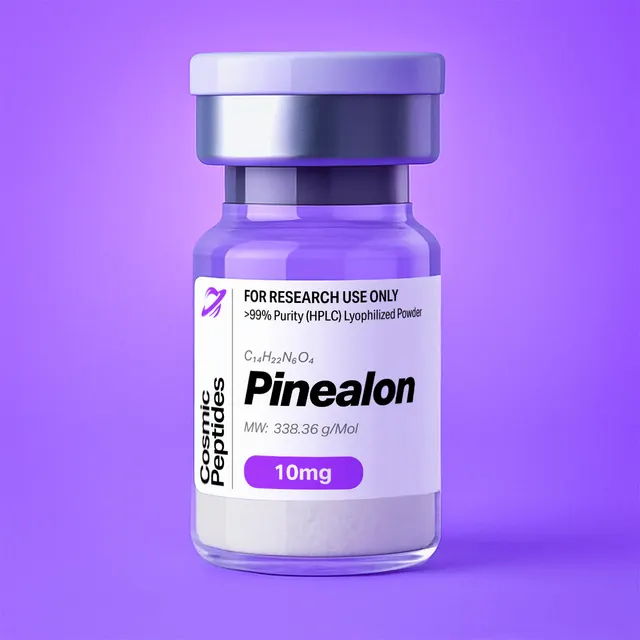
Pinealon
Commonly researched with these items
Patent-pending lot tracking: Complete transparency from production to vial.
Pinealon (Glu-Asp-Arg): An Anionic Tripeptide Used as a Model for Sequence-Selective Peptide–Nucleic Acid Interaction
Disclaimer
Products described here are supplied for research use only and are not intended for diagnostic, therapeutic, or clinical application. All statements regarding biological activity reflect preclinical and in vitro findings exclusively and have not been evaluated by the U.S. Food and Drug Administration. These materials and related content are provided for educational and investigational purposes only and are intended solely for qualified researchers in laboratory and academic settings.
Key Facts
- Compound class
- Linear, unprotected tripeptide; member of the ultrashort (two- to four-residue) peptide bioregulator series, where it is named pinealon and abbreviated EDR
- Sequence
H-Glu-Asp-Arg-OH(one-letter code: EDR), all three residues in the L-configuration- Molecular formula
- C15H26N6O8
- Molecular weight
- 418.40 g/mol (average, free acid)
- Monoisotopic mass
- 418.1812 Da
- CAS number
- 175175-23-2
- PubChem CID
- 10273502 (ChEBI:156374)
- Ionization
- Three carboxylic acids against one guanidinium group and one α-amino group; net charge approximately −1 at pH 7.0; calculated isoelectric point near 4.1
- Physical form as supplied
- Lyophilized powder
- Lot traceability
- Every unit is tied to a tracked lot; the lot number, recorded purity and verified quantity of the lot on sale are shown live on this page, with its certificate of analysis.
Molecular Identity and Classification
Pinealon is the tripeptide L-glutamyl-L-aspartyl-L-arginine, written H-Glu-Asp-Arg-OH and almost always abbreviated EDR in the primary literature. It is fully unprotected — free α-amino group, free C-terminal acid, no acetylation, amidation, cyclisation or non-natural residue. Its formula is C15H26N6O8, average molecular weight 418.40 g/mol, monoisotopic mass 418.1812 Da; it carries CAS number 175175-23-2 and appears as PubChem compound 10273502 and ChEBI:156374.
Its systematic name, (4S)-4-amino-5-[[(2S)-3-carboxy-1-[[(1S)-1-carboxy-4-(diaminomethylideneamino)butyl]amino]-1-oxopropan-2-yl]amino]-5-oxopentanoic acid, encodes the L-configuration at all three α-carbons, free γ- and β-carboxyls on glutamate and aspartate, and arginine's guanidine side chain as the only basic group.
Formula, mass and sequence can be checked against each other. Summing the monoisotopic residue masses of glutamate (129.04259 Da), aspartate (115.02694 Da) and arginine (156.10111 Da) gives 400.17064 Da; adding one water molecule (18.01056 Da) for the two amide condensations gives 418.18120 Da, matching the computed monoisotopic mass of C15H26N6O8 (418.18121 Da) to within 0.00001 Da. The elemental tally agrees too: glutamate contributes C5, aspartate C4 and arginine C6 for the fifteen carbons, and arginine's four nitrogens plus two backbone nitrogens account for all six.
Among ultrashort peptides — oligopeptides of two to four residues — EDR is unusual for being strongly anionic: two of its three side chains are carboxylic acids and the free C-terminus adds a third. Most short peptides with a nucleic-acid-binding literature are cationic, so an anionic tripeptide reported to interact with polyanionic DNA poses a non-obvious physical-chemistry problem — which is why it recurs in the biophysical literature.
Salt form therefore matters more than it would for a basic peptide, and more than one form is on record: PubChem lists "pinealon acetate salt form" among the synonyms of CID 10273502, trifluoroacetate can pair with the guanidinium and N-terminal ammonium after purification, and sodium or ammonium salts of the carboxylates are possible. Because salt form changes the mass of powder needed for a given molar concentration, each lot's analytical record is the basis for quantitative work.
Structure and Chemical Features
EDR has no aromatic ring, no sulfur, no proline and no secondary structure. What it has is an unusually dense set of well-separated charges, from which nearly every distinctive property follows. The N-terminal glutamate contributes an α-amino group and a γ-carboxyl three carbons away. The central aspartate contributes a β-carboxyl one carbon closer to the backbone, which makes it both the more acidic side chain and the one able to reach its own backbone amide — the prerequisite for the aspartimide chemistry below. The C-terminal arginine contributes the guanidinium group, a planar cation that delocalises its charge over three nitrogens and can donate up to five hydrogen bonds in a characteristic twin-donor geometry, carried on the molecule's only flexible aliphatic segment.
Charge State and Ionization
pK values measured for these groups in model compounds and unfolded polypeptides place an aspartate side-chain carboxyl near 3.5, a glutamate side-chain carboxyl near 4.2, a C-terminal carboxyl between about 3.3 and 3.7 and an α-amino group between 7.5 and 8.0; a guanidinium sits far higher, near 12.5. Below pH 2 EDR is a dication; between roughly pH 5 and pH 10 all three carboxylates are deprotonated and the guanidinium protonated, a 2+/3− zwitterion whose net charge is approximately −1 at pH 7.0; above pH 10 it approaches −2. Summing those ionization fractions by Henderson–Hasselbalch and solving for zero net charge (taking 3.5 for the C-terminal acid) puts the calculated isoelectric point near 4.1, a model value rather than a measurement: another pK compilation moves it by up to about 0.3 unit. It has three practical consequences: solubility passes through a minimum around pH 4, buffers at pH 3–5 give the least reproducible retention, and ion-exchange purification needs an anion exchanger above pH 5 rather than the cation exchanger most short peptides use.
With no proline and no ring, EDR is conformationally unconstrained: 14 rotatable bonds in a molecule of only 29 heavy atoms. It populates a broad solution ensemble, and its 1H NMR spectrum is expected to show a single set of resonances rather than the doubled peaks that proline-containing peptides such as KPV display from slow cis–trans isomerisation. Such flexibility is entropically costly on binding a structured partner, one reason reported binding constants are modest.
The recognition geometry is nonetheless specific. A guanidinium flanked by two carboxylates is a bifunctional hydrogen-bonding module: the guanidinium donates a pair of parallel N–H bonds to the O6 and N7 acceptors on the major-groove face of guanine, or to a phosphate oxygen, while a nearby carboxylate accepts hydrogen bonds from the exocyclic amino groups of cytosine (N4) or guanine (N2). That arrangement — arginine paired with acidic residues — recurs throughout protein–DNA interfaces and in histone tails, and is the structural argument offered in the literature for how a net-anionic tripeptide might read a base sequence rather than simply be repelled by the phosphate backbone. Two absences shape the laboratory work that follows: no aromatic residue, hence no absorbance near 280 nm, and no cysteine or methionine, hence no disulfide or oxidation chemistry.
Synthesis and Manufacturing Chemistry
EDR is assembled by solid-phase peptide synthesis, the method Merrifield introduced in 1963, on an insoluble resin from the C-terminus toward the N-terminus. Current practice uses Fmoc chemistry, built on the base-labile 9-fluorenylmethoxycarbonyl group described by Carpino and Han in 1972: Fmoc on each incoming α-amino group comes off with 20% piperidine in dimethylformamide before the next coupling, while side chains carry acid-labile protection removed at the end. The building blocks are Fmoc-Arg(Pbf)-OH, Fmoc-Asp(OtBu)-OH and Fmoc-Glu(OtBu)-OH, on an acid-labile linker such as 2-chlorotrityl chloride or Wang resin for the free C-terminal acid. Short as it is, the sequence concentrates four well-known side reactions, and knowing them is what lets a laboratory read an EDR chromatogram intelligently.
Aspartimide formation is the dominant risk. Aspartate protected as its tert-butyl ester has a side-chain ester positioned for attack by the adjacent backbone amide nitrogen, and piperidine catalyses that cyclisation during every Fmoc removal, giving a five-membered succinimide ring that opens to a mixture of the α-peptide and the β-linked isoaspartyl peptide; its acidic α-proton also makes the pathway a route to the D-aspartate epimer. Karlström and Undén introduced the hindered 3-methylpent-3-yl (OMpe) ester in 1996 to suppress it; HOBt or formic acid in the piperidine solution, shorter deprotection and lower temperature are the other measures. Because the isoaspartyl and D-aspartyl products are isobaric with the target, this family is why an EDR chromatogram deserves a long shallow gradient.
Arginine chemistry contributes two more. Activating the carboxyl of Fmoc-Arg(Pbf)-OH invites intramolecular attack by the guanidine δ-nitrogen, forming an unreactive δ-lactam; loading arginine onto the resin first, as is natural for a C-terminal residue, avoids it. At final cleavage the bulky Pbf group can migrate onto the guanidine instead of leaving cleanly — the arginine sulfonation side reaction documented by Beck-Sickinger and colleagues in 1991 — giving a persistent, more hydrophobic adduct; carbocation scavengers such as triisopropylsilane, water and thioanisole keep it low, and Pbf removal is the slowest step of the cleavage.
Pyroglutamate formation is specific to the N-terminal residue: once the final Fmoc group is removed, a free N-terminal glutamate can cyclise onto its own γ-carboxyl with loss of water, giving a pyroglutamyl peptide 18 Da lighter than the target. Acid, heat and prolonged storage in solution all promote it, which makes a signal 18 Da below the expected mass the most useful stress marker for this peptide.
Cleavage with trifluoroacetic acid containing water and a silane scavenger removes the tert-butyl esters and the Pbf group in one step. Purification is then the hard part, for the reasons set out below, and lyophilization from water gives the powder, with trifluoroacetate the expected counterion unless exchanged by lyophilization from dilute acetic acid.
Analytical Characterization
Reversed-phase HPLC with ultraviolet detection at 210–220 nm is the workhorse for purity as percentage of total peak area; with no signal at 280 nm the amide band is the only option, so additives must have low background there. Retention is the real obstacle: at a computed XLogP of −6.1 the peptide elutes at or near the column void on conventional C18 material. An aqueous-stable or polar-embedded C18 phase run from 0% to about 10% acetonitrile in 0.1% trifluoroacetic acid gives workable retention, the acid ion-pairing with the guanidinium; heptafluorobutyric acid pairs more strongly still. Hydrophilic-interaction chromatography inverts the problem, retaining EDR on an amide or zwitterionic phase run from high to lower acetonitrile with 5–10 mM ammonium formate precisely because the analyte is polar; Guo and Baran's assessment of partitioning versus surface adsorption guides method development in that mode.
Mass spectrometry confirms identity in both polarities: the guanidinium ionises readily in positive mode, while three carboxylates make negative mode unusually sensitive. The values below follow from the monoisotopic mass of 418.1812 Da and a proton mass of 1.00728 Da.
| Ion | Calculated m/z | Notes |
|---|---|---|
| [M+H]+ | 419.1885 | Base peak in positive-mode electrospray |
| [M+2H]2+ | 210.0979 | Minor; only one strongly basic site |
| [M+Na]+ | 441.1704 | Common with carboxylate-rich peptides |
| [M−H]− | 417.1739 | Negative mode is often the more sensitive choice |
| [M−2H]2− | 208.0833 | Seen at higher pH in the infusion solvent |
| y1 (Arg) | 175.1190 | The classic arginine y1 ion; strong diagnostic |
| y2 (Asp-Arg) | 290.1459 | Confirms the C-terminal dipeptide |
| b2 (Glu-Asp) | 245.0768 | Confirms the N-terminal dipeptide |
| a2 | 217.0819 | b2 minus CO |
| Arginine side-chain ions | 70.0651, 112.0869, 129.1135 | Low-mass markers for an arginine-containing peptide |
| [M+H−H2O]+ | 401.1779 | Pyroglutamyl species; marker of acid or thermal stress |
Two fragmentation features are specific to this sequence. The arginine y1 ion at m/z 175.119 is among the most recognisable signals in peptide tandem mass spectrometry and makes a good qualifier transition. An N-terminal glutamate also steers fragmentation: Godugu and colleagues showed in 2010 that N-terminal glutamic acid and glutamine promote water loss and alter backbone fragment intensities, so an EDR spectrum should not be expected to show the even b/y ladder of a neutral sequence. The M+1 isotope peak should sit near 19% of the monoisotopic peak (15 × 1.07% for 13C gives 16.1 points, 6 × 0.36% for 15N a further 2.2, 2H and 17O under 1), the expectation against which an apparent isotope ratio flags a co-eluting species one dalton away.
Purity, Peptide Content and Worked Calculations
Purity is the fraction of ultraviolet-absorbing material that is the target peptide. Peptide content is the fraction of the weighed powder that is peptide at all, as opposed to counterions, bound water and residual salts. A powder can be 99% pure by HPLC and still be under 90% peptide by weight. A 10 mM solution of the free acid in 1.00 mL needs 10 µmol, or 4.184 mg; as a trisodium salt, each acidic proton replaced by sodium adding 21.982 g/mol, the formula weight is 484.35 g/mol and the same 10 µmol needs 4.844 mg — a 16% error if the free-acid weight were used. At 85% peptide content, 10 mg of powder holds 8.5 mg of peptide, or 20.3 µmol — 10.2 mM in 2.00 mL rather than the 12.0 mM the gross weight suggests.
Amino-acid analysis after acid hydrolysis should give 1:1:1, reported as Asx:Glx:Arg because hydrolysis cannot distinguish aspartate from asparagine or glutamate from glutamine; chiral analysis quantifies any D-aspartate from the aspartimide pathway. Ion chromatography (or 19F NMR for trifluoroacetate) measures the counterion, Karl Fischer titration the water, and quantitative NMR against a certified standard the peptide content.
Physicochemical Properties
| Property | Value | Notes |
|---|---|---|
| Molecular formula | C15H26N6O8 | Free acid, without counterions |
| Average molecular weight | 418.40 g/mol | PubChem CID 10273502 |
| Monoisotopic mass | 418.1812 Da | For high-resolution mass spectrometry |
| Net charge at pH 7.0 | approximately −1 | Two cationic, three anionic groups |
| Isoelectric point | approximately 4.1 (calculated) | Acidic, in contrast to most short peptides |
| XLogP3 | −6.1 | Among the most hydrophilic tripeptides |
| Topological polar surface area | 261 Å2 | Computed |
| Hydrogen-bond donors / acceptors | 8 / 10 | Computed |
| Rotatable bonds | 14 | No proline, no ring; fully flexible backbone |
| Aqueous solubility | Freely soluble in water and aqueous buffers | Minimum near the isoelectric point, around pH 4 |
A polar surface area of 261 Å2 and an XLogP of −6.1 place EDR well outside the range in which passive diffusion across a lipid bilayer is plausible, and predict negligible non-specific binding to hydrophobic surfaces. Having no cysteine or methionine, it lacks the oxidation pathways that dominate degradation of larger peptides, leaving only the three sequence-specific liabilities described under laboratory handling below.
Research Context
The published work on EDR turns on one question well suited to in vitro study: can an ultrashort, net-anionic peptide interact sequence-selectively with nucleic acids and chromatin proteins, and can that be detected in cultured cells?
Interaction With DNA and Oligonucleotides
The most-cited experimental work is the 2011 study by Fedoreyeva, Kireev, Khavinson and Vanyushin. Using fluorescein isothiocyanate-labelled peptides, including EDR, they saw fluorescence in the cytoplasm, nucleus and nucleolus of cultured HeLa cells, showing that a tripeptide of this size reaches the nuclear compartment. In the cell-free part of the work they measured Stern–Volmer constants for quenching of 5,6-carboxyfluorescein-labelled single- and double-stranded deoxyribooligonucleotides, and of DNA–ethidium bromide complexes, by each unlabelled peptide; the constants differed systematically with peptide sequence — the operational definition of specific rather than purely electrostatic binding. EDR, with the tetrapeptide Epitalon (Ala-Glu-Asp-Gly) and Ala-Glu-Asp-Leu, bound preferentially to oligonucleotides containing CNG and particularly CAG sequences, and discriminated between otherwise identical sequences differing in cytosine methylation. CNG cytosines are methylation targets in many eukaryotes, which is why the authors framed the result in epigenetic terms.
A second assay tested occupancy functionally rather than spectroscopically: Khavinson, Fedoreyeva and Vanyushin reported in 2011 that site-specific binding of short peptides to DNA modulated a eukaryotic endonuclease's activity on that DNA — an approach needing no fluorescent label and reporting on a defined site. How an anionic peptide approaches a polyanion at all was discussed earlier by Malinin and Khavinson, who reviewed spectroscopic and viscometric work on small-peptide–DNA systems and emphasised divalent cations: magnesium screens phosphate charge and lowers the electrostatic penalty, so magnesium concentration is a variable any EDR binding measurement should fix and report.
The computational literature complements these measurements. Kolchina and colleagues published a docking and molecular-dynamics survey in Nucleic Acids Research in 2019, testing all 400 dipeptides against every tetranucleotide central site in B-form duplex DNA: most could not bind at all, 57 formed selective low-energy complexes, and selectivity rose sharply with peptide length. Khavinson and colleagues applied the same machinery to EDR against duplexes covering all hexanucleotide combinations, reporting lowest-energy complexes at sequences found in the promoter regions of CASP3, NES, GAP43, APOE, SOD2, PPARA and PPARG. These are docking energies from a modelled duplex, not measured affinities: no high-resolution structure of an EDR–DNA complex has been reported, and the published binding data are apparent constants from fluorescence quenching rather than site-resolved thermodynamics.
Interaction With Histone Proteins
Naked DNA is not the only candidate partner. In 2013 Fedoreyeva, Smirnova, Kolomijtseva and co-workers extended the quenching approach to isolated histones, showing that EDR and five related short peptides bind fluorescein-labelled wheat histones H1, H2B, H3 and H4. Binding mapped to the N-terminal tails of the core histones, and since no homologous linear sequences were found the authors proposed a conserved local conformation as the site. The same study found core histones bind methylated duplexes preferentially, and that DNA in a histone complex can either enhance or inhibit peptide binding — so a chromatin-level system, not an isolated duplex, may be the more faithful in vitro model.
Cell-Based In Vitro Read-Outs
The most frequently cited cell-culture study is the 2011 report by Khavinson, Ribakova, Kulebiakin and co-workers. In three systems — primary cerebellar granule cell cultures, isolated neutrophils and the PC12 pheochromocytoma line — they measured concentration-dependent restriction of reactive-oxygen-species accumulation under both receptor-dependent and receptor-independent oxidative challenge, with reduced necrotic cell death scored by propidium iodide uptake, and a delayed time course of ERK1/2 activation measurable by immunoblotting for the phosphorylated kinases. The concentration dependences did not coincide: restriction of radical accumulation and of cell mortality saturated at lower concentrations while cell-cycle modification continued at higher ones — a dissociation the authors read as a second mode of action alongside radical scavenging.
The review literature maps the gene products examined in this context: a 2020 review by Khavinson, Linkova, Kozhevnikova and Trofimova collects reported relationships between EDR and the MAPK/ERK pathway, caspase-3 and p53, the antioxidant enzymes SOD2 and GPX1, and PPARA and PPARG, while Ilina and colleagues set out in 2022 the candidate epigenetic mechanisms: binding to histone tails, to cis- and trans-regulatory DNA elements, or to DNA- and RNA-binding effector proteins.
A typical in vitro design therefore layers: a concentration series of roughly 1 nM to 100 µM, since the published read-outs separate across concentration ranges; time-course sampling; binding measurements at fixed, reported magnesium and ionic strength; scrambled-sequence, single-residue-variant and free-amino-acid controls, to show the effect needs the intact tripeptide; a counterion-matched vehicle control, since Cornish and colleagues found trifluoroacetate alone alters cell proliferation in culture; and an uptake control, since the partition coefficient excludes passive diffusion and no transporter is established for a tripeptide carrying three carboxylates.
Discovery and Nomenclature
EDR belongs to a series of two- to four-residue peptides described from the 1990s onward by Khavinson and co-workers in Saint Petersburg, and that series explains the compound's two names. Each member was given a trivial name — epithalon, testagen, bronchogen, pinealon — while the molecule itself is defined only by its sequence, and as the chemistry became the focus of publication the one-letter abbreviations took over: the literature now calls this compound EDR far more often than pinealon, so a literature search on the trivial name alone will miss most of the relevant work. CAS registry number 175175-23-2 places formal registration in the mid-1990s. Read as chemistry the series is consistent: molecules of 300 to 450 g/mol built from small charged residues, with very negative partition coefficients and no secondary structure, so the only recognition chemistry available to them is hydrogen bonding and electrostatics against a polar partner such as a nucleic acid or a histone tail.
Comparison With Related Short Peptides
| Peptide | Formula and molecular weight | Monoisotopic mass | XLogP | Net charge at pH 7 / calculated pI |
|---|---|---|---|---|
| EDR (Glu-Asp-Arg), this product | C15H26N6O8, 418.40 g/mol | 418.1812 Da | −6.1 | approximately −1 / 4.1 |
| AEDG (Ala-Glu-Asp-Gly), Epitalon | C14H22N4O9, 390.35 g/mol | 390.1387 Da | −5.5 | approximately −2 / 3.3 |
| KED (Lys-Glu-Asp) | C15H26N4O8, 390.39 g/mol | 390.1751 Da | −7.4 | approximately −1 / 4.1 |
| KPV (Lys-Pro-Val) | C16H30N4O4, 342.43 g/mol | 342.2267 Da | −3.5 | approximately +1 / 9.7–10 |
The near-isobaric pair in this set is not EDR but Epitalon (AEDG) and KED: average weights of 390.35 and 390.39 g/mol are indistinguishable at nominal resolution, and even monoisotopically they differ by only 0.0364 Da (390.1387 against 390.1751), about 93 ppm — trivial for a time-of-flight or orbital-trap instrument, impossible for a single quadrupole. EDR itself sits 28.006 Da above KED, which is exactly the arginine-minus-lysine residue difference (156.10111 − 128.09496), and 28.043 Da above Epitalon, so it is never confused with either by mass alone. EDR is also the only one of the four whose basic side chain is a guanidinium rather than an amine, which is what gives it the diagnostic y1 ion at m/z 175.119. All three anionic peptides have XLogP below −5, making HILIC or ion-pairing chromatography effectively mandatory, whereas KPV retains on C18. Since Epitalon and EDR were studied side by side in the quenching work, that pair is a convenient internal comparison; a wider panel adds GHK-Cu or a multi-component preparation such as Thymalin.
Laboratory Handling and Storage
Storing the solid. Keep lyophilized EDR tightly closed at −20 °C, away from light and moisture. A carboxylate-rich lyophilizate is distinctly hygroscopic, so let a container equilibrate to room temperature inside a desiccator before opening, or moisture condenses on the cold powder and inflates the weighed mass.
Preparing solutions. EDR dissolves readily in high-purity water and common aqueous buffers. Prepare stocks in water or near-neutral buffer rather than acid, since solubility passes through its minimum near the isoelectric point around pH 4, and correct the weighed mass for peptide content and counterion: a 10 mM stock of the free acid is 4.184 mg/mL. Filtration through a 0.22 µm membrane suits cell-culture work; divide into one-use aliquots at −80 °C and avoid repeated freeze–thaw cycles.
Buffer and surface considerations. The three carboxylates make EDR a weak chelator of divalent cations. Magnesium is an intended variable in binding work, since it screens DNA phosphate charge, so fix and report it rather than inherit it from a general-purpose buffer; and where an assay depends on free calcium or magnesium, a millimolar peptide stock can perturb the available cation pool. Being anionic, EDR adsorbs less to polypropylene than cationic peptides do, but can bind positively charged membranes and a chromatograph's steel flow path, so a recovery check on new consumables is worthwhile.
Stability and re-verification. With no cysteine or methionine, EDR needs no reducing additive. Its three characteristic degradation routes are all detectable: pyroglutamate formation at the N-terminal glutamate, 18 Da lighter at m/z 401.18; aspartate-promoted backbone cleavage in strong acid; and succinimide-mediated isoaspartate formation at neutral to mildly alkaline pH. Avoid prolonged storage below pH 3 or above pH 8, or of dilute solutions at room temperature. A short LC-MS check — [M+H]+ at m/z 419.19 or [M−H]− at m/z 417.17, and no significant 401.18 signal — verifies integrity after long storage. Each lot's certificate of analysis, shown live on this page, records the analytical results for the material on sale.
Frequently Asked Research Questions
- What is Pinealon?
- Pinealon is the tripeptide L-glutamyl-L-aspartyl-L-arginine,
H-Glu-Asp-Arg-OH, abbreviated EDR in the literature: a fully unprotected linear peptide with free termini, molecular formula C15H26N6O8, average molecular weight 418.40 g/mol and monoisotopic mass 418.1812 Da. - What are the CAS number and PubChem CID of EDR?
- EDR carries CAS number 175175-23-2 and appears as PubChem compound 10273502, where "pinealon", "Glu-Asp-Arg" and ChEBI:156374 are all recorded. The literature uses "EDR" far more often than "pinealon", so a search should cover both names and the sequence.
- What masses should EDR give by mass spectrometry?
- In positive-mode electrospray [M+H]+ appears at m/z 419.1885 and the sodium adduct at 441.1704; negative mode gives [M−H]− at m/z 417.1739 and is often more sensitive. In tandem mass spectrometry the arginine y1 ion at m/z 175.119 is the strongest diagnostic signal, with y2 at 290.1459 and b2 at 245.0768.
- Why does EDR show no absorbance at 280 nm?
- It contains no tryptophan, tyrosine or phenylalanine, the residues responsible for absorbance near 280 nm, and has no intrinsic fluorescence, so detection uses the amide band near 214 nm and absolute quantitation is better done by quantitative NMR or amino-acid analysis.
- Why does EDR barely retain on a C18 column, and what should be used instead?
- Its computed XLogP is −6.1 and its polar surface area 261 Å2, so it elutes close to the void volume on conventional reversed-phase material. Workable options are a polar-embedded or aqueous-stable C18 phase with a very shallow gradient from 0% organic plus an ion-pairing acid, hydrophilic-interaction chromatography, or anion exchange above pH 5.
- What is the net charge and isoelectric point of EDR?
- Three carboxylic acids — the glutamate and aspartate side chains and the C-terminal acid — against one guanidinium and one α-amino group give a net charge of approximately −1 at pH 7.0, with a calculated isoelectric point near 4.1. EDR is therefore acidic, unlike most short research peptides.
- Which synthesis-related impurities are characteristic of EDR?
- Aspartimide with its ring-opened α- and β-linked products and the D-aspartate epimer; a pyroglutamyl species 18 Da lighter; an arginine-sulfonated adduct from Pbf migration during cleavage; and deletion sequences such as Glu-Arg or Asp-Arg. The isoaspartyl and epimeric impurities are isobaric with the target, so a long shallow gradient or chiral amino-acid analysis is needed to see them.
- What in vitro evidence exists that EDR interacts with DNA?
- Fedoreyeva and colleagues reported in 2011 that Stern–Volmer quenching constants for labelled oligonucleotides and for DNA–ethidium bromide complexes depend on the sequence of the added peptide, with EDR preferring CNG- and particularly CAG-containing oligonucleotides and discriminating cytosine methylation status; a companion study showed that such binding alters the activity of a eukaryotic endonuclease on the same DNA. Docking and molecular-dynamics studies, including a 2019 survey in Nucleic Acids Research, provide modelled complexes but not measured affinities, and no high-resolution structure of an EDR–DNA complex has been published.
- Does EDR interact with proteins as well as with DNA?
- Yes. In a 2013 fluorescence-quenching study, EDR and five related short peptides bound labelled histones H1, H2B, H3 and H4, with binding localised to the N-terminal tail regions of the core histones. The same work found that DNA in the complex can either enhance or suppress peptide binding — an argument for chromatin-like rather than naked-DNA systems.
- How does EDR enter cells in culture, and what controls does that imply?
- Its partition coefficient rules out passive diffusion, yet fluorescently labelled EDR has been detected in the cytoplasm, nucleus and nucleolus of HeLa cells. No transporter is established for a peptide carrying three carboxylates, and PepT1, which carries many di- and tripeptides, favours neutral or zwitterionic substrates. Include an uptake measurement, transporter competition or knock-down arms, and a counterion-matched vehicle control.
Sources & Further Reading
- National Center for Biotechnology Information. PubChem Compound Summary for CID 10273502, Glu-Asp-Arg (pinealon). PubChem CID 10273502.
- Fedoreyeva LI, Kireev II, Khavinson VKh, Vanyushin BF. Penetration of short fluorescence-labeled peptides into the nucleus in HeLa cells and in vitro specific interaction of the peptides with deoxyribooligonucleotides and DNA. Biochemistry (Moscow) 2011;76(11):1210–1219. doi:10.1134/S0006297911110022
- Khavinson VKh, Fedoreyeva LI, Vanyushin BF. Site-specific binding of short peptides with DNA modulated eukaryotic endonuclease activity. Bulletin of Experimental Biology and Medicine 2011;151(1):66–70. doi:10.1007/s10517-011-1261-8
- Fedoreyeva LI, Smirnova TA, Kolomijtseva GY, et al. Interaction of short peptides with FITC-labeled wheat histones and their complexes with deoxyribooligonucleotides. Biochemistry (Moscow) 2013;78(2):166–175. doi:10.1134/S0006297913020053
- Khavinson V, Ribakova Y, Kulebiakin K, et al. Pinealon increases cell viability by suppression of free radical levels and activating proliferative processes. Rejuvenation Research 2011;14(5):535–541. doi:10.1089/rej.2011.1172
- Kolchina N, Khavinson V, Linkova N, et al. Systematic search for structural motifs of peptide binding to double-stranded DNA. Nucleic Acids Research 2019;47(20):10553–10563. doi:10.1093/nar/gkz850
- Khavinson V, Ilina A, Kraskovskaya N, et al. Neuroprotective effects of tripeptides—epigenetic regulators in mouse model of Alzheimer's disease. Pharmaceuticals 2021;14(6):515. doi:10.3390/ph14060515
- Khavinson V, Linkova N, Kozhevnikova E, Trofimova S. EDR peptide: possible mechanism of gene expression and protein synthesis regulation involved in the pathogenesis of Alzheimer's disease. Molecules 2020;26(1):159. doi:10.3390/molecules26010159
- Khavinson V, Popovich I, Linkova N, et al. Peptide regulation of gene expression: a systematic review. Molecules 2021;26(22):7053. doi:10.3390/molecules26227053
- Ilina A, Khavinson V, Linkova N, Petukhov M. Neuroepigenetic mechanisms of action of ultrashort peptides in Alzheimer's disease. International Journal of Molecular Sciences 2022;23(8):4259. doi:10.3390/ijms23084259
- Malinin VV, Khavinson VKh. Interaction between small peptides and DNA double helix. In: Gerontological Aspects of Genome Peptide Regulation. Karger, 2005:64–87. doi:10.1159/000085321
- Merrifield RB. Solid phase peptide synthesis. I. The synthesis of a tetrapeptide. Journal of the American Chemical Society 1963;85(14):2149–2154. doi:10.1021/ja00897a025
- Carpino LA, Han GY. 9-Fluorenylmethoxycarbonyl amino-protecting group. The Journal of Organic Chemistry 1972;37(22):3404–3409. doi:10.1021/jo00795a005
- Karlström A, Undén A. A new protecting group for aspartic acid that minimizes piperidine-catalyzed aspartimide formation in Fmoc solid phase peptide synthesis. Tetrahedron Letters 1996;37(24):4243–4246. doi:10.1016/0040-4039(96)00807-6
- Beck-Sickinger AG, Schnorrenberg G, Metzger J, et al. Sulfonation of arginine residues as side reaction in Fmoc-peptide synthesis. International Journal of Peptide and Protein Research 1991;38(1):25–31. doi:10.1111/j.1399-3011.1991.tb01405.x
- Thurlkill RL, Grimsley GR, Scholtz JM, Pace CN. pK values of the ionizable groups of proteins. Protein Science 2006;15(5):1214–1218. doi:10.1110/ps.051840806
- Godugu B, Neta P, Simón-Manso Y, Stein SE. Effect of N-terminal glutamic acid and glutamine on fragmentation of peptide ions. Journal of the American Society for Mass Spectrometry 2010;21(7):1169–1176. doi:10.1016/j.jasms.2010.03.027
- Guo Y, Baran D. Hydrophilic partitioning or surface adsorption? A quantitative assessment of retention mechanisms for hydrophilic interaction chromatography (HILIC). Molecules 2023;28(18):6459. doi:10.3390/molecules28186459
- Cornish J, Callon KE, Lin CQ, et al. Trifluoroacetate, a contaminant in purified proteins, inhibits proliferation of osteoblasts and chondrocytes. American Journal of Physiology-Endocrinology and Metabolism 1999;277(5):E779–E783. doi:10.1152/ajpendo.1999.277.5.E779
Last reviewed:
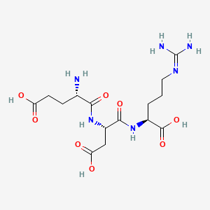
| CAS Number | 175175-23-2 |
|---|---|
| Molecular Formula | C15H26N6O8 |
| Molecular Weight | 418.4 g/Mol |
| Purity | 99.73% |
| Lot Number | 25042 |
| Quantity | 11.30mg |
| Sequence | Glu-Asp-Arg |
Cosmic Peptides Certificate of Analysis
Unable to load certificate. Download COA