Sermorelin
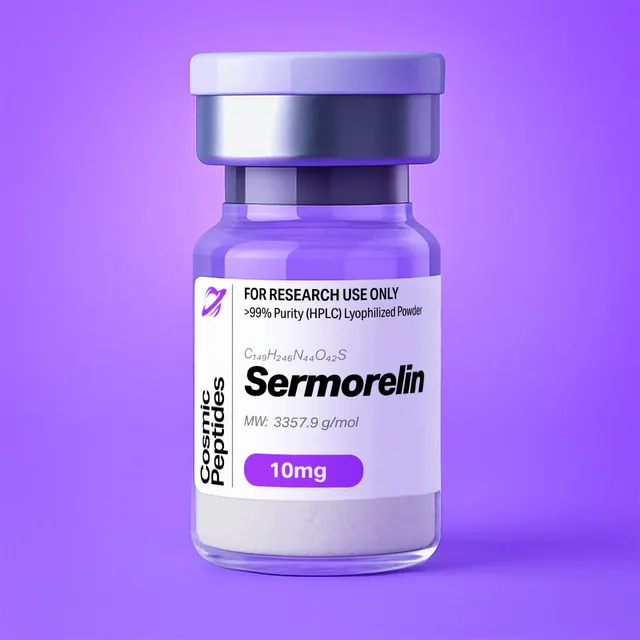
Sermorelin
Commonly researched with these items
Patent-pending lot tracking: Complete transparency from production to vial.
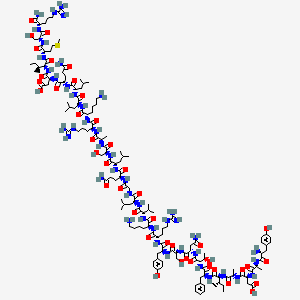
| CAS Number | 86168-78-7 |
|---|---|
| Molecular Formula | C149H246N44O42S |
| Molecular Weight | 3357.9 g/Mol |
| Purity | 99.7% |
| Lot Number | 26085 |
| Quantity | 11.05mg |
| Sequence | Tyr-Ala-Asp-Ala-Ile-Phe-Thr-Asn-Ser-Tyr-Arg-Lys-Val-Leu-Gly-Gln-Leu-Ser-Ala-Arg-Lys-Leu-Leu-Gln-Asp-Ile-Met-Ser-Arg-NH₂ |
Sermorelin: The 29-Residue N-Terminal Amide Fragment of Human Growth Hormone-Releasing Hormone
Disclaimer
Products described here are supplied for research use only and are not intended for diagnostic, therapeutic, or clinical application. All statements regarding biological activity reflect preclinical and in vitro findings exclusively and have not been evaluated by the U.S. Food and Drug Administration. These materials and related content are provided for educational and investigational purposes only and are intended solely for qualified researchers in laboratory and academic settings.
Key Facts
- Compound class
- Linear 29-residue peptide amide; the N-terminal fragment (residues 1–29) of human growth hormone-releasing hormone (GHRH), written GRF(1–29)NH2 or hGHRH(1–29)NH2
- Sequence / structure
H-Tyr-Ala-Asp-Ala-Ile-Phe-Thr-Asn-Ser-Tyr-Arg-Lys-Val-Leu-Gly-Gln-Leu-Ser-Ala-Arg-Lys-Leu-Leu-Gln-Asp-Ile-Met-Ser-Arg-NH2(YADAIFTNSYRKVLGQLSARKLLQDIMSR-NH2), all L-configuration, C-terminus amidated- Molecular formula
- C149H246N44O42S
- Molecular weight
- 3357.9 g/mol (average, free base); monoisotopic mass 3355.819 Da
- CAS number
- 86168-78-7
- PubChem CID
- 16132413 (also UNII 89243S03TE, ChEBI 9118)
- Charge and isoelectric point
- Net charge approximately +4 at pH 7.0; calculated isoelectric point 10.95 (calculated, EMBOSS pKa set)
- Chromophores
- Two Tyr, one Phe; calculated molar extinction coefficient 2980 M−1cm−1 at 280 nm
- Physical form as supplied
- Lyophilized powder
- Lot traceability
- Every unit is tied to a tracked lot; the lot number, recorded purity and verified quantity of the lot on sale are shown live on this page, with its certificate of analysis.
Molecular Identity and Classification
Sermorelin is the synthetic peptide amide corresponding to residues 1 through 29 of human growth hormone-releasing hormone, a 44-residue hypothalamic peptide of the secretin structural superfamily. It appears in the literature as GRF(1–29)NH2, hGRF(1–29)NH2 and hGHRH(1–29)NH2, and under its Chemical Abstracts index name as somatoliberin 29-L-argininamide with residues 30–44 deleted — all the same covalent entity, YADAIFTNSYRKVLGQLSARKLLQDIMSR-NH2, registered as CAS 86168-78-7 and PubChem CID 16132413. It is not somatorelin, the name of the full-length 44-residue amide (C215H358N72O66S, about 5040 g/mol, PubChem CID 16132353): two letters apart in name, fifteen residues and some 1680 g/mol apart in substance.
The parent hormone is encoded as a 108-residue precursor, somatoliberin (UniProt P01286), in which the mature hormone begins at Tyr32 immediately after an Arg30-Arg31 dibasic processing site and ends at Leu75, whose amide is generated enzymatically from a glycine extension by peptidylglycine α-amidating monooxygenase. Sermorelin reproduces only the first 29 of those 44 residues and its amide is installed synthetically on Arg29. The amide matters when comparing data across sources: GRF(1–29)NH2 and GRF(1–29)OH are different compounds whose monoisotopic masses differ by only 0.984 Da, which a nominal-mass instrument cannot resolve.
The choice of residue 29 as the truncation point came out of systematic structure–activity chemistry. Ling and colleagues synthesized a graded series of C-terminally shortened hGRF analogues with free-acid or amidated termini and measured growth hormone release from anterior pituitary cells in monolayer culture. Potency fell to about 23% of the full-length hormone at hGRF(1–34)OH, but the amidated fragments (1–31), (1–30) and (1–29) all held at roughly 50% of the potency of hGRF; full intrinsic activity persisted down to (1–21)NH2 at much lower potency and was lost only at (1–19)NH2, placing the minimal active core at residues 3–21. Sermorelin is thus the shortest chain that behaves like the hormone in cell culture — hence its role as the reference ligand in GHRH receptor work and as the scaffold for later analogues.
Structure and Chemical Features
Sermorelin contains 236 non-hydrogen atoms and fifteen amino acid types: Ala3, Arg3, Asn1, Asp2, Gln2, Gly1, Ile2, Leu4, Lys2, Met1, Phe1, Ser3, Thr1, Tyr2, Val1. Three features of that list dominate its behaviour: there is no cysteine, so the molecule is a single linear chain with no disulfide chemistry and no thiol to oxidize; the only sulfur is the Met27 thioether, the principal oxidation-sensitive site; and the single Asn, at position 8 between Thr7 and Ser9, is the principal chemically labile site in aqueous storage.
The charge pattern is strongly basic. Five cationic side chains (Arg11, Lys12, Arg20, Lys21, Arg29) and the free N-terminal α-amine are opposed by only two anionic ones (Asp3, Asp25), and the C-terminal carboxylate is replaced by a carboxamide, removing the negative charge the free acid would carry. Summing Henderson–Hasselbalch contributions over the EMBOSS pKa set gives +3.98 at pH 7.0, +5.98 at pH 2 and an isoelectric point of 10.95; other pKa tables place the latter between about 10.5 and 11.5. Sermorelin is therefore a cation across the working range of reversed-phase and ion-exchange chromatography, binds readily to anionic surfaces, and separates cleanly from acidic impurities on cation exchangers.
Helical Architecture in Solution
Although a 29-mer has no tertiary fold, sermorelin is not a random chain. Clore, Martin and Gronenborn studied two hGHRF analogues, including 27-norleucine-hGHRF(1–29)NH2, by circular dichroism and two-dimensional nuclear magnetic resonance: ordered structure appeared at about 30% 2,2,2-trifluoroethanol with roughly 23 to 25 residues helical, and full proton assignment with nuclear Overhauser effect analysis located two α-helical segments, residues 6 to 13 and 16 to 29, interrupted near Gly15.
That architecture explains much of the molecule's chemistry. The helix presents Leu, Ile, Val and Phe along one face and Arg, Lys, Asp and Ser along the other, so the GRAVY index of −0.22 describes a peptide whose polar and apolar residues are segregated by face, not averaged along the chain. Amphipathicity is why it adsorbs to hydrophobic surfaces and air–water interfaces, and why the helix-destabilizing Gly15 is the position most often replaced by Ala in engineered analogues. Residues 1 to 5, outside both helices and flexible in solution, carry the determinants that insert deepest into the receptor.
Synthesis and Manufacturing Chemistry
Sermorelin is assembled by solid-phase peptide synthesis, introduced by Merrifield in 1963, in the Fmoc/tert-butyl strategy that follows from the 9-fluorenylmethoxycarbonyl group described by Carpino and Han in 1972. Because the target carries a C-terminal carboxamide, the chain is built on an amide-forming linker such as Rink amide, from which trifluoroacetic acid cleavage delivers the amide directly with no separate amidation step.
Assembly runs from Arg29 toward Tyr1, each cycle pairing Fmoc removal by a mild base (commonly 20% piperidine in dimethylformamide) with coupling of the next Fmoc-amino acid activated by DIC/Oxyma, HBTU or HATU. Side-chain protection is standard acid-labile practice: tert-butyl esters on the two aspartates, tert-butyl ethers on the three serines, Thr7 and both tyrosines, trityl on the Asn8, Gln16 and Gln24 amides, Boc on Lys12 and Lys21, and a pentamethyldihydrobenzofuransulfonyl group on the three arginines.
Four sequence-specific difficulties shape the process chemistry. The hydrophobic Leu22-Leu23 stretch and the Leu14-Gly15-Gln16 region favour on-resin aggregation, which slows deprotection and coupling; low-loading resins, elevated temperature or a more polar solvent mixture are the usual fixes. The three arginines are the slowest couplings and the most likely to need double coupling, because the bulky sulfonyl group shields the incoming α-amine. Aspartimide formation, the base-catalysed cyclization of an aspartate ester onto the following backbone nitrogen, is a lesser risk here than in Asp-Gly or Asp-Ser sequences because both aspartates are followed by bulkier residues (Asp3-Ala4, Asp25-Ile26); but Asp25 is installed fifth and then sits through two dozen piperidine exposures, so a bulky aspartate ester or a weak acid additive is cheap insurance. And the Met27 thioether must be shielded during cleavage, hence thiol or silane scavengers in the cocktail.
After cleavage the crude peptide is precipitated in cold ether, purified by preparative reversed-phase chromatography on C18 or C8 silica with acetonitrile/water gradients containing trifluoroacetic acid, and lyophilized. Because trifluoroacetic acid is used throughout, the isolated solid is normally a trifluoroacetate salt unless the counterion is exchanged for acetate or chloride. With six protonatable basic sites (three Arg, two Lys and the N-terminal amine) the peptide can carry up to six counterions, and the effect on the mass balance is large enough to tabulate. Enzyme-assisted routes, including α-amidating enzyme catalysis of the C-terminal amide from a glycine-extended precursor, are also described in the literature.
| Counterion | 4 equivalents | 6 equivalents (all basic sites) |
|---|---|---|
| Chloride (as HCl) | 3503.7 g/mol; 95.8% peptide | 3576.6 g/mol; 93.9% peptide |
| Acetate (as acetic acid) | 3598.1 g/mol; 93.3% peptide | 3718.2 g/mol; 90.3% peptide |
| Trifluoroacetate (as TFA) | 3814.0 g/mol; 88.0% peptide | 4042.0 g/mol; 83.1% peptide |
The actual stoichiometry depends on the final purification and exchange steps and is measured, not assumed: ion chromatography or 19F nuclear magnetic resonance gives the trifluoroacetate content directly, and residual water, typically several percent by mass, lowers the peptide fraction further.
Analytical Characterization
Reversed-phase chromatography. Sermorelin separates well on C18 or C8 columns of 100–300 Å pore size with a water/acetonitrile gradient containing 0.1% trifluoroacetic acid. Detection at 214–220 nm reads the amide backbone; the two tyrosines add a 275–280 nm signal whose ratio to the backbone trace is a useful identity check. A gradient of roughly 20–45% acetonitrile over 20–30 minutes at 1 mL/min on a 4.6 × 250 mm column places the main peak mid-gradient, with the deamidated and oxidized forms eluting close to it; gradient slope and a column temperature held at 30–40 °C decide whether a method resolves them. Formic acid can replace trifluoroacetic acid when the eluent feeds a mass spectrometer, at some cost in peak sharpness.
Mass spectrometry. The average mass of the free base is 3357.88 g/mol and the monoisotopic mass 3355.819 Da. At this size the monoisotopic peak is no longer the tallest in the envelope: isotope intensities calculated from natural abundances run approximately 55 : 100 : 98 : 68 : 37 for A, A+1, A+2, A+3 and A+4, so the most abundant isotopologue carries one 13C at 3356.822 Da. Reporting an observed mass without saying whether it is monoisotopic, most-abundant or average is the commonest way identity data for peptides of this size get miscompared.
Expected Charge States and Peptide Map
With six basic sites, sermorelin ionizes efficiently in positive electrospray and spreads over several charge states, the behaviour described in Fenn's foundational work on electrospray ionization. Calculated m/z values from the monoisotopic mass are 3356.83 [M+H]+, 1678.92 [M+2H]2+, 1119.61 [M+3H]3+, 839.96 [M+4H]4+ and 672.17 [M+5H]5+, with the 3+ and 4+ ions usually dominant. Tryptic digestion gives orthogonal confirmation: trypsin cleaves after Arg11, Lys12, Arg20 and Lys21, yielding YADAIFTNSYR (monoisotopic 1319.615, [M+2H]2+ 660.81), VLGQLSAR (842.497, [M+H]+ 843.50) and the C-terminal amide LLQDIMSR-NH2 (973.538, [M+H]+ 974.55) plus two free lysines. The third fragment is the one to watch, since it reports at once on the C-terminal amide and on the oxidation state of Met27 (+15.995 Da).
Content, counterion and water. Purity and peptide content answer different questions: purity is the fraction of ultraviolet-absorbing material that is the target peptide, content the fraction of the weighed powder that is peptide at all. Amino-acid analysis after acid hydrolysis is the reference method for content, returning molar ratios Ala 3 : Arg 3 : Asx 3 : Glx 2 : Gly 1 : Ile 2 : Leu 4 : Lys 2 : Phe 1 : Ser 3 : Thr 1 : Tyr 2 : Val 1, with Met partly destroyed and Asn/Gln converted to Asp/Glu. Quantitative nuclear magnetic resonance and A280 are faster, the calculated 2980 M−1cm−1 giving A280 = 0.89 at 1 mg/mL; ion chromatography quantifies the counterion and Karl Fischer titration the water, routinely several percent by mass.
Worked molar calculation. Weigh 10.0 mg of material whose certificate records 93% peptide content: the peptide mass is 9.3 mg, which divided by 3357.9 g/mol is 2.77 µmol, so a 1.00 mL stock is 2.77 mM and not the 2.98 mM implied by the gross mass. Conversely a 1.00 mM solution holds 3.358 mg of peptide per millilitre, about 3.61 mg of weighed solid at 93% content.
Physicochemical Properties
| Property | Value | Notes |
|---|---|---|
| Molecular formula | C149H246N44O42S | Free base, no counterions |
| Average molecular weight | 3357.9 g/mol | 3357.88 calculated from the sequence |
| Monoisotopic mass | 3355.819 Da | All-12C isotopologue |
| Most abundant isotopologue | 3356.822 Da | One 13C; A+1 is the tallest peak |
| C-terminus | Carboxamide (−NH2) | Free acid differs by +0.984 Da |
| [M+3H]3+ / [M+4H]4+ | m/z 1119.61 / 839.96 | Usually the base peaks in electrospray |
| Net charge | +3.98 at pH 7.0; +5.98 at pH 2 | Six cationic sites against two Asp; no cysteine in the chain |
| Isoelectric point | 10.95 (calculated) | Strongly basic peptide |
| Molar extinction at 280 nm | 2980 M−1cm−1 | Two Tyr; A280 = 0.89 at 1 mg/mL |
| GRAVY (Kyte–Doolittle) | −0.22 | Amphipathic rather than uniformly polar |
| XLogP3 | −12.1 | Computed, PubChem |
| Topological polar surface area | 1470 Å2 | Computed, PubChem |
| Hydrogen-bond donors / acceptors | 52 / 49 | Computed, PubChem |
| Rotatable bonds / heavy atoms | 118 / 236 | Computed, PubChem |
| Helical content | 23–25 residues in about 30% trifluoroethanol | Helices at 6–13 and 16–29 by nuclear magnetic resonance |
| Labile sites | Asn8 (deamidation), Met27 (oxidation), Ala2-Asp3 (peptidase cleavage) | See degradation chemistry below |
| Solubility | Water, dilute aqueous acid, dimethyl sulfoxide | Lowest near the isoelectric point |
Two entries are routinely misread. The XLogP3 value of −12.1 is a whole-molecule descriptor and says little about how an amphipathic helix partitions; the GRAVY index of −0.22 and the apolar helical face describe the surface chemistry better, and predict adsorption to plastics and mid-gradient C18 retention. The topological polar surface area of 1470 Å2 is a sum over 86 heteroatoms and carries no conformational information.
Solubility follows the charge profile: in water or dilute acetic acid the peptide is strongly cationic and dissolves readily, while near the calculated isoelectric point of about 11 the net charge approaches zero and both solubility and resistance to aggregation fall. Near-neutral low-ionic-strength buffers are the usual analytical compromise, with the caveat that neutral pH is where Asn8 deamidation is fastest.
Research Context
Sermorelin serves as the reference agonist for the GHRH receptor and as the starting point for analogue design. Mayo cloned the receptor in 1992 by amplifying G protein-coupled receptor sequences from pituitary messenger RNA; expressed in a kidney cell line, the human homologue gave membranes that bound GHRH with high affinity and cells that responded with cyclic AMP production. Gaylinn and colleagues isolated it from a pituitary complementary DNA library the following year: a 423-amino-acid protein with seven putative transmembrane domains, a secretin-family receptor sharing 47%, 42%, 35% and 28% sequence identity with the receptors for vasoactive intestinal peptide, secretin, calcitonin and parathyroid hormone, which on transient expression in COS cells gave saturable, GHRH-specific binding and cyclic AMP accumulation at low ligand concentrations, both blocked by a GHRH antagonist. Those papers moved GHRH pharmacology from tissue extracts to transfected cells, and sermorelin is the ligand that calibrates the resulting assays.
Structural Basis of Receptor Recognition
Cryo-electron microscopy completed the picture. Zhou and colleagues solved the GHRH receptor bound to its endogenous ligand and the stimulatory G protein at 2.6 Å. The ligand is α-helical in the complex and forms a continuous interaction network with all of the receptor's extracellular loops, every transmembrane helix except TM4, and the extracellular domain, the ligand N-terminus making an especially broad set of contacts deep in the helical bundle. This is the canonical two-domain binding mode of class B receptors: the peptide's C-terminal helix is captured by the extracellular domain while its N-terminal residues insert into the transmembrane core to trigger activation.
Read against the truncation series, the structure makes the chemistry intelligible: residues 1–5, unstructured in free solution, are the activation determinants, residues 6–29 grip the extracellular domain, and residues 30–44 of the full hormone are largely dispensable for receptor engagement. It also explains why substitutions at positions 1 and 2 are so consequential, altering the deepest receptor contacts and removing the peptidase recognition motif at once.
In vitro work uses a few standard formats: competition binding in membranes from receptor-expressing cell lines against a labelled GHRH tracer; cyclic AMP accumulation or cyclic AMP response element reporter assays in transfected cells; immunoblot for phosphorylation of the cyclic AMP response element-binding protein at serine 133; calcium mobilization with fluorescent indicators; and immunoassay of growth hormone in the medium of cultured anterior pituitary cells, the classical read-out in which GHRH activity was first demonstrated by Brazeau and colleagues in 1982. Sermorelin is also the reference substrate in enzymology on GHRH-processing peptidases.
Degradation Chemistry: Three Reactions Worth Knowing
Asn8 deamidation and isomerization. Asparagine followed by a small residue deamidates through a cyclic succinimide intermediate, the mechanism worked out by Geiger and Clarke in 1987, giving α-aspartyl and rearranged β-aspartyl (isoaspartyl) products with a mass increase of 0.984 Da. Friedman and colleagues showed this is the dominant route for GRF peptides in neutral aqueous solution: [Leu27]hGRF(1–32)NH2 at pH 7.4 and 37 °C gave β-Asp8 and α-Asp8 as the major products with a parent half-life of 202 hours, extended to 1550 hours when Asn8 was replaced by serine. The consequence is severe: in cultured anterior pituitary cells the β-Asp8 product was 400 to 500 times less potent than the Asn8 parent and the α-Asp8 product about 25 times less potent. A solution held at neutral pH can therefore lose most of its activity with almost no change in nominal mass or retention time.
Met27 oxidation. The single thioether is oxidized to the sulfoxide by dissolved oxygen, peroxide impurities and light, adding 15.995 Da. The sulfoxide is more polar and elutes slightly earlier on reversed-phase columns; in a tryptic map it is read from LLQDIMSR-NH2, whose [M+H]+ moves from 974.55 to 990.54. Analogues replacing Met27 with Leu or norleucine were designed to remove this liability, which is also why the norleucine analogue was used in the original nuclear magnetic resonance study.
Ala2-Asp3 cleavage by dipeptidyl peptidase IV. Bongers and colleagues characterized the kinetics and selectivity of this cleavage with purified placental dipeptidyl peptidase IV, following the reaction by chromatography. Initial rates at pH 7.8 and 37 °C with 0.15 mM substrate were approximately 5 µmol min−1 mg−1 and essentially identical for GRF(1–44)NH2, GRF(1–29)NH2 and GRF(1–20)NH2, while much shorter fragments were cleaved more slowly, indicating substrate binding beyond the scissile bond. A D-configured residue at P2, P1 or P1′, and N-methylated or desamino Tyr1, abolished cleavage altogether, and kcat/Km was maximal for a hydrophobic P1 side chain about 0.25 nm long, close to the ethyl group of α-aminobutyric acid. The product is GRF(3–29)NH2, C137H232N42O39S, average mass 3123.63 g/mol, monoisotopic 3121.718 Da — a loss of the N-terminal Tyr-Ala dipeptide, 234.10 Da monoisotopic (234.25 average), unmistakable in a mass spectrum and worth checking for whenever the medium carries peptidase activity.
| Species | Monoisotopic mass (Da) | Shift from parent | [M+3H]3+ m/z |
|---|---|---|---|
| Sermorelin, intact | 3355.819 | — | 1119.61 |
| α-Asp8 or β-Asp8 (deamidated) | 3356.803 | +0.984 | 1119.94 |
| Met27 sulfoxide | 3371.814 | +15.995 | 1124.95 |
| GRF(3–29)NH2 (peptidase product) | 3121.718 | −234.100 | 1041.58 |
Two of the three are obvious even at low resolution: on the 3+ ion the sulfoxide moves the peak by 5.33 m/z and the peptidase product by 78.03 m/z. Deamidation is the hard one — 0.984 Da on a 3+ ion is a displacement of 0.33 m/z, inside the isotope envelope — so the two isomers are resolved chromatographically rather than by mass, and told apart by the isoaspartate-specific methyltransferase assay or by electron-transfer dissociation, which yields diagnostic c and z fragment ions across the modified bond.
Related Compounds and Analogue Design
The three reactions above map onto the substitutions in engineered GHRH analogues, a logic set out by Campbell and colleagues in 1994: modify the amino terminus to block peptidase cleavage, residue 8 to slow isomerization and residue 27 to stop oxidation, and put Ala in place of Gly15 to strengthen receptor binding. Replacing Tyr1-Ala2 with His1-Val2 in [Ala15,Leu27]hGRF(1–32)OH abolished dipeptidyl peptidase IV cleavage over 24 hours, while Thr8, Ser8 or Gln8 for Asn8 slowed isomerization in aqueous solution.
| Compound | Chain / modification | Formula | Average mass (g/mol) |
|---|---|---|---|
| Sermorelin, GRF(1–29)NH2 | Residues 1–29 of GHRH, C-terminal amide, native side chains | C149H246N44O42S | 3357.9 |
| CJC-1295 (no DAC) | GRF(1–29)NH2 with D-Ala2, Gln8, Ala15, Leu27; no sulfur because Met27 is replaced | C152H252N44O42 | 3367.9 |
| Tesamorelin | Full-length GRF(1–44)NH2 bearing a trans-3-hexenoyl group on the N-terminal Tyr | C221H366N72O67S | 5136.0 |
| Ipamorelin | Unrelated pentapeptide amide acting at the ghrelin receptor, not the GHRH receptor | C38H49N9O5 | 711.9 |
CJC-1295 in its unconjugated form is sermorelin's backbone carrying those four substitutions. It contains no sulfur, so sulfur analysis alone distinguishes it, while its average mass differs by only about 10 Da — within the isotope envelope of a low-resolution spectrum, and therefore a real analytical trap; high-resolution measurement separates them cleanly (monoisotopic 3365.894 against 3355.819), as does the tryptic map, where LLQDIMSR-NH2 (monoisotopic 973.538) is replaced by LLQDILSR-NH2 at 955.581, exactly 17.956 Da lighter. Tesamorelin keeps the full 44-residue chain and blocks the peptidase site with an N-terminal acyl group rather than by substitution. Ipamorelin is the contrast case: a pentapeptide amide acting at the ghrelin receptor, and running the two in the same cell system is the standard way to separate GHRH receptor-mediated from ghrelin receptor-mediated signalling in vitro.
Discovery as a Problem in Chemistry
GHRH resisted isolation from hypothalamic tissue for two decades; the material that broke the problem open in 1982 came from a far richer source, pancreatic islet tumour extracts. Guillemin, Brazeau, Böhlen, Esch, Ling and Wehrenberg reported the purification and sequence of a 44-residue amidated peptide in Science; Rivier, Spiess, Thorner and Vale characterized a growth hormone-releasing factor from an islet tumour in Nature the same month; and Brazeau and colleagues showed that year that the isolated material released pituitary growth hormone in vitro.
The C-terminal deletion series followed within two years, and it is why sermorelin exists as a distinct entity: the shortest fragment that behaves like the hormone in cell culture, and at 29 residues accessible by routine synthesis where the 44-mer is markedly harder to make. Structural work in the mid-1980s added the conformational picture, receptor cloning the molecular target, and the 2020 cryo-electron microscopy structure the atomic detail.
Laboratory Handling and Storage
The lyophilized solid. Store the sealed container at −20 °C or below, protected from light and moisture; −80 °C is preferable for multi-year holdings. The powder is hygroscopic, so let it reach room temperature in a desiccator before opening, or water condenses onto the cold solid and compromises the weighing. A desiccant and an inert headspace slow moisture uptake and Met27 oxidation alike — residual water acts as a mobility plasticizer in the glassy solid, as Manning and colleagues review.
Preparing solutions. Sermorelin dissolves in high-purity water and, more readily, in dilute aqueous acetic acid (0.01–0.1%), where its high positive charge keeps it monomeric. Add solvent gently down the wall of the container and let the solid wet before mixing; high-speed vortexing drives an amphipathic peptide to the air–water interface, promoting aggregation and surface loss. Dimethyl sulfoxide serves for concentrated stocks diluted into buffer, provided the final organic content suits the assay. Calculate concentrations from peptide content, not gross weighed mass.
Solution stability. Neutral aqueous solutions are not for storage, since Asn8 deamidation is fastest there; mildly acidic stocks near pH 4–5 deamidate far more slowly. Divide stocks into single-use aliquots in low-binding polypropylene, freeze at −20 °C or −80 °C, and avoid repeated freeze–thaw cycles. A carrier protein such as 0.1% bovine serum albumin, where the assay allows it, markedly reduces adsorptive loss from dilute solutions, and a 0.22 µm low-binding membrane should be pre-wetted: the first millilitre through a dry membrane can strip much of a dilute peptide.
Controls and re-verification. Peptides isolated from trifluoroacetic acid-containing eluents carry trifluoroacetate, so a counterion-matched vehicle control is good practice: Cornish and colleagues showed that trifluoroacetate alone inhibits proliferation of cultured osteoblasts and chondrocytes, and an unmatched vehicle can confound a concentration–response series. After long storage, a short run at 214 nm plus a mass check of the 3+ ion at m/z 1119.6 confirms integrity; where resolution allows, look for a +0.98 Da shoulder (deamidation) and a +15.99 Da satellite (Met27 oxidation) rather than trusting the main peak.
Frequently Asked Research Questions
- What is sermorelin, chemically?
- A synthetic 29-residue peptide amide identical to residues 1–29 of human growth hormone-releasing hormone, sequence
YADAIFTNSYRKVLGQLSARKLLQDIMSR-NH2, molecular formula C149H246N44O42S, average molecular weight 3357.9 g/mol, CAS 86168-78-7, PubChem CID 16132413. - Why the 1–29 fragment rather than the full 44-residue hormone?
- A C-terminal deletion series showed that the amidated (1–31), (1–30) and (1–29) fragments all keep about 50% of the potency of full-length hGRF in anterior pituitary cell culture, that fragments down to (1–21) retain full intrinsic activity at lower potency, and that activity is lost only at (1–19). The (1–29) amide is the shortest chain that behaves like the hormone, and far easier to synthesize than the 44-mer.
- What masses and ions should I expect by mass spectrometry?
- Monoisotopic 3355.819 Da, average 3357.88 g/mol, most abundant isotopologue (one 13C) 3356.822 Da. Calculated m/z: 1119.61 [M+3H]3+, 839.96 [M+4H]4+, 1678.92 [M+2H]2+, the 3+ and 4+ ions usually dominant.
- How do I distinguish GRF(1–29)NH2 from GRF(1–29)OH?
- The free acid is heavier by 0.984 Da, which a nominal-mass instrument cannot resolve. Use high-resolution mass spectrometry on the intact peptide, or digest with trypsin and measure the C-terminal fragment
LLQDIMSR-NH2at [M+H]+ 974.55 against 975.53 for the free acid. - Which positions are chemically labile, and which degrades fastest?
- Three sites: Asn8 deamidates and isomerizes through a succinimide intermediate (+0.984 Da), Met27 oxidizes to the sulfoxide (+15.995 Da), and dipeptidyl peptidase IV cleaves Ala2-Asp3 to give GRF(3–29)NH2 (−234.10 Da monoisotopic). Deamidation near neutral pH is the fastest chemical route: for the closely related [Leu27]hGRF(1–32)NH2 the measured half-life was 202 hours at pH 7.4 and 37 °C, rising to 1550 hours with serine at position 8, and the β-aspartyl product was 400–500 times less potent in cultured anterior pituitary cells — so activity can be lost with almost no change in nominal mass.
- What is the net charge and isoelectric point?
- Five basic side chains (Arg11, Lys12, Arg20, Lys21, Arg29) plus the N-terminal amine are opposed by only two aspartates, and the C-terminus is amidated rather than acidic: the calculated net charge is +3.98 at pH 7.0 and +5.98 at pH 2, with a calculated isoelectric point of 10.95. The peptide is a cation throughout normal reversed-phase and ion-exchange conditions.
- How is sermorelin distinguished analytically from CJC-1295 without DAC?
- They share the same backbone length and differ by about 10 Da in average mass, which low-resolution spectra cannot separate. Three orthogonal checks work: the substituted analogue contains no sulfur, so sulfur analysis or an oxidation stress test is diagnostic; high-resolution mass spectrometry resolves monoisotopic 3355.819 from 3365.894; and the tryptic C-terminal fragment is
LLQDIMSR-NH2at 973.538 for sermorelin againstLLQDILSR-NH2at 955.581 for the Leu27 analogue. - Does sermorelin absorb at 280 nm?
- Yes, weakly: two tyrosines give a calculated molar extinction coefficient of 2980 M−1cm−1, an absorbance of 0.89 for 1 mg/mL in a 1 cm cell. Chromatographic quantitation is still normally done at 214–220 nm, where the 28 backbone amides absorb far more strongly.
- What solvents are suitable, and at what pH?
- High-purity water and dilute aqueous acetic acid (0.01–0.1%) are the usual choices, with dimethyl sulfoxide for concentrated stocks to be diluted into buffer. Mildly acidic conditions help both solubility, since the peptide is far from its isoelectric point, and stability, since deamidation accelerates toward neutrality.
- Which receptor does sermorelin act at in vitro, and what is measured?
- The GHRH receptor, a 423-amino-acid class B (secretin-family) G protein-coupled receptor sharing 47% identity with the vasoactive intestinal peptide receptor and signalling through Gs and cyclic AMP. Standard in vitro read-outs: competition binding in membranes from receptor-expressing cells, cyclic AMP accumulation or reporter assays in transfected cells, phosphorylation of the cyclic AMP response element-binding protein, calcium mobilization, and growth hormone immunoassay in pituitary cell culture medium.
- How is the peptide recognized by its receptor at the structural level?
- A 2.6 Å cryo-electron microscopy structure of the receptor with its endogenous ligand and the stimulatory G protein shows the ligand bound as an α-helix contacting all extracellular loops, every transmembrane helix except TM4, and the extracellular domain, with the ligand N-terminus making the deepest contacts — so residues 1–5 are the activation determinants and residues 6–29 the affinity grip.
- Why does my weighed mass not match the molar concentration I expected?
- Because chromatographic purity is not peptide content. A lyophilized basic peptide from trifluoroacetic acid-containing eluent carries counterions and several percent water: four equivalents of trifluoroacetate add about 456 g/mol to the 3357.9 g/mol free base (roughly 88% peptide by mass), four of acetate about 240 g/mol (roughly 93%). Calculate molarity from the content figure on the lot's certificate of analysis, shown live on this page.
Sources & Further Reading
- National Center for Biotechnology Information. PubChem Compound Summary for CID 16132413, Sermorelin. PubChem CID 16132413.
- UniProt Consortium. Somatoliberin (GHRH), Homo sapiens. UniProtKB accession P01286.
- Guillemin R, Brazeau P, Böhlen P, Esch F, Ling N, Wehrenberg WB. Growth hormone-releasing factor from a human pancreatic tumor that caused acromegaly. Science 1982;218(4572):585–587. doi:10.1126/science.6812220
- Rivier J, Spiess J, Thorner M, Vale W. Characterization of a growth hormone-releasing factor from a human pancreatic islet tumour. Nature 1982;300(5889):276–278. doi:10.1038/300276a0
- Brazeau P, Ling N, Böhlen P, Esch F, Ying SY, Guillemin R. Growth hormone releasing factor, somatocrinin, releases pituitary growth hormone in vitro. Proceedings of the National Academy of Sciences 1982;79(24):7909–7913. doi:10.1073/pnas.79.24.7909
- Ling N, Baird A, Wehrenberg WB, Ueno N, Munegumi T, Brazeau P. Synthesis and in vitro bioactivity of C-terminal deleted analogs of human growth hormone-releasing factor. Biochemical and Biophysical Research Communications 1984;123(2):854–861. doi:10.1016/0006-291X(84)90309-7
- Clore GM, Martin SR, Gronenborn AM. Solution structure of human growth hormone releasing factor. Combined use of circular dichroism and nuclear magnetic resonance spectroscopy. Journal of Molecular Biology 1986;191(3):553–561. doi:10.1016/0022-2836(86)90147-6
- Mayo KE. Molecular cloning and expression of a pituitary-specific receptor for growth hormone-releasing hormone. Molecular Endocrinology 1992;6(10):1734–1744. doi:10.1210/mend.6.10.1333056
- Gaylinn BD, Harrison JK, Zysk JR, Lyons CE, Lynch KR, Thorner MO. Molecular cloning and expression of a human anterior pituitary receptor for growth hormone-releasing hormone. Molecular Endocrinology 1993;7(1):77–84. doi:10.1210/mend.7.1.7680413
- Zhou F, Zhang H, Cong Z, Zhao LH, Zhou Q, Mao C, et al. Structural basis for activation of the growth hormone-releasing hormone receptor. Nature Communications 2020;11(1):5205. doi:10.1038/s41467-020-18945-0
- Geiger T, Clarke S. Deamidation, isomerization, and racemization at asparaginyl and aspartyl residues in peptides. Succinimide-linked reactions that contribute to protein degradation. Journal of Biological Chemistry 1987;262(2):785–794. doi:10.1016/S0021-9258(19)75855-4
- Friedman AR, Ichhpurani AK, Brown DM, Hillman RM, Krabill LF, et al. Degradation of growth hormone releasing factor analogs in neutral aqueous solution is related to deamidation of asparagine residues. International Journal of Peptide and Protein Research 1991;37(1):14–20. doi:10.1111/j.1399-3011.1991.tb00727.x
- Bongers J, Lambros T, Ahmad M, Heimer EP. Kinetics of dipeptidyl peptidase IV proteolysis of growth hormone-releasing factor and analogs. Biochimica et Biophysica Acta 1992;1122(2):147–153. doi:10.1016/0167-4838(92)90317-7
- Campbell RM, Stricker P, Miller R, Bongers J, Liu W, et al. Enhanced stability and potency of novel growth hormone-releasing factor (GRF) analogues derived from rodent and human GRF sequences. Peptides 1994;15(3):489–495. doi:10.1016/0196-9781(94)90211-9
- Merrifield RB. Solid phase peptide synthesis. I. The synthesis of a tetrapeptide. Journal of the American Chemical Society 1963;85(14):2149–2154. doi:10.1021/ja00897a025
- Carpino LA, Han GY. 9-Fluorenylmethoxycarbonyl amino-protecting group. The Journal of Organic Chemistry 1972;37(22):3404–3409. doi:10.1021/jo00795a005
- Mant CT, Chen Y, Yan Z, Popa TV, Kovacs JM, et al. HPLC analysis and purification of peptides. Methods in Molecular Biology 2007;386:3–55. doi:10.1007/978-1-59745-430-8_1
- Fenn JB, Mann M, Meng CK, Wong SF, Whitehouse CM. Electrospray ionization for mass spectrometry of large biomolecules. Science 1989;246(4926):64–71. doi:10.1126/science.2675315
- Manning MC, Chou DK, Murphy BM, Payne RW, Katayama DS. Stability of protein pharmaceuticals: an update. Pharmaceutical Research 2010;27(4):544–575. doi:10.1007/s11095-009-0045-6
- Cornish J, Callon KE, Lin CQ, Xiao CL, Mulvey TB, et al. Trifluoroacetate, a contaminant in purified proteins, inhibits proliferation of osteoblasts and chondrocytes. American Journal of Physiology-Endocrinology and Metabolism 1999;277(5):E779–E783. doi:10.1152/ajpendo.1999.277.5.E779
Last reviewed:
In the research library
Everything the library holds on Sermorelin, and the methods behind the numbers on its certificate.
- Sermorelin reference entry Identity, calculated properties, the mass spectrometry ions to expect and analytical notes, from the structure.
- Check a certificate against this sequence Paste the masses from a spectrum or a certificate; the mass-match tool compares them with what this sequence should give.
- Peptide calculator, with this sequence Formula, average and monoisotopic mass, charge across pH, isoelectric point and absorbance, recalculated as you edit.
- Growth hormone–releasing hormone analogs The family side by side: sequences aligned, each member's relation to the reference peptide, and how analysis tells them apart.
- Sermorelin and CJC-1295 (no DAC) compared Both are 29-residue C-terminal amides built on the first 29 residues of growth hormone–releasing hormone. CJC-1295 without DAC carries four substitutions in that chain; sermorelin is the unmodified sequence.
- Sermorelin and Tesamorelin compared Sermorelin is the first 29 residues of growth hormone–releasing hormone as an amide. Tesamorelin is the full 44-residue amide with a trans-3-hexenoyl group on its N-terminus, so sermorelin's whole sequence is contained in it.
- How to read a certificate of analysis What a peptide certificate of analysis should contain, how to check that it belongs to the material in hand, how to read its identity and purity results, and the signs that it deserves a second look.
- HPLC purity analysis of peptides How reversed-phase HPLC measures the chromatographic purity of a peptide, what the figure includes, and what it leaves out.
- Mass spectrometry for peptide identity How mass spectrometry confirms that a peptide is the intended sequence: electrospray and MALDI, working out the expected mass, reading charge states and adducts, sequence confirmation, and the limits of the method.
- Net peptide content and purity: two different numbers Why a peptide that is 99% pure by HPLC can be only 75–85% peptide by weight: what the powder contains, how each part is measured, and how the two figures combine.
- Preparing peptide solutions in the laboratory From a freeze-dried vial to a solution of known concentration: the arithmetic, the solvent, the charge chemistry and the handling, with the errors each step can introduce and the article that covers it.
- Peptide stability and storage How peptides degrade, chemically and physically, why the dry solid lasts far longer than a solution, which sequences are most at risk, and the storage and handling that slow it down.