SS-31
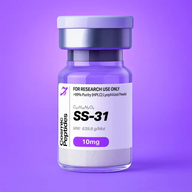
SS-31
Commonly researched with these items
Patent-pending lot tracking: Complete transparency from production to vial.
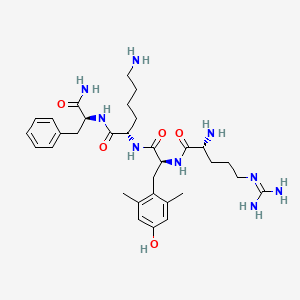
| CAS Number | 736992-21-5 |
|---|---|
| Molecular Formula | C32H49N9O5 |
| Molecular Weight | 639.8 g/Mol |
| Purity | 99.78% |
| Lot Number | 26080 |
| Quantity | 10.12mg |
| Sequence | H-D-Arg-Tyr(2,6-diMe)-Lys-Phe-NH2 |
SS-31 (D-Arg-Dmt-Lys-Phe-NH2): An Aromatic-Cationic Tetrapeptide That Partitions Into Cardiolipin-Rich Membranes
Disclaimer
Products described here are supplied for research use only and are not intended for diagnostic, therapeutic, or clinical application. All statements regarding biological activity reflect preclinical and in vitro findings exclusively and have not been evaluated by the U.S. Food and Drug Administration. These materials and related content are provided for educational and investigational purposes only and are intended solely for qualified researchers in laboratory and academic settings.
Key Facts
- Compound class
- Synthetic linear tetrapeptide amide of the Szeto-Schiller (SS) aromatic-cationic series; contains one D-amino acid and one non-proteinogenic amino acid
- Sequence / structure
H-D-Arg-Dmt-Lys-Phe-NH2, where Dmt is 2′,6′-dimethyl-L-tyrosine; IUPAC-style name D-arginyl-2,6-dimethyl-L-tyrosyl-L-lysyl-L-phenylalaninamide- Molecular formula
- C32H49N9O5 (free base)
- Molecular weight
- 639.8 g/mol (average, free base); monoisotopic mass 639.3857 Da
- CAS number
- 736992-21-5
- PubChem CID
- 11764719
- Formal charge at pH 7
- +3 (N-terminal amine, arginine guanidinium, lysine ε-ammonium; the C-terminus is an uncharged amide)
- Physical form as supplied
- Lyophilized powder
- Lot traceability
- Every unit is tied to a tracked lot; the lot number, recorded purity and verified quantity of the lot on sale are shown live on this page, with its certificate of analysis.
Molecular Identity and Classification
SS-31 is the tetrapeptide amide H-D-Arg-Dmt-Lys-Phe-NH2. Reading from the N-terminus it contains D-arginine, the synthetic aromatic amino acid 2′,6′-dimethyl-L-tyrosine (Dmt), L-lysine and L-phenylalanine, with the C-terminal carboxyl converted to a primary carboxamide. Its systematic name is D-arginyl-2,6-dimethyl-L-tyrosyl-L-lysyl-L-phenylalaninamide; PubChem gives the full IUPAC name as (2S)-6-amino-2-[[(2S)-2-[[(2R)-2-amino-5-(diaminomethylideneamino)pentanoyl]amino]-3-(4-hydroxy-2,6-dimethylphenyl)propanoyl]amino]-N-[(2S)-1-amino-1-oxo-3-phenylpropan-2-yl]hexanamide. The four defined stereocentres are one R (the D-arginine α-carbon) and three S (Dmt, lysine and phenylalanine).
The molecular formula is C32H49N9O5, the average molecular weight 639.8 g/mol (639.80 from standard atomic weights) and the monoisotopic mass 639.3857 Da, under CAS number 736992-21-5 and PubChem CID 11764719. The letters "SS" stand for Szeto and Schiller, whose laboratories designed the series; the same entity appears in the literature and in chemical databases under the synonym elamipretide and as the shorthand D-Arg-Dmt-Lys-Phe-NH2.
What sets SS-31 apart from ordinary tetrapeptides is the deliberate alternation of basic and aromatic residues, a design its originators called the aromatic-cationic motif: three positive charges in water, yet two bulky aromatic side chains able to insert into the non-polar region of a lipid bilayer. That makes it a widely used tool compound in membrane biophysics and in in vitro studies of the mitochondrial inner membrane, where cardiolipin is concentrated. Both termini are also blocked against exopeptidase chemistry: an N-terminal D-residue is a poor aminopeptidase substrate, and the C-terminal amide removes the carboxylate carboxypeptidases recognize.
Structure and Chemical Features
Each residue contributes a defined chemical function. D-Arginine (position 1) supplies the most basic group, a guanidinium ion with a pKa near 12.5, plus the free N-terminal α-amine. Dmt (position 2) is a tyrosine analogue with methyl groups on both ring carbons adjacent to the benzylic attachment point. Lysine (position 3) adds a primary ε-ammonium group on a flexible four-methylene side chain. Phenylalanine (position 4) provides an unsubstituted benzyl side chain, the most hydrophobic element of the molecule, and its amidated C-terminus.
The 2′,6′-Dimethyltyrosine Residue
Dmt is the signature residue of the SS series. Relative to tyrosine it adds two methyl groups (net C2H4, +28.031 Da) at the 2′ and 6′ positions of the aromatic ring, that is, ortho to the CH2 linking the ring to the backbone and meta to the phenolic hydroxyl. The methyls have three consequences: they restrict rotation about the side-chain χ1 and χ2 torsions, so the ring samples fewer orientations than in tyrosine; they add hydrophobic surface, favouring burial in a membrane interface; and they make the phenol a sterically hindered phenol of the kind used as a radical-trapping motif in antioxidant chemistry, able to donate its phenolic hydrogen to a peroxyl or hydroxyl radical and leave a stabilized phenoxyl radical behind. The residue also carries the molecule's main near-ultraviolet chromophore and its intrinsic fluorescence, which Mitchell and colleagues used in 2020 to follow membrane partitioning label-free (excitation 283 nm, emission 290–400 nm).
Charge Distribution and Amphipathicity
SS-31 is an unusually dense package of charge for its size. The arginine guanidinium (pKa ≈ 12.5), the lysine ε-ammonium (pKa ≈ 10.5) and the N-terminal α-amine (pKa ≈ 8) are all protonated at neutral pH, while the single ionizable acid-type group, the Dmt phenol (pKa ≈ 10.3), is neutral there and the C-terminal amide bears no charge. The formal charge is therefore +3 at pH 7, as the biophysical literature states, and the calculated isoelectric point lies above pH 11.
Two of the four side chains are nonetheless aromatic hydrocarbons. The molecule has no hydrophobic and hydrophilic face as an amphipathic helix would; the basic and aromatic groups simply alternate along a backbone too short to fold. Molecular dynamics simulations reported by Mitchell and colleagues show how that geometry behaves at a bilayer: the basic side chains sit in the interfacial region, arginine 1.75 nm from the bilayer centre, just below the average depth of the lipid phosphates (1.98 nm), and lysine slightly more distal among the headgroups (1.99 nm), while the aromatic side chains bury in the non-polar core, phenylalanine deeper (1.12 nm) than Dmt (1.37 nm). The same depths were found with dianionic and monoanionic lipids alike, so the insertion geometry belongs to the peptide rather than to one lipid. Arginine adsorbed within about 50 ns and phenylalanine took roughly 180 ns to localize: the peptide docks electrostatically first and inserts its aromatic residues second.
Conformation in Solution
With four residues and no disulfide, SS-31 has no stable secondary structure in water: it is a conformational ensemble, and that matters energetically, because a peptide needing no pre-organization into a helix or sheet pays little conformational entropy penalty on binding a membrane. Isothermal titration calorimetry of the peptide binding anionic liposomes gave only small favourable enthalpies (ΔH of −4.2, −4.7 and −2.1 kJ mol−1 for bilayers containing tetraoleoyl cardiolipin, monolysocardiolipin and phosphatidylglycerol) against much larger favourable entropic terms (−TΔS of −22.7, −20.8 and −21.5 kJ mol−1). The interaction is dominated by burial of the aromatic side chains and release of ordered water, not by hydrogen bonding or folding.
Synthesis and Manufacturing Chemistry
SS-31 is made by solid-phase peptide synthesis (SPPS), the strategy Merrifield introduced in 1963, in which the chain is assembled on an insoluble polymeric support so that excess reagents and soluble by-products are washed away; published biophysical work records material prepared this way and used as an aqueous stock.
Because the C-terminus is a primary carboxamide rather than an acid, synthesis starts on an amide-forming resin, classically a Rink amide or 4-methylbenzhydrylamine-type support, so that acidolytic cleavage liberates the C-terminal amide directly. Assembly runs from the C-terminus towards the N-terminus — phenylalanine, lysine, Dmt, D-arginine — using the base-labile 9-fluorenylmethoxycarbonyl (Fmoc) α-amino protecting group described by Carpino and Han in 1972 with acid-labile side-chain protection: tert-butyloxycarbonyl on the lysine ε-amine, a sulfonyl-type group on the arginine guanidinium, a tert-butyl ether on the Dmt phenol.
Three points are specific to this sequence. First, the non-proteinogenic residue has to be made before the peptide can be: enantiomerically pure 2,6-dimethyl-L-tyrosine is itself a synthetic target, and an asymmetric route to it was published by Dygos and co-workers in 1992; Fmoc-protected Dmt dominates the raw-material cost. Second, the Dmt α-carbon sits next to a doubly ortho-substituted ring, so couplings onto and from it are slower than for tyrosine and often need a double coupling or a stronger activator. Third, the finished peptide must be shown not to contain its epimers, which are isobaric and so invisible to mass spectrometry alone.
Final cleavage with trifluoroacetic acid and scavengers removes the side-chain protection and releases the peptide amide in one operation; the crude material is precipitated, purified by preparative reversed-phase HPLC and lyophilized. Because trifluoroacetic acid is present throughout, the isolated powder is normally a trifluoroacetate salt unless the counterion is exchanged for acetate or chloride — a choice that changes the mass of salt per milligram of powder, as the counterion table below makes explicit.
Analytical Characterization
Reversed-phase HPLC. SS-31 behaves on a C18 column as a moderately retained, strongly ion-pairing analyte. Mobile phases of water and acetonitrile with 0.05–0.1% trifluoroacetic acid are conventional: trifluoroacetate pairs with the guanidinium and ammonium groups, suppresses interaction with residual silanols and sharpens what would otherwise be a tailing peak. Detection at 214–220 nm reports the amide backbone and gives the area-percent purity figure; a second channel at 275–280 nm reports the Dmt phenol, and the ratio of the two is a quick identity check, since a deletion impurity lacking Dmt loses the 280 nm signal but keeps most of the 214 nm signal. Changing the ion-pairing agent to formic acid shortens retention and can resolve impurities that co-elute under trifluoroacetic acid, so an orthogonal second method is worth running before a lot is used quantitatively.
Mass spectrometry. The monoisotopic mass of the free base is 639.3857 Da, so positive-mode electrospray gives [M+H]+ at m/z 640.393. Because there are three basic sites, the doubly and triply charged ions at m/z 320.700 and 214.136 are usually the most abundant species at low pH; an analyst looking only near 640 may wrongly conclude the sample did not ionize. With 32 carbons the first isotope peak is intense, about 39% of the monoisotopic peak, and in the 3+ cluster the isotope spacing collapses to 0.33 m/z.
| Species | Calculated m/z | What it tells the analyst |
|---|---|---|
| [M+H]+ | 640.393 | Intact tetrapeptide amide |
| [M+2H]2+ | 320.700 | Usually prominent; isotope spacing 0.5 m/z |
| [M+3H]3+ | 214.136 | Three basic sites; isotope spacing 0.33 m/z |
| [M+Na]+ | 662.375 | Sodium adduct from glassware or buffer salts |
| b2 (D-Arg-Dmt) | 348.203 | Confirms the N-terminal pair |
| b3 (D-Arg-Dmt-Lys) | 476.298 | Confirms position of Lys |
| y1 (Phe-NH2) | 165.102 | Diagnostic of the C-terminal amide; 166.086 if the amide has hydrolysed |
| y2 (Lys-Phe-NH2) | 293.197 | Common base peak in tandem MS of this sequence |
| Dmt immonium | 164.107 | Side-chain reporter; absent in des-Dmt impurities |
| [M+H]+ of the free-acid form | 641.377 | +0.984 Da: C-terminal amide hydrolysed to the acid |
| [M+H]+ of a des-Dmt deletion | 449.298 | −191.095 Da: missed coupling at the hindered residue |
| [M+H]+ of a des-Lys deletion | 512.298 | −128.095 Da |
Stereochemical purity. Mass spectrometry cannot distinguish SS-31 from its epimers, and a routine C18 gradient need not resolve them either. Because the design depends on the first residue being D-arginine, complete characterization includes a chiral method: hydrolysis with chiral gas or liquid chromatography, or Marfey-type derivatization and reversed-phase separation of the diastereomers. The expected result is D-Arg with L-Dmt, L-Lys and L-Phe.
Composition. Total hydrolysis gives a 1:1:1 molar ratio of arginine, lysine and phenylalanine plus one equivalent of 2,6-dimethyltyrosine, which needs its own reference standard because it is absent from calibration mixtures. Elemental analysis is an independent check: C32H49N9O5 is 60.07% C, 7.72% H, 19.70% N and 12.50% O by mass, and that nitrogen fraction is itself characteristic.
Purity is not content. An area-percent purity figure describes the fraction of ultraviolet-absorbing material that is the target peptide; peptide content describes the fraction of the weighed powder that is peptide at all, the remainder being counterions, water and residual salts. For a molecule with three basic sites that distinction is large: with all three paired with trifluoroacetate the salt has a formula weight of about 982 g/mol and the powder is roughly 65% peptide by mass, so a 1 mg/mL "solution" is 1.02 mM, not the 1.56 mM a free-base calculation predicts. Quantitative work therefore needs peptide content from the certificate of analysis or an in-house determination by quantitative NMR or amino-acid analysis, with water by Karl Fischer titration and counterion by ion chromatography or 19F NMR.
Physicochemical Properties
| Property | Value | Notes |
|---|---|---|
| Molecular formula | C32H49N9O5 | Free base, no counterions |
| Average molecular weight | 639.8 g/mol | 639.80 from standard atomic weights |
| Monoisotopic mass | 639.3857 Da | Exact mass, PubChem CID 11764719 |
| Heavy atoms / defined stereocentres | 46 / 4 | One R (D-Arg) and three S centres |
| Formal charge at pH 7 | +3 | Guanidinium, ε-ammonium, α-amine |
| Isoelectric point | >11 (calculated) | Only ionizable acid-type group is the phenol |
| XLogP | 0 | Computed; charge-balanced amphipathic character |
| Topological polar surface area | 267 Å2 | Computed |
| Hydrogen-bond donors / acceptors | 9 / 8 | Computed |
| Rotatable bonds | 19 | Computed; no stable fold in water |
| Chromophores | Dmt phenol (275–280 nm), Phe (~257 nm) | Dmt also gives intrinsic fluorescence |
| Solubility | Freely soluble in water and aqueous buffers | Polycation; prepared as aqueous stocks in published biophysical work |
| Partition into 20% cardiolipin bilayers | KP = 5.07 ± 0.76 × 10⁴ | Molar partition coefficient, Mitchell et al. 2020 |
| Partition into 20% monolysocardiolipin bilayers | KP = 5.90 ± 0.44 × 10⁴ | Same study |
| Partition into 20% phosphatidylglycerol bilayers | KP = 1.28 ± 0.63 × 10⁴ | Monoanionic lipid; ~4-fold weaker |
| Partition into pure phosphatidylcholine bilayers | Negligible | Anionic lipid is required for binding |
The descriptor set reads oddly at first glance: a computed XLogP of zero alongside a polar surface area of 267 Å2 and nine hydrogen-bond donors. That is the aromatic-cationic design in numbers. The polar surface and the donors come from the guanidinium, the ammonium groups and the amide backbone, and the near-zero partition coefficient reflects two aromatic side chains offsetting them: freely water-soluble, yet with enough hydrophobic surface to partition into a lipid interface once electrostatics have brought it there.
Counterion identity changes every gravimetric calculation made with the powder.
| Salt form | Formula weight of the salt | Peptide (free base) by mass | 1 mg/mL corresponds to |
|---|---|---|---|
| Free base | 639.80 g/mol | 100% | 1.563 mM |
| Tri-trifluoroacetate | 981.87 g/mol | 65.2% | 1.018 mM |
| Tri-acetate | 819.96 g/mol | 78.0% | 1.220 mM |
| Tri-hydrochloride | 749.18 g/mol | 85.4% | 1.335 mM |
A worked example: 10 mg of the free base is 15.63 µmol, a 15.6 mM stock in 1.00 mL of water, while 10 mg of the tri-trifluoroacetate salt contains about 6.5 mg of peptide, or 10.2 µmol, giving a 10.2 mM stock. The factor of 1.5 between the two conventions is larger than the concentration steps in most titration series, so the convention used belongs in the methods.
Chemical liabilities follow from the composition. There is no cysteine, methionine or tryptophan, so the oxidation pathways that dominate degradation of larger peptides are absent; the Dmt phenol is itself oxidizable, which is central to the radical-trapping chemistry but also means solutions left in air and light develop phenol-derived oxidation products, including the dimer expected from coupling of two phenoxyl radicals. The C-terminal carboxamide hydrolyses to the free acid under prolonged acidic or basic conditions, a +0.984 Da change easily seen by high-resolution mass spectrometry.
Research Context
SS-31 is studied almost entirely as a probe of one chemical relationship: how a small polycationic peptide interacts with the anionic phospholipid cardiolipin and with the proteins cardiolipin organizes. The work divides into membrane biophysics, redox chemistry at the lipid-protein interface, and interaction mapping.
The Chemistry of the Binding Partner
Cardiolipin is not an ordinary phospholipid. It is dimeric: two phosphatidyl units joined through a central glycerol, giving a headgroup with two phosphates and four acyl chains instead of two. As Paradies and colleagues set out in 2014, it is largely restricted to energy-transducing membranes and, in mitochondria, to the inner membrane, where it participates in the assembly and activation of respiratory complexes and, through its conical shape, favours negative membrane curvature.
The ionization of that two-phosphate headgroup was argued over for decades, and the answer matters for the binding chemistry of a tripositive peptide. Sathappa and Alder settled it in model bilayers in 2016 with electrokinetic and spectroscopic measurements analysed by Gouy-Chapman-Stern formalism: both phosphates ionize as strong acids with low pKa values, so the headgroup is fully ionized as a dianion rather than a monoanion with one protonated phosphate. Cardiolipin is therefore a dominant contributor to the surface charge density of the inner membrane, and it is that surface charge, not a lock-and-key pocket, that SS-31 recognizes. Mitchell and colleagues made the point quantitatively in 2020 by comparing bilayers containing a dianionic lipid (cardiolipin) with a monoanionic one (phosphatidylglycerol) at the same mole fraction: partitioning was roughly four-fold stronger into the dianionic membranes, the enthalpic contribution scaled with the formal charge of the lipid, and binding to a purely zwitterionic bilayer was negligible. Their binding densities make the electrostatic bookkeeping explicit: at 20 mol% cardiolipin each peptide occupies about 560–580 Å2 and associates with 1.4–1.5 dianionic lipids, while at 20 mol% phosphatidylglycerol the footprint is roughly 1080 Å2 and about three monoanionic lipids. Both come to approximately three lipid negative charges per tripositive peptide, which is what a surface-charge mechanism predicts and a stereospecific binding site would not.
Binding also did what a surface-adsorbed polycation should: it reduced the magnitude of the zeta potential of anionic liposomes in a saturable manner and raised the emission of 1-anilinonaphthalene-8-sulfonate, a reporter whose fluorescence at anionic bilayers rises as the surface potential becomes less negative. Both read-outs indicate that the peptide tunes surface electrostatics rather than disrupting the bilayer, which stayed lamellar even at the highest binding densities, with only saturable changes in lipid packing; the same study saw altered distribution of divalent calcium in the interfacial region of isolated mitochondria.
Redox Chemistry at the Lipid-Protein Interface
Cardiolipin is also the phospholipid most vulnerable to oxidation in mitochondria, and the oxidant is a protein it binds. Kagan and colleagues showed in 2005, using oxidative lipidomics, that cardiolipin is the only mitochondrial phospholipid that undergoes early oxidation during apoptosis, catalysed by a cardiolipin-specific peroxidase activity of cytochrome c itself once that protein is bound to cardiolipin: a conditional peroxidase whose chemistry depends on its lipid environment.
Birk and colleagues placed SS-31 in that picture. Their 2013 study used a polarity-sensitive fluorescent analogue to show high-affinity binding to cardiolipin and reported that the peptide-cardiolipin complex inhibited the peroxidase activity of cytochrome c by shielding its heme iron. Their 2014 study moved to defined lipid systems: the peptide interacted only with liposomes and bicelles containing cardiolipin, at roughly 1:1 with the lipid, and nuclear magnetic resonance showed its aromatic residues penetrating deep into those bilayers. Added cardiolipin slowed cytochrome c reduction and oxygen consumption by mitoplasts and frozen mitochondria and the peptide reversed both effects, while in freshly isolated mitochondria it raised state 3 respiration and the efficiency of adenosine triphosphate synthesis — the peroxidase chemistry of the cytochrome c/cardiolipin complex inhibited, the electron-carrier function of the same protein intact.
The role of the Dmt residue was tested by subtraction. Zhao and colleagues reported in 2004 that analogues on the same alternating backbone but lacking dimethyltyrosine failed to inhibit reactive oxygen species generation or swelling in isolated mitochondria, whereas Dmt-containing peptides reduced reactive oxygen species, inhibited the permeability transition and prevented calcium-induced cytochrome c release; in N2A cells exposed to tert-butyl hydroperoxide they lowered intracellular reactive oxygen species at nanomolar half-maximal effective concentrations. The 2005 follow-up in N2A and SH-SY5Y cells reported effects below 1 nM and the partitioning that explains them: tritium-labelled SS-31 reached intracellular concentrations about six-fold above extracellular, and concentrated roughly 5000-fold into the pellet of isolated mitochondria. Szeto's 2006 review attributed the series' scavenging chemistry to the tyrosine or dimethyltyrosine residue.
A chemical caveat follows. Mitchell and colleagues argued in 2020 that radical trapping cannot be the whole account, on two grounds: a phenolic antioxidant is consumed stoichiometrically rather than acting catalytically, so one phenol quenches only one or two radicals; and analogues carrying no scavenging side chain at all, such as SS-20, are studied in the same systems. Hence the move towards surface electrostatics and lipid packing, where a bound peptide is not consumed by what it does.
Which proteins the peptide sits near was addressed by Chavez and colleagues in 2020, by chemical cross-linking with mass spectrometry in isolated mitochondria. The partners they identified were all known cardiolipin binders, in two groups: components of oxidative phosphorylation and enzymes of 2-oxoglutarate metabolism, with cross-linked residues often close to regions where those proteins contact cardiolipin. Read with the liposome work, the picture is of a peptide that occupies cardiolipin-rich interfacial regions and so reports on, and perturbs, the lipid environment of the proteins embedded there.
Three kinds of system recur: liposomes or bicelles of defined composition for fluorescence titrations, zeta potential, calorimetry and solid-state 31P nuclear magnetic resonance; isolated mitochondria and mitoplasts for respirometry, membrane-potential dyes and cross-linking; and cultured cell lines for uptake with labelled analogues.
Design Lineage: Where the Sequence Came From
SS-31 was not discovered in an extract; it was built, and its ancestry is traceable through peptide chemistry. The starting point was DALDA, the tetrapeptide amide H-Tyr-D-Arg-Phe-Lys-NH2, a deliberately polar, strongly cationic peptide amide studied as a ligand in isolated membrane binding assays. In 2000 Schiller and colleagues published DALDA analogues in which the N-terminal tyrosine was replaced by ring-substituted surrogates — 2′,6′-dimethyltyrosine (Dmt), N,2′,6′-trimethyltyrosine, 2′-methyltyrosine and 2′-hydroxy-6′-methyltyrosine — and characterized the resulting [Dmt1]DALDA as combining a net charge of 3+ with resistance to enzymatic degradation. That paper is where the Dmt residue, the D-amino acid and the C-terminal amide first appear together.
The property that redirected the chemistry came next. Zhao and colleagues reported in 2003 that [Dmt1]DALDA, despite three positive charges, translocated across Caco-2 monolayers with a permeability coefficient near 1.24 × 10⁻⁵ cm/s, was detectable inside cells within five minutes, and entered SH-SY5Y, HEK293 and CRFK cells alike. Uptake was concentration-dependent but independent of temperature and pH, and diethylpyrocarbonate, which suppressed uptake of glycyl-sarcosine, raised peptide uptake 34-fold — evidence that the proton-coupled transporter PEPT1 was not the route. A tripositive tetrapeptide that crosses membranes without a transporter is the chemical basis of everything the series does.
SS-31 is the analogue in which those four building blocks are arranged as H-D-Arg-Dmt-Lys-Phe-NH2. Szeto and Schiller reviewed the series in 2011, and Szeto's 2014 article set out the cardiolipin-centred account of its chemistry that the later biophysical work tested in model membranes. The series is best read as a set of single-variable comparisons on one scaffold.
| Peptide | Sequence | Formula / monoisotopic mass | Formal charge at pH 7 | What changes relative to SS-31 |
|---|---|---|---|---|
| SS-31 | H-D-Arg-Dmt-Lys-Phe-NH2 | C32H49N9O5 / 639.3857 Da | +3 | Reference compound |
| DALDA | H-Tyr-D-Arg-Phe-Lys-NH2 | C30H45N9O5 / 611.3544 Da | +3 | Parent scaffold: unmethylated phenol, different residue order |
| [Dmt1]DALDA | H-Dmt-D-Arg-Phe-Lys-NH2 | C32H49N9O5 / 639.3857 Da | +3 | Sequence isomer: same composition, different order |
| SS-20 | H-Phe-D-Arg-Phe-Lys-NH2 | C30H45N9O4 / 595.3594 Da | +3 | No phenol at all: separates surface-charge chemistry from radical trapping |
| [ald]SS-31 | Aladan in place of the C-terminal Phe | Not calculated here | +3 | Environment-sensitive fluorophore for binding measurements in spectrally crowded samples |
The third row is an analytical trap. Because SS-31 and [Dmt1]DALDA are built from the same four residues in a different order, they share the formula C32H49N9O5, the same average and monoisotopic masses, and even the same b2 and y2 fragments, which contain the same residue pairs; accurate mass on the intact molecule cannot tell them apart. Two calculated fragments can: y1 is m/z 165.102 for SS-31 (Phe-NH2) against 146.129 for [Dmt1]DALDA (Lys-NH2), and b3 is 476.298 against 495.271. Identity confirmation therefore rests on tandem mass spectrometry or chromatographic comparison with a reference standard.
Related Research Compounds
Work on mitochondrial biochemistry in vitro often sets chemically unrelated tool compounds side by side. MOTS-c is a short peptide whose open reading frame lies within the mitochondrial genome; it contrasts with SS-31 in being an ordinary all-L peptide with no engineered charge motif. NAD is the redox cofactor at the entry point of the respiratory chain, and the absorbance of its reduced form at 340 nm is the standard read-out in the dehydrogenase assays that accompany respirometry. Within the SS series the comparators are the chemical ones tabulated above.
Laboratory Handling and Storage
The solid. Lyophilized SS-31 is kept tightly closed at −20 °C, protected from light and moisture. Like most salted polycationic peptides the powder is hygroscopic, so a container taken from the freezer is brought to room temperature in a desiccator before opening; opening it cold condenses atmospheric water onto the powder and inflates the weighed mass. The balance reading is recorded together with the peptide content figure from the certificate of analysis.
Preparing solutions. The peptide dissolves readily in high-purity water, and published biophysical studies prepared it as aqueous stocks. Water is the better solvent for a concentrated primary stock: it avoids precipitating the polycation with multivalent anions and leaves buffer composition to the working dilution. Organic co-solvents are unnecessary and, in membrane work, undesirable, since dimethyl sulfoxide alters lipid packing. Concentrations are calculated from the free-base molecular weight corrected for peptide content, not from the gross powder mass.
Buffer chemistry. High ionic strength screens the attraction that drives membrane partitioning, so binding measured at 150 mM salt is weaker than at 25 mM, and the salt concentration belongs with any reported binding constant. Polyanions such as heparin or polyphosphate sequester the peptide and concentrated phosphate forms sparingly soluble complexes with polycations, so a zwitterionic buffer is the common choice; Mitchell and colleagues worked in 20 mM HEPES-KOH at pH 7.5.
Adsorption losses. A tripositive peptide adsorbs to negatively charged surfaces, including borosilicate glass and untreated plastic ware, and at sub-micromolar concentrations the loss is enough to shift an apparent binding constant. Low-binding polypropylene ware and pre-rinsed tips reduce the effect; in liposome titrations the usual safeguard is to confirm that the isotherm does not depend on the order of addition.
Storage of solutions and re-verification. Aqueous stocks are divided into single-use aliquots and frozen at −20 °C or −80 °C, since repeated freeze-thaw cycles concentrate solutes in the unfrozen fraction and promote adsorption and phenol oxidation. A short LC-MS run on a stored stock — looking for [M+2H]2+ at m/z 320.700, the +0.984 Da amide-hydrolysis product and phenol oxidation products — together with the 214/280 nm ratio from the same run, confirms that the material is still what the label says.
Controls specific to this chemistry. Because the powder is normally a trifluoroacetate salt, a counterion-matched vehicle is a meaningful control: Cornish and colleagues showed in 1999 that trifluoroacetate carried over from peptide purification is not inert in cell culture. In membrane work the matched controls are a zwitterionic-lipid-only bilayer and a monoanionic-lipid bilayer at the same mole fraction.
Frequently Asked Research Questions
- What is SS-31?
- SS-31 is a synthetic tetrapeptide amide,
H-D-Arg-Dmt-Lys-Phe-NH2, where Dmt is 2′,6′-dimethyl-L-tyrosine. It belongs to the Szeto-Schiller series of aromatic-cationic peptides, has the formula C32H49N9O5 and an average molecular weight of 639.8 g/mol, and is used in vitro as a probe of how polycationic peptides interact with cardiolipin-containing membranes. - What are the CAS number and PubChem CID of SS-31?
- CAS number 736992-21-5 and PubChem CID 11764719. The same structure is catalogued under the synonym elamipretide and written in the literature as D-Arg-Dmt-Lys-Phe-NH2.
- What is Dmt and why is it in the sequence?
- Dmt is 2′,6′-dimethyl-L-tyrosine, a tyrosine analogue methylated on both ring positions flanking the benzylic carbon. The methyls add hydrophobic surface, restrict side-chain rotation and make the phenol a sterically hindered, radical-trapping phenol. In the 2004 study by Zhao and colleagues, peptides of the same series without dimethyltyrosine did not suppress reactive oxygen species generation or swelling in isolated mitochondria.
- Why does the sequence contain a D-amino acid?
- D-arginine is a poor substrate for aminopeptidases, and the C-terminal carboxamide removes the carboxylate carboxypeptidases recognize, so both termini are blocked against exopeptidase chemistry. The stereochemistry needs a chiral method to verify, since mass spectrometry cannot distinguish D- from L-arginine.
- What masses should I expect by mass spectrometry?
- The monoisotopic mass of the free base is 639.3857 Da. In positive-mode electrospray, [M+H]+ appears at m/z 640.393, [M+2H]2+ at 320.700 and [M+3H]3+ at 214.136, the multiply charged ions often being the most abundant. Characteristic fragments are y1 (Phe-NH2) 165.102, y2 293.197, b2 348.203, b3 476.298 and the Dmt immonium ion 164.107.
- What is the net charge of SS-31 and its isoelectric point?
- The formal charge is +3 at neutral pH, from the N-terminal α-amine, the arginine guanidinium and the lysine ε-ammonium; the C-terminus is an uncharged amide. The only ionizable acid-type group is the Dmt phenol, so the calculated isoelectric point lies above pH 11 and the molecule is a polycation across the ordinary working range.
- Why does SS-31 bind cardiolipin-containing membranes and not phosphatidylcholine membranes?
- Binding is driven by surface electrostatics. Cardiolipin carries a fully ionized dianionic headgroup, as Sathappa and Alder established in model bilayers in 2016, so a cardiolipin-rich bilayer presents a strongly negative surface to a tripositive peptide; once adsorbed, the aromatic side chains bury in the acyl region. A zwitterionic phosphatidylcholine bilayer offers no such surface, and Mitchell and colleagues found binding to it negligible.
- What binding constants have been reported in model membranes?
- Mitchell and colleagues reported molar partition coefficients of 5.07 ± 0.76 × 10⁴ (20 mol% tetraoleoyl cardiolipin), 5.90 ± 0.44 × 10⁴ (20 mol% monolysocardiolipin) and 1.28 ± 0.63 × 10⁴ (20 mol% phosphatidylglycerol), with association dominated by a favourable entropy term rather than enthalpy.
- Does SS-31 absorb at 280 nm?
- Yes, weakly but usefully: the Dmt phenol absorbs near 275–280 nm and fluoresces, which is how Mitchell and colleagues followed membrane partitioning without a label. Purity is still read at 214–220 nm from the amide backbone, and the 214/280 nm ratio is a quick identity check.
- How do I convert a weighed mass into a molar concentration?
- Use the free-base molecular weight (639.80 g/mol) and correct for the peptide content on the certificate of analysis. If all three basic sites are paired with trifluoroacetate the salt weighs about 982 g/mol and the powder is roughly 65% peptide by mass, so 1 mg/mL is about 1.02 mM rather than 1.56 mM.
- What should I avoid in the buffer?
- Polyanions such as heparin or polyphosphate, which sequester the peptide; high phosphate concentrations, which form poorly soluble complexes with polycations; and unnecessarily high ionic strength, which screens the attraction that drives membrane binding. Zwitterionic buffers such as HEPES are the common choice.
- Where do I see the purity and lot details of the material on sale?
- This page shows the lot number, recorded purity and verified quantity of the lot currently on sale, taken live from the shop's lot-tracking database, together with that lot's certificate of analysis.
Sources & Further Reading
- National Center for Biotechnology Information. PubChem Compound Summary for CID 11764719, Elamipretide. PubChem CID 11764719.
- Schiller PW, Nguyen TMD, Berezowska I, et al. Synthesis and in vitro opioid activity profiles of DALDA analogues. European Journal of Medicinal Chemistry 2000;35(10):895–901. doi:10.1016/S0223-5234(00)01171-5
- Zhao K, Luo G, Zhao GM, Schiller PW, Szeto HH. Transcellular transport of a highly polar 3+ net charge opioid tetrapeptide. The Journal of Pharmacology and Experimental Therapeutics 2003;304(1):425–432. doi:10.1124/jpet.102.040147
- Zhao K, Zhao GM, Wu D, et al. Cell-permeable peptide antioxidants targeted to inner mitochondrial membrane inhibit mitochondrial swelling, oxidative cell death, and reperfusion injury. Journal of Biological Chemistry 2004;279(33):34682–34690. doi:10.1074/jbc.M402999200
- Zhao K, Luo G, Giannelli S, Szeto HH. Mitochondria-targeted peptide prevents mitochondrial depolarization and apoptosis induced by tert-butyl hydroperoxide in neuronal cell lines. Biochemical Pharmacology 2005;70(12):1796–1806. doi:10.1016/j.bcp.2005.08.022
- Kagan VE, Tyurin VA, Jiang J, et al. Cytochrome c acts as a cardiolipin oxygenase required for release of proapoptotic factors. Nature Chemical Biology 2005;1(4):223–232. doi:10.1038/nchembio727
- Szeto HH. Cell-permeable, mitochondrial-targeted, peptide antioxidants. The AAPS Journal 2006;8(2):E277–E283. doi:10.1007/BF02854898
- Szeto HH, Schiller PW. Novel therapies targeting inner mitochondrial membrane — from discovery to clinical development. Pharmaceutical Research 2011;28(11):2669–2679. doi:10.1007/s11095-011-0476-8
- Birk AV, Liu S, Soong Y, et al. The mitochondrial-targeted compound SS-31 re-energizes ischemic mitochondria by interacting with cardiolipin. Journal of the American Society of Nephrology 2013;24(8):1250–1261. doi:10.1681/ASN.2012121216
- Birk AV, Chao WM, Bracken C, Warren JD, Szeto HH. Targeting mitochondrial cardiolipin and the cytochrome c/cardiolipin complex to promote electron transport and optimize mitochondrial ATP synthesis. British Journal of Pharmacology 2014;171(8):2017–2028. doi:10.1111/bph.12468
- Szeto HH. First-in-class cardiolipin-protective compound as a therapeutic agent to restore mitochondrial bioenergetics. British Journal of Pharmacology 2014;171(8):2029–2050. doi:10.1111/bph.12461
- Paradies G, Paradies V, De Benedictis V, Ruggiero FM, Petrosillo G. Functional role of cardiolipin in mitochondrial bioenergetics. Biochimica et Biophysica Acta (BBA) – Bioenergetics 2014;1837(4):408–417. doi:10.1016/j.bbabio.2013.10.006
- Sathappa M, Alder NN. The ionization properties of cardiolipin and its variants in model bilayers. Biochimica et Biophysica Acta (BBA) – Biomembranes 2016;1858(6):1362–1372. doi:10.1016/j.bbamem.2016.03.007
- Mitchell W, Ng EA, Tamucci JD, et al. The mitochondria-targeted peptide SS-31 binds lipid bilayers and modulates surface electrostatics as a key component of its mechanism of action. Journal of Biological Chemistry 2020;295(21):7452–7469. doi:10.1074/jbc.RA119.012094
- Chavez JD, Tang X, Campbell MD, et al. Mitochondrial protein interaction landscape of SS-31. Proceedings of the National Academy of Sciences 2020;117(26):15363–15373. doi:10.1073/pnas.2002250117
- Dygos JH, Yonan EE, Scaros MG, et al. A convenient asymmetric synthesis of the unnatural amino acid 2,6-dimethyl-L-tyrosine. Synthesis 1992;1992(8):741–743. doi:10.1055/s-1992-26212
- Merrifield RB. Solid phase peptide synthesis. I. The synthesis of a tetrapeptide. Journal of the American Chemical Society 1963;85(14):2149–2154. doi:10.1021/ja00897a025
- Carpino LA, Han GY. 9-Fluorenylmethoxycarbonyl amino-protecting group. The Journal of Organic Chemistry 1972;37(22):3404–3409. doi:10.1021/jo00795a005
- Cornish J, Callon KE, Lin CQ, et al. Trifluoroacetate, a contaminant in purified proteins, inhibits proliferation of osteoblasts and chondrocytes. American Journal of Physiology-Endocrinology and Metabolism 1999;277(5):E779–E783. doi:10.1152/ajpendo.1999.277.5.E779
Last reviewed:
In the research library
Everything the library holds on SS-31, and the methods behind the numbers on its certificate.
- SS-31 reference entry Identity, calculated properties, the mass spectrometry ions to expect and analytical notes, from the structure.
- Check a certificate against this sequence Paste the masses from a spectrum or a certificate; the mass-match tool compares them with what this sequence should give.
- Peptide calculator, with this sequence Formula, average and monoisotopic mass, charge across pH, isoelectric point and absorbance, recalculated as you edit.
- How to read a certificate of analysis What a peptide certificate of analysis should contain, how to check that it belongs to the material in hand, how to read its identity and purity results, and the signs that it deserves a second look.
- HPLC purity analysis of peptides How reversed-phase HPLC measures the chromatographic purity of a peptide, what the figure includes, and what it leaves out.
- Mass spectrometry for peptide identity How mass spectrometry confirms that a peptide is the intended sequence: electrospray and MALDI, working out the expected mass, reading charge states and adducts, sequence confirmation, and the limits of the method.
- Net peptide content and purity: two different numbers Why a peptide that is 99% pure by HPLC can be only 75–85% peptide by weight: what the powder contains, how each part is measured, and how the two figures combine.
- Preparing peptide solutions in the laboratory From a freeze-dried vial to a solution of known concentration: the arithmetic, the solvent, the charge chemistry and the handling, with the errors each step can introduce and the article that covers it.
- Peptide stability and storage How peptides degrade, chemically and physically, why the dry solid lasts far longer than a solution, which sequences are most at risk, and the storage and handling that slow it down.