TB-500
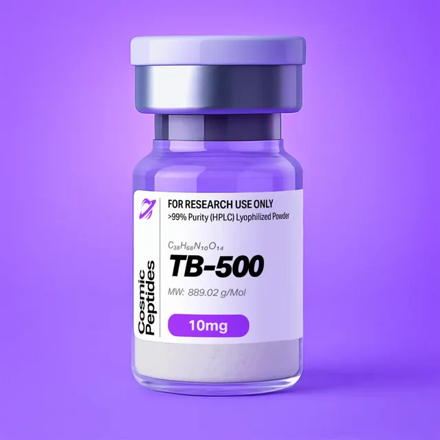
TB-500
Commonly researched with these items



Patent-pending lot tracking: Complete transparency from production to vial.
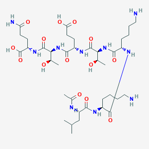
| CAS Number | 885340-08-9 |
|---|---|
| Molecular Formula | C38H68N10O14 |
| Molecular Weight | 889.2 g/Mol |
| Purity | 99.8% |
| Lot Number | 25058 |
| Quantity | 9.97mg |
| Sequence | Ac-Leu-Lys-Lys-Thr-Glu-Thr-Gln-OH |
TB-500 (Ac-LKKTETQ): The N-Acetylated 17–23 Actin-Binding Fragment of Thymosin β4
Disclaimer
Products described here are supplied for research use only and are not intended for diagnostic, therapeutic, or clinical application. All statements regarding biological activity reflect preclinical and in vitro findings exclusively and have not been evaluated by the U.S. Food and Drug Administration. These materials and related content are provided for educational and investigational purposes only and are intended solely for qualified researchers in laboratory and academic settings.
Key Facts
- Compound class
- Linear, N-terminally acetylated heptapeptide; residues 17–23 of thymosin β4 (Tβ4), the segment that contains the conserved LKKTET actin-binding motif of the β-thymosin/WH2 family
- Sequence / structure
Ac-Leu-Lys-Lys-Thr-Glu-Thr-Gln-OH(one-letter code: Ac-LKKTETQ), all residues in the L-configuration, free C-terminal acid- Molecular formula
- C38H68N10O14
- Molecular weight
- 889.0 g/mol (average; 889.01–889.02 depending on the atomic-weight table); monoisotopic mass 888.4916 Da
- CAS number
- 885340-08-9
- PubChem CID
- 62707662
- Physical form as supplied
- Lyophilized powder
- Lot traceability
- Every unit is tied to a tracked lot; the lot number, recorded purity and verified quantity of the lot on sale are shown live on this page, with its certificate of analysis.
Molecular Identity and Classification
The compound sold here as TB-500 is the heptapeptide Ac-Leu-Lys-Lys-Thr-Glu-Thr-Gln-OH: N-acetyl-L-leucyl-L-lysyl-L-lysyl-L-threonyl-L-α-glutamyl-L-threonyl-L-glutamine. Its seven residues are identical to positions 17 to 23 of thymosin β4, a 43-residue, N-terminally acetylated polypeptide that is one of the most abundant actin-monomer-binding proteins of vertebrate cells. The synthetic fragment carries an acetyl group on the α-amine of Leu17 and a free carboxylic acid on Gln23.
The molecular formula is C38H68N10O14, the average molecular weight 889.0 g/mol and the monoisotopic mass 888.4916 Da. The peptide is registered under CAS number 885340-08-9 and as PubChem compound 62707662, whose synonyms include TB-500, TB 500 and the systematic peptide name given above; the Global Substance Registration System lists it under UNII QHK6Z47GTG. The full IUPAC name, (2S)-2-[[(2S,3R)-2-[[(2S)-2-[[(2S,3R)-2-[[(2S)-2-[[(2S)-2-[[(2S)-2-acetamido-4-methylpentanoyl]amino]-6-aminohexanoyl]amino]-6-aminohexanoyl]amino]-3-hydroxybutanoyl]amino]-4-carboxybutanoyl]amino]-3-hydroxybutanoyl]amino]-5-amino-5-oxopentanoic acid, encodes nine stereocentres: the seven α-carbons (all S) and the two threonine β-carbons (both R).
What the Name TB-500 Refers To
"TB-500" is a trade name, not a systematic one, and it has been used for two different molecules: the short acetylated fragment described on this page, and synthetic full-length thymosin β4 (CAS 77591-33-4, PubChem CID 16132341, C212H350N56O78S, about 4963 g/mol, monoisotopic mass 4960.4863 Da). The two differ by a factor of about 5.6 in mass and are distinguished at a glance by mass spectrometry. In this catalog TB-500 means the heptapeptide, and the verified sequence of the lot on sale is shown in the product information box on this page. Most structural and biochemical work on β-thymosins used intact Tβ4, which bears on the fragment only as far as the fragment reproduces part of the parent's binding surface.
Within the parent protein, the heptapeptide sits between two regions that are helical when Tβ4 is bound to actin: an N-terminal segment (roughly residues 4–16) and a C-terminal segment (roughly residues 30–40). The full Tβ4 sequence is Ac-SDKPDMAEIEKFDKSKLKKTETQEKNPLPSKETIEQEKQAGES, with the TB-500 segment shown in bold. Another well-known piece of the same protein is the N-terminal tetrapeptide Ac-Ser-Asp-Lys-Pro (Ac-SDKP, C20H33N5O9, 487.5 g/mol, PubChem CID 65938), whose generation from Tβ4 Hannappel's review attributes to prolyl oligopeptidase. The same review covers the parent's other post-translational modifications: removal of the initiator methionine and N-terminal acetylation during biosynthesis, transglutaminase cross-linking, and proteomic detections of acetylated lysines and two phosphorylated threonines. The 17–23 segment contains both residue types, so modified versions of the fragment are chemically conceivable; a lysine ε-acetyl adds 42.0106 Da (the same shift as a missing N-terminal acetyl, but at a different position in the fragment ladder) and a phosphothreonine adds 79.9663 Da.
Structure and Chemical Features
Reading from the N-terminus, each residue contributes a distinct chemical feature:
- Ac-Leu17: the acetyl cap removes the positive charge of the N-terminal amine and turns the first residue into a secondary amide, so the fragment's N-terminus resembles the peptide bond that joins Leu17 to Lys16 in the parent protein. The isobutyl side chain is the only substantially hydrophobic group in the molecule.
- Lys18 and Lys19: two ε-amines (pKa about 10.5) that are protonated across the whole pH range used in biochemistry. In Tβ4 these lysines are part of the conserved LKKTET motif, and mutational analysis by Van Troys and colleagues showed that this motif is central to actin binding.
- Thr20 and Thr22: β-branched residues with secondary hydroxyl groups, each adding a hydrogen-bond donor and acceptor and a second stereocentre.
- Glu21: a side-chain carboxylic acid (pKa about 4.2) that is negatively charged at neutral pH.
- Gln23: a side-chain primary amide and, in the fragment, the free C-terminal carboxylate. In Tβ4 this residue is followed by Glu24; the carboxylate is a new charge that exists only in the excised peptide.
Counting charges at pH 7.0 gives two positive charges (Lys18, Lys19) and two negative charges (Glu21 side chain, C-terminal carboxylate), so the molecule is a zwitterion with a net charge close to zero. The net charge stays near zero from about pH 6 to pH 9, and a calculation with textbook pKa values places the isoelectric point near 7.2. Below pH 3 the carboxyl groups are protonated and the peptide carries a net charge approaching +2; above pH 11 the lysines lose their protons and the charge approaches −2.
The heptapeptide contains no aromatic residue, cysteine, methionine or proline: it has no 280 nm absorbance, cannot form disulfides, and escapes the methionine oxidation that converts intact Tβ4 (Met6) into its sulfoxide. With eight amide groups (six backbone amides, the acetyl-cap amide and the Gln side-chain amide), two hydroxyls, two amines and two carboxyl groups, it is an extremely polar molecule: PubChem computes 14 hydrogen-bond donors, 16 acceptors, 32 rotatable bonds, a topological polar surface area of 414 Å2 and an XLogP3 of −8.6.
Conformation in Solution and on Actin
In aqueous solution, a seven-residue linear peptide of this composition is expected to sample a broad ensemble of extended and turn-like conformations rather than a single fold. The same is true of the parent protein: NMR studies by Domanski and colleagues showed that free Tβ4 is largely unstructured and acquires its helical segments only when it binds G-actin, a coupled folding-and-binding process. Irobi and colleagues stabilized the complex by fusing gelsolin domain 1 to the C-terminal half of Tβ4 and solved the hybrid bound to actin at 2 Å, showing that Tβ4 sequesters by capping both ends of the monomer. Taken together with the NMR work and the later structures of Xue and colleagues, the picture is that the N-terminal helix packs into the hydrophobic cleft between subdomains 1 and 3 at the barbed face of the actin monomer, the LKKTET segment runs in an extended conformation across the monomer, and the C-terminal helix reaches the pointed face near subdomains 2 and 4. The fragment therefore corresponds to the linker that connects the two capping elements, and in the bound protein it is the part that crosses the monomer surface.
Synthesis and Manufacturing Chemistry
Ac-LKKTETQ is made by solid-phase peptide synthesis, the approach introduced by Merrifield, using Fmoc chemistry built on the base-labile protecting group of Carpino and Han. For a C-terminal free acid the chain is started from glutamine on an acid-labile resin such as Wang or 2-chlorotrityl chloride resin and extended towards the N-terminus in six coupling cycles, each consisting of Fmoc removal (commonly 20% piperidine in dimethylformamide), washing and coupling of the next activated amino acid.
The standard building blocks are Fmoc-Gln(Trt)-OH, Fmoc-Thr(tBu)-OH, Fmoc-Glu(OtBu)-OH, Fmoc-Lys(Boc)-OH and Fmoc-Leu-OH. The trityl group on glutamine suppresses dehydration of the side-chain amide to a nitrile during activation and improves the solubility of the building block; the tert-butyl ether and ester protect the threonine hydroxyls and the glutamate carboxyl; the Boc groups protect the lysine ε-amines. After the final Fmoc removal, the free N-terminal amine of Leu17 is acetylated on the resin, typically with acetic anhydride and a tertiary amine base. A single cleavage step in trifluoroacetic acid (TFA) and scavengers such as water and triisopropylsilane then releases the peptide and removes all side-chain protecting groups at once.
The sequence is short and lacks the residues that cause the most common side reactions in Fmoc synthesis: it has no Asp (so no aspartimide formation), no Cys or Met (no oxidation or alkylation of sulfur), no Trp, no Arg and no N-terminal Gln (the acetyl cap is on Leu, so pyroglutamate formation cannot occur). The points that deserve attention are the two β-branched threonines, whose couplings are slower than average; the two adjacent lysines, which make a des-Lys deletion a plausible minor impurity; completeness of the on-resin acetylation, since any unacetylated material survives as a free-amine variant; and complete removal of the trityl and tert-butyl groups during cleavage. The crude peptide is precipitated in cold ether, purified by preparative reversed-phase HPLC and lyophilized. Because TFA is present during cleavage and purification, the two lysine amines usually leave the column as trifluoroacetate salts unless the material is exchanged into acetate or chloride.
| Variant | Mass change (Da) | Neutral monoisotopic mass (Da) | Origin |
|---|---|---|---|
| Target peptide | 0 | 888.4916 | Ac-LKKTETQ-OH |
| Des-acetyl (free N-terminal amine) | −42.0106 | 846.4811 | Incomplete on-resin acetylation |
| Des-Lys deletion | −128.0950 | 760.3967 | Incomplete coupling of one of the two lysines |
| Des-Thr deletion | −101.0477 | 787.4440 | Incomplete coupling of a β-branched threonine |
| Gln23 → Glu (deamidation) | +0.9840 | 889.4757 | Hydrolysis of the side-chain amide on storage |
| tert-Butyl retained | +56.0626 | 944.5542 | Incomplete removal of a Thr or Glu protecting group |
| Trityl retained | +242.1096 | 1130.6012 | Incomplete removal of the Gln protecting group |
| D-epimer at any residue | 0 | 888.4916 | Racemization during activation; isobaric, needs chiral analysis or orthogonal HPLC |
Analytical Characterization
Reversed-Phase HPLC and Mass Spectrometry
Reversed-phase HPLC. With an XLogP3 of −8.6, Ac-LKKTETQ is weakly retained on C18 columns. Analysts generally start the gradient at 0–2% acetonitrile in water with 0.1% TFA and run a shallow ramp (for example to 30% acetonitrile over 20–30 minutes). TFA is useful here for more than pH control: it pairs with the two protonated lysines and increases retention, so the same peptide elutes noticeably earlier with formic acid as the modifier. Detection is at 210–220 nm, where the amide bonds absorb; there is no 280 nm signal because the sequence has no Trp, Tyr or Phe. Mant and colleagues' method review covers the effect of ion-pairing reagents and gradient slope on the separation of small polar peptides. HILIC is a useful orthogonal method.
Intact mass. In positive-mode electrospray the peptide gives [M+H]+ at m/z 889.4989, [M+2H]2+ at m/z 445.2531 and, less abundantly, [M+3H]3+ at m/z 297.1712. The doubly charged ion is usually prominent because the two lysine amines are the most basic sites. A sodium adduct [M+Na]+ appears at m/z 911.4809, and in negative mode [M−H]− appears at m/z 887.4844. For a molecule of this size the monoisotopic peak is the most intense in the isotope cluster, with the M+1 peak at roughly 46% of its height, almost all of that from 13C in its 38 carbons.
A worked resolution problem: telling deamidation from the 13C isotope. Deamidation of Gln23 to Glu adds 0.9840 Da, almost exactly one mass unit. The deamidated product's [M+H]+ falls at m/z 890.4830, while the 13C1 isotope peak of the intact peptide falls at m/z 890.5023. The two are 0.0193 units apart, so merely resolving them at half height needs a resolving power (m/Δm, FWHM) of about 890 / 0.0193 ≈ 46,000, and clean baseline separation roughly twice that (the same ratio applies to the 2+ ions at m/z 445.745 and 445.755). A unit-resolution instrument sees a deamidated sample only as a distorted isotope pattern, so low-level deamidation is better quantified by high-resolution MS or by chromatography: in TFA-containing reversed-phase systems the Glu-23 variant usually resolves from the parent (typically eluting slightly later), and at neutral pH it carries one extra negative charge, which ion-exchange separations exploit.
Tandem MS. Collision-induced dissociation of the protonated peptide produces b- and y-ion series; because the N-terminus is acetylated, the b-ions include the acetyl group and a b1 ion (acetyl-Leu acylium) can be observed. The expected singly charged fragments are listed below. Immonium ions in the low-mass region provide composition checks: Leu 86.0964, Thr 74.0600, Glu 102.0550, Lys 101.1073 (with its characteristic ammonia-loss ion at 84.0808) and Gln 101.0709. Note that the Lys and Gln residue masses (128.0950 and 128.0586 Da) differ by only 0.0364 Da, so the order of the Lys-Lys pair and the C-terminal Gln is established from the fragment ladder and accurate mass rather than from nominal mass.
| Cleavage after | b-ion | m/z | y-ion | m/z |
|---|---|---|---|---|
| Leu17 | b1 (Ac-L) | 156.1019 | y6 (KKTETQ) | 734.4043 |
| Lys18 | b2 (Ac-LK) | 284.1969 | y5 (KTETQ) | 606.3093 |
| Lys19 | b3 (Ac-LKK) | 412.2918 | y4 (TETQ) | 478.2144 |
| Thr20 | b4 (Ac-LKKT) | 513.3395 | y3 (ETQ) | 377.1667 |
| Glu21 | b5 (Ac-LKKTE) | 642.3821 | y2 (TQ) | 248.1241 |
| Thr22 | b6 (Ac-LKKTET) | 743.4298 | y1 (Q) | 147.0764 |
Purity, peptide content and counterions. HPLC purity and peptide content answer different questions. Purity is the fraction of the UV-absorbing material that is the target peptide. Peptide content is the fraction of the weighed powder that is peptide at all, as opposed to counterions, bound water and residual salts. With two basic lysines, Ac-LKKTETQ can carry up to two counterions per molecule, and the effect on the weighed mass is substantial: the bis-trifluoroacetate salt has a formula weight of about 1117.1 g/mol (peptide fraction 79.6%), the bis-acetate about 1009.1 g/mol (88.1%) and the mono-acetate about 949.1 g/mol (93.7%), before any allowance for water. Amino-acid analysis after acid hydrolysis is the usual way to measure peptide content; for this sequence the expected molar ratio is Leu 1 : Lys 2 : Thr 2 : Glu+Gln 3 (the glutamine side-chain amide is hydrolysed to glutamate, so Glu and Gln are reported together as Glx), with threonine recoveries usually corrected for partial destruction during hydrolysis. Ion chromatography or 19F NMR quantifies trifluoroacetate, and Karl Fischer titration measures water.
Physicochemical Properties
| Property | Value | Notes |
|---|---|---|
| Molecular formula | C38H68N10O14 | Free peptide, without counterions |
| Average molecular weight | 889.0 g/mol | PubChem CID 62707662; 889.02 with current standard atomic weights |
| Monoisotopic mass | 888.4916 Da | For high-resolution MS |
| [M+H]+ / [M+2H]2+ / [M+3H]3+ | m/z 889.4989 / 445.2531 / 297.1712 | Positive-mode ESI |
| Heavy atoms | 62 | Computed |
| Stereocentres | 9 (7 α-carbons S; 2 Thr β-carbons R) | All L-amino acids |
| Net charge at pH 7 | about 0 (+2 / −2) | Two Lys; Glu side chain and C-terminal carboxylate |
| Isoelectric point | about 7.2 (calculated) | Midpoint of a broad near-neutral charge plateau |
| Net charge at pH 2 / pH 12 | about +1.9 / −1.9 (calculated) | Relevant to ESI and ion-exchange behaviour |
| XLogP3 | −8.6 | Computed; extremely hydrophilic |
| Topological polar surface area | 414 Å2 | Computed |
| Hydrogen-bond donors / acceptors | 14 / 16 | Computed |
| Rotatable bonds | 32 | Computed |
| UV absorbance | Amide bonds only (about 214 nm) | No aromatic residues; no 280 nm signal |
| Oxidation-prone residues | None | No Met, Cys or Trp |
| Main chemical liability | Deamidation of Gln23 | +0.984 Da; slow at neutral pH and low temperature |
The profile is that of a small, highly charged, highly polar oligopeptide. The four ionizable groups, together with eight amides and two hydroxyls, make Ac-LKKTETQ readily water-soluble and give it no tendency to partition into lipid phases; the strongly negative XLogP3 means it would not be expected to cross lipid bilayers by passive diffusion. Because the isoelectric point lies close to neutral, the peptide carries little net charge in unbuffered water or in pH 7 buffers. Cation-exchange chromatography is therefore best run at acidic pH, where the peptide carries a clear positive charge.
The chemical stability of the sequence is dominated by one reaction. Deamidation of glutamine proceeds through a six-membered glutarimide intermediate and is generally much slower than deamidation of asparagine, which goes through a five-membered succinimide. Robinson and Robinson developed a mass-spectrometric method for measuring glutaminyl and asparaginyl deamidation rates and reported rates for 306 asparaginyl sequences in model peptides at pH 7.4 and 37.0 °C in 0.15 M Tris·HCl, showing how strongly the neighbouring sequence controls the rate; glutaminyl residues such as Gln23 sit at the slow end of that scale. In the frozen lyophilized solid the reaction is negligible.
Research Context
Thymosin β4 and Actin Monomer Sequestration
Thymosin β4 was sequenced in 1981 by Low, Hu and Goldstein from calf thymus extracts, where it was first studied as a thymic factor. Ten years later Safer, Elzinga and Nachmias showed that an abundant actin-sequestering peptide from platelets, called Fx, was identical to Tβ4, which recast the protein as a cytoskeletal regulator present in most nucleated cells. Tβ4 forms a 1:1 complex with monomeric (G-) actin and prevents the bound monomer from joining filaments. Its affinity is sub-micromolar to low-micromolar for Mg-ATP-actin and considerably weaker for ADP-actin, as characterized in the early binding studies of Weber and colleagues and in later work from the Carlier laboratory. Because Tβ4 occurs in many cell types at concentrations of tens to hundreds of micromolar, as reviewed by Huff and colleagues, it can hold a large fraction of a cell's unpolymerized actin in a reserve that other proteins, notably profilin, can draw on. Xue and colleagues solved structures showing how a Tβ4–actin complex hands its monomer to profilin for filament assembly.
Carlier and colleagues showed in 1996 that the picture is more nuanced than simple sequestration: at high concentrations Tβ4 also interacts with filamentous actin. Hertzog and colleagues then placed the β-thymosins within the wider family of WH2 (WASP-homology 2) domains, short actin-binding modules found in many cytoskeletal proteins, and showed that closely related sequences can either inhibit or promote filament assembly depending on what lies on either side of the core motif. The LKKTET motif present in TB-500 is the hallmark of that family.
What the 17–23 Fragment Represents
Van Troys and colleagues mapped the actin-binding site of Tβ4 by systematic mutation and identified the central LKKTET sequence, together with the N-terminal helical region, as the main determinants of binding. The isolated heptapeptide therefore reproduces the sequence of the motif but not the two helices that cap the barbed and pointed faces of the monomer in the full complex. In actin-binding terms it is a minimal motif peptide rather than a sequestering protein in miniature: experiments that compare it with intact Tβ4, with N-terminal and C-terminal fragments and with scrambled or alanine-substituted versions of the motif are the standard way to measure how much of a measured effect the motif alone can account for.
A 2010 review by Sosne and colleagues collected the work that assigns different cellular activities of Tβ4 to short sequences. It separates the N-terminal Ac-SDKP and 1–15 regions from the central LKKTETQ segment (the actin-binding domain, residues 17–22, plus Gln23), which it associates with cell-migration read-outs. That assignment is the reason the heptapeptide exists as a research tool: it lets a cell-migration effect be tested against the motif in isolation.
In Vitro Methods Used With the Fragment
The assays applied to Ac-LKKTETQ and to Tβ4 fragments are those of actin biochemistry and cell motility:
- Pyrene-actin polymerization assays, in which the fluorescence of pyrene-labelled actin rises as monomers join filaments, to measure whether a peptide slows nucleation or elongation, or shifts the steady-state concentration of unpolymerized actin.
- Sedimentation and DNase I inhibition assays, which separate or quantify unpolymerized actin and so report on monomer sequestration.
- Binding measurements by fluorescence titration, isothermal titration calorimetry or NMR chemical-shift perturbation, to estimate the affinity of a motif peptide and the actin residues it contacts. Domanski and colleagues used NMR to follow how Tβ4 folds as it binds actin, an approach that can be applied to fragments.
- Cell-migration assays in culture, such as transwell (Boyden chamber) migration and closure of a scratch made in a confluent monolayer, with read-outs including migration distance, closure rate and staining of filamentous actin with fluorescent phalloidin.
Cell-based designs commonly span about 10 nM to 10 µM, whereas binding titrations of a motif peptide that lacks both capping helices may need concentrations in the hundreds of micromolar, where the pH and counterion content of the stock start to matter. Good designs compare the fragment with equimolar intact Tβ4, include a scrambled-sequence control of identical composition and mass, and match the vehicle for counterion content. Cornish and colleagues showed that trifluoroacetate on its own can alter cell proliferation in culture, which is why a TFA-matched vehicle or an acetate-exchanged peptide is good practice for cell-based work.
TB-500 Compared With Intact Thymosin β4 and Ac-SDKP
| Feature | TB-500 (Ac-Tβ4 17–23) | Thymosin β4 (1–43) | Ac-SDKP (Ac-Tβ4 1–4) |
|---|---|---|---|
| Sequence | Ac-LKKTETQ-OH | Ac-SDKPDMAEIEKFDKSKLKKTETQEKNPLPSKETIEQEKQAGES-OH | Ac-SDKP-OH |
| Residues | 7 | 43 | 4 |
| Molecular formula | C38H68N10O14 | C212H350N56O78S | C20H33N5O9 |
| Average MW (g/mol) | 889.0 | 4963 | 487.5 |
| Monoisotopic mass (Da) | 888.4916 | 4960.4863 | 487.2278 |
| PubChem CID | 62707662 | 16132341 | 65938 |
| CAS number | 885340-08-9 | 77591-33-4 | 120081-14-3 (PubChem synonym) |
| Contains LKKTET motif | Yes | Yes | No |
| Helical capping segments | None | N-terminal and C-terminal, formed on binding actin | None |
| Methionine (oxidation site) | None | Met6 | None |
| Net charge at pH 7 | about 0 | about −3 (calculated; 9 Lys against 11 Asp/Glu plus the C-terminus) | about −1 (calculated) |
The fragment and the full protein are therefore not interchangeable: the heptapeptide is a near-neutral, sulfur-free, flexible motif peptide one-fifth the mass of Tβ4; by ESI-MS the full protein appears as a broad envelope of multiply charged ions, the fragment as 1+, 2+ and 3+ ions.
Related Compounds
TB-500 is also offered in combination with another research peptide as BPC-157/TB-500, and as a component of the three-peptide blend GLOW (BPC-157, TB-500, GHK-Cu); the pentadecapeptide BPC-157 is available on its own.
The similar names of the thymosins cause frequent confusion. Thymosin Alpha-1 is a 28-residue acetylated peptide (C129H215N33O55, about 3108 g/mol) derived from prothymosin α; despite the shared name it is unrelated in sequence and structure to the β-thymosins and has no actin-binding motif. Both names come from the same historical source, the thymus extract "thymosin fraction 5" from which a series of peptides was purified in the 1970s and 1980s and named according to their behaviour on isoelectric focusing.
Glossary
- G-actin and F-actin
- Globular (monomeric) actin and filamentous (polymerized) actin. The balance between them is regulated by monomer-binding proteins such as thymosin β4 and profilin.
- Barbed end and pointed end
- The two ends of an actin filament, named from the arrowhead pattern seen when filaments are decorated with myosin fragments. The barbed end grows faster. The same terms describe the corresponding faces of an actin monomer.
- Sequestration
- Binding of an actin monomer in a complex that cannot add to filaments, keeping it in a reserve pool.
- WH2 domain
- WASP-homology 2 domain: a short actin-binding module, related to the β-thymosins, found in many cytoskeletal proteins.
- LKKTET motif
- The conserved Leu-Lys-Lys-Thr-Glu-Thr core of β-thymosin and WH2 actin-binding sequences; residues 17–22 of Tβ4 and the first six residues of TB-500.
Laboratory Handling and Storage
Storage of the solid. Keep lyophilized Ac-LKKTETQ tightly closed at −20 °C, protected from light and moisture. Like most lyophilized peptide salts it is hygroscopic, so allow a cold vial to reach room temperature in a desiccator before opening it, to keep atmospheric water from condensing on the powder and adding to the weighed mass.
Preparing solutions. The peptide dissolves readily in high-purity water and in common aqueous buffers. If dissolution is slow, a slightly acidic aqueous medium such as 0.1% acetic acid moves the solution away from the isoelectric point near pH 7.2. For accurate molar concentrations, correct the weighed mass for peptide content. As a worked example, 10.0 mg of the free peptide in 1.00 mL gives 11.25 mM, but if the solid is the bis-trifluoroacetate salt with no other impurities, the same 10.0 mg contains 7.96 mg of peptide and gives 8.95 mM; for the bis-acetate the figure is 9.91 mM. Conversely, a 1.00 mM solution contains 0.889 mg of peptide per mL. Stock solutions are commonly made at 1–10 mM, passed through a 0.22 µm membrane when destined for cell culture, divided into single-use aliquots and frozen at −20 °C or −80 °C, avoiding repeated freeze–thaw cycles.
Solution stability. Neutral to slightly acidic buffers (pH 5–7) and low temperature minimize deamidation of Gln23. Alkaline buffers and elevated temperature accelerate it.
Working at low concentrations. Because the sequence carries two lysines, losses through adsorption to glass and some plastics can become significant at nanomolar concentrations. Low-binding polypropylene tubes and tips, and a carrier protein in assay buffers where the design permits, limit these losses.
Re-verification. A quick LC-MS check for the [M+H]+ ion at m/z 889.50 and the [M+2H]2+ ion at m/z 445.25 confirms identity after long storage. Growth of a later-eluting, one-unit-heavier species, or of an apparent change in the isotope ratio at m/z 890.5, points to deamidation. Each lot's certificate of analysis, linked on this page, records the analytical results for that lot.
Frequently Asked Research Questions
- What is TB-500?
- In this catalog, TB-500 is the synthetic heptapeptide Ac-Leu-Lys-Lys-Thr-Glu-Thr-Gln-OH (Ac-LKKTETQ), residues 17–23 of thymosin β4 with an acetylated N-terminus. Its formula is C38H68N10O14, its molecular weight 889.0 g/mol and its CAS number 885340-08-9 (PubChem CID 62707662).
- Is TB-500 the same as thymosin β4?
- No. Thymosin β4 is a 43-residue protein of about 4963 g/mol (CAS 77591-33-4). TB-500 as defined here is a seven-residue fragment of it, about 889 g/mol, containing the LKKTET actin-binding motif but neither of the helical segments that cap the actin monomer in the full complex. The trade name has also been used for full-length Tβ4, so check the sequence and mass of any material.
- What mass should I see for TB-500 by mass spectrometry?
- In positive electrospray, [M+H]+ at m/z 889.4989, [M+2H]2+ at m/z 445.2531 and [M+3H]3+ at m/z 297.1712. The monoisotopic mass of the neutral peptide is 888.4916 Da.
- Which fragment ions confirm the sequence?
- The acetylated b-series (b2 284.1969, b3 412.2918, b4 513.3395) and the y-series (y2 248.1241, y3 377.1667, y4 478.2144, y5 606.3093) together cover every residue. Immonium ions at m/z 86.096 (Leu), 101.107 (Lys) and 74.060 (Thr) confirm composition.
- Why is the N-terminus acetylated?
- Leu17 follows Lys16 in intact Tβ4, so in the parent protein its amine is part of a peptide bond. The acetyl cap reproduces that neutral amide, removes a positive charge that would not exist in the protein, and protects the N-terminus from aminopeptidases in biological media.
- Does TB-500 bind actin as strongly as thymosin β4?
- It should not be assumed to. In Tβ4, high-affinity sequestration depends on the LKKTET motif together with the N-terminal helix that occupies the cleft between actin subdomains 1 and 3 and the C-terminal helix at the pointed face. The isolated motif peptide lacks both helices, so it is used to measure what the motif alone contributes, compared side by side with intact Tβ4.
- What is the main degradation pathway in storage?
- Deamidation of the C-terminal glutamine to glutamate (+0.984 Da). Glutamine deamidates much more slowly than asparagine and the reaction is negligible in the frozen solid, but it accelerates in solution at high pH and temperature. The sequence has no methionine, cysteine or tryptophan, so oxidation is not a concern.
- How do counterions affect concentration calculations?
- Each of the two lysines can carry a counterion. A bis-trifluoroacetate salt weighs about 1117.1 g/mol per mole of peptide, so 10.0 mg in 1.00 mL gives 8.95 mM rather than the 11.25 mM calculated for the free peptide. Peptide content from amino-acid analysis gives the exact correction.
- How is TB-500 different from thymosin alpha-1?
- They share only a historical name. Thymosin α1 is a 28-residue peptide from prothymosin α with no sequence relationship to the β-thymosins and no actin-binding motif; TB-500 is a fragment of thymosin β4.
- Where can I see the purity of the TB-500 I am buying?
- This page shows the lot number, recorded purity and verified quantity of the lot currently on sale, taken live from the shop's lot-tracking database, together with that lot's certificate of analysis.
Sources & Further Reading
- National Center for Biotechnology Information. PubChem Compound Summary for CID 62707662, TB-500 (Ac-Leu-Lys-Lys-Thr-Glu-Thr-Gln-OH). PubChem CID 62707662.
- National Center for Biotechnology Information. PubChem Compound Summary for CID 16132341, thymosin β4. PubChem CID 16132341.
- National Center for Biotechnology Information. PubChem Compound Summary for CID 65938, N-acetyl-Ser-Asp-Lys-Pro (Ac-SDKP). PubChem CID 65938.
- Low TL, Hu SK, Goldstein AL. Complete amino acid sequence of bovine thymosin beta 4: a thymic hormone that induces terminal deoxynucleotidyl transferase activity in thymocyte populations. Proceedings of the National Academy of Sciences USA 1981;78(2):1162–1166. doi:10.1073/pnas.78.2.1162
- Safer D, Elzinga M, Nachmias VT. Thymosin β4 and Fx, an actin-sequestering peptide, are indistinguishable. Journal of Biological Chemistry 1991;266(7):4029–4032. doi:10.1016/S0021-9258(20)64278-8
- Weber A, Nachmias VT, Pennise CR, Pring M, Safer D. Interaction of thymosin β4 with muscle and platelet actin: implications for actin sequestration in resting platelets. Biochemistry 1992;31(27):6179–6185. doi:10.1021/bi00142a002
- Van Troys M, Dewitte D, Goethals M, Carlier MF, Vandekerckhove J, Ampe C. The actin binding site of thymosin β4 mapped by mutational analysis. The EMBO Journal 1996;15(2):201–210. doi:10.1002/j.1460-2075.1996.tb00350.x
- Carlier MF, Didry D, Erk I, et al. Tβ4 is not a simple G-actin sequestering protein and interacts with F-actin at high concentration. Journal of Biological Chemistry 1996;271(16):9231–9239. doi:10.1074/jbc.271.16.9231
- Huff T, Müller CSG, Otto AM, Netzker R, Hannappel E. β-Thymosins, small acidic peptides with multiple functions. The International Journal of Biochemistry & Cell Biology 2001;33(3):205–220. doi:10.1016/S1357-2725(00)00087-X
- Domanski M, Hertzog M, Coutant J, et al. Coupling of folding and binding of thymosin β4 upon interaction with monomeric actin monitored by nuclear magnetic resonance. Journal of Biological Chemistry 2004;279(22):23637–23645. doi:10.1074/jbc.M311413200
- Hertzog M, van Heijenoort C, Didry D, et al. The β-thymosin/WH2 domain: structural basis for the switch from inhibition to promotion of actin assembly. Cell 2004;117(5):611–623. doi:10.1016/S0092-8674(04)00403-9
- Irobi E, Aguda AH, Larsson M, et al. Structural basis of actin sequestration by thymosin-β4: implications for WH2 proteins. The EMBO Journal 2004;23(18):3599–3608. doi:10.1038/sj.emboj.7600372
- Xue B, Leyrat C, Grimes JM, Robinson RC. Structural basis of thymosin-β4/profilin exchange leading to actin filament polymerization. Proceedings of the National Academy of Sciences USA 2014;111(43). doi:10.1073/pnas.1412271111
- Hannappel E. Thymosin β4 and its posttranslational modifications. Annals of the New York Academy of Sciences 2010;1194:27–35. doi:10.1111/j.1749-6632.2010.05485.x
- Sosne G, Qiu P, Goldstein AL, Wheater M. Biological activities of thymosin β4 defined by active sites in short peptide sequences. The FASEB Journal 2010;24(7):2144–2151. doi:10.1096/fj.09-142307
- Merrifield RB. Solid phase peptide synthesis. I. The synthesis of a tetrapeptide. Journal of the American Chemical Society 1963;85(14):2149–2154. doi:10.1021/ja00897a025
- Carpino LA, Han GY. 9-Fluorenylmethoxycarbonyl amino-protecting group. The Journal of Organic Chemistry 1972;37(22):3404–3409. doi:10.1021/jo00795a005
- Mant CT, Chen Y, Yan Z, et al. HPLC analysis and purification of peptides. Methods in Molecular Biology 2007;386:3–55. doi:10.1007/978-1-59745-430-8_1
- Robinson NE, Robinson AB. Molecular clocks. Proceedings of the National Academy of Sciences USA 2001;98(3):944–949. doi:10.1073/pnas.98.3.944
- Cornish J, Callon KE, Lin CQ, et al. Trifluoroacetate, a contaminant in purified proteins, inhibits proliferation of osteoblasts and chondrocytes. American Journal of Physiology-Endocrinology and Metabolism 1999;277(5):E779–E783. doi:10.1152/ajpendo.1999.277.5.E779
Last reviewed:
In the research library
Everything the library holds on TB-500, and the methods behind the numbers on its certificate.
- TB-500 reference entry Identity, calculated properties, the mass spectrometry ions to expect and analytical notes, from the structure.
- Check a certificate against this sequence Paste the masses from a spectrum or a certificate; the mass-match tool compares them with what this sequence should give.
- Peptide calculator, with this sequence Formula, average and monoisotopic mass, charge across pH, isoelectric point and absorbance, recalculated as you edit.
- Thymic peptides and fragments The family side by side: sequences aligned, each member's relation to the reference peptide, and how analysis tells them apart.
- Thymosin β4 and TB-500 compared TB-500 is residues 17–23 of thymosin β4, LKKTETQ, with an acetyl group on its N-terminal leucine. Thymosin β4 is the full 43-residue, N-acetylated peptide.
- BPC-157 and TB-500 compared Unrelated in sequence and origin. BPC-157 is a linear 15-residue peptide, rich in proline, with free termini. TB-500 is a seven-residue N-acetylated peptide, residues 17–23 of thymosin β4, with a free C-terminal acid. They are compared because the two are often paired in research-material blends, where each must be identified and measured on its own.
- How to read a certificate of analysis What a peptide certificate of analysis should contain, how to check that it belongs to the material in hand, how to read its identity and purity results, and the signs that it deserves a second look.
- HPLC purity analysis of peptides How reversed-phase HPLC measures the chromatographic purity of a peptide, what the figure includes, and what it leaves out.
- Mass spectrometry for peptide identity How mass spectrometry confirms that a peptide is the intended sequence: electrospray and MALDI, working out the expected mass, reading charge states and adducts, sequence confirmation, and the limits of the method.
- Net peptide content and purity: two different numbers Why a peptide that is 99% pure by HPLC can be only 75–85% peptide by weight: what the powder contains, how each part is measured, and how the two figures combine.
- Preparing peptide solutions in the laboratory From a freeze-dried vial to a solution of known concentration: the arithmetic, the solvent, the charge chemistry and the handling, with the errors each step can introduce and the article that covers it.
- Peptide stability and storage How peptides degrade, chemically and physically, why the dry solid lasts far longer than a solution, which sequences are most at risk, and the storage and handling that slow it down.