Tesamorelin
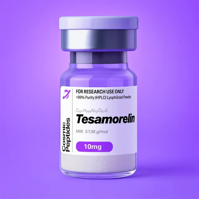
Tesamorelin
Commonly researched with these items
Patent-pending lot tracking: Complete transparency from production to vial.
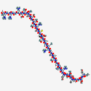
| CAS Number | 218949-48-5 |
|---|---|
| Molecular Formula | C221H366N72O67S |
| Molecular Weight | 5136.0 g/Mol |
| Purity | 99.74% |
| Lot Number | 26116 |
| Quantity | 10.22mg |
| Sequence | Unk-Tyr-Ala-Asp-Ala-Ile-Phe-Thr-Asn-Ser-Tyr-Arg-Lys-Val-Leu-Gly-Gln-Leu-Ser-Ala-Arg-Lys-Leu-Leu-Gln-Asp-Ile-Met-Ser-Arg-Gln-Gln-Gly-Glu-Ser-Asn-Gln-Glu-Arg-Gly-Ala-Arg-Ala-Arg-Leu-NH₂ |
Tesamorelin: trans-3-Hexenoyl-Modified Human GHRH(1–44) Amide — Chemistry, Analytics and In Vitro Receptor Pharmacology
Disclaimer
Products described here are supplied for research use only and are not intended for diagnostic, therapeutic, or clinical application. All statements regarding biological activity reflect preclinical and in vitro findings exclusively and have not been evaluated by the U.S. Food and Drug Administration. These materials and related content are provided for educational and investigational purposes only and are intended solely for qualified researchers in laboratory and academic settings.
Key Facts
- Compound class
- Synthetic 44-residue peptide amide; N-terminally acylated analogue of human GHRH(1–44)-NH2; class B1 G protein-coupled receptor agonist in vitro
- Sequence / structure
(E)-hex-3-enoyl-Tyr-Ala-Asp-Ala-Ile-Phe-Thr-Asn-Ser-Tyr-Arg-Lys-Val-Leu-Gly-Gln-Leu-Ser-Ala-Arg-Lys-Leu-Leu-Gln-Asp-Ile-Met-Ser-Arg-Gln-Gln-Gly-Glu-Ser-Asn-Gln-Glu-Arg-Gly-Ala-Arg-Ala-Arg-Leu-NH2; one-letter codeYADAIFTNSYRKVLGQLSARKLLQDIMSRQQGESNQERGARARL, with a trans-3-hexenoyl group on the Tyr1 α-amine and a C-terminal carboxamide; all residues L-configured- Molecular formula
- C221H366N72O67S
- Molecular weight
- 5136.0 g/mol (average); monoisotopic mass 5132.7166 Da
- CAS number
- 218949-48-5
- PubChem CID
- 16137828
- Development code
- TH9507 (Theratechnologies Inc.)
- Physical form as supplied
- Lyophilized powder
- Lot traceability
- Every unit is tied to a tracked lot; the lot number, recorded purity and verified quantity of the lot on sale are shown live on this page, with its certificate of analysis.
Molecular Identity and Classification
Tesamorelin is the trans-3-hexenoyl derivative of human growth hormone-releasing hormone 1–44 amide. It is a single linear polypeptide of 44 residues whose sequence is identical to the mature human GHRH 44-mer, carrying two features that are not encoded genetically: a C-terminal carboxamide in place of a free carboxylic acid, and a six-carbon unsaturated acyl group bonded to the α-amino group of the N-terminal tyrosine. Its molecular formula is C221H366N72O67S, its average molecular weight 5136.0 g/mol and its monoisotopic mass 5132.7166 Da; it is indexed as PubChem CID 16137828 under CAS registry number 218949-48-5, with 804475-66-9 circulating for the acetate salt. The development code TH9507 appears throughout the older literature.
The parent hormone is the product of the gene GHRH, whose 108-residue precursor somatoliberin is catalogued as UniProtKB P01286. The mature 44-mer occupies residues 32–75 of that precursor, and the next residue, Gly76, is the amide donor: peptidylglycine α-amidating monooxygenase converts the Leu-Gly pair into a C-terminal leucinamide, which is why both the natural hormone and this analogue end in -Arg-Leu-NH2. GHRH was isolated and sequenced in 1982 by two groups independently, reported by Guillemin and co-workers in Science and by Rivier and colleagues in Nature; the 44-residue amide is the longest naturally occurring form, while the 40-residue amide and the synthetic 1–29 amide retain receptor activity in vitro.
In the numbering used here, residue 1 is the tyrosine bearing the acyl cap, and the hexenoyl group is a substituent on it rather than an extra residue, so tesamorelin is a 44-mer and not a 45-mer. The point is worth stating because some catalogue and database records encode the acylated N-terminus as an unspecified or “unknown” residue placed before Tyr, making the chain appear one position longer than it is. The correct representation is (E)-hex-3-enoyl-Tyr1…Leu44-NH2.
By ligand chemistry tesamorelin belongs to the secretin superfamily, whose members share a two-part architecture: a short N-terminal activation segment that inserts into the transmembrane core of a class B1 G protein-coupled receptor, and a longer C-terminal helical segment that binds the receptor’s extracellular domain. By its modification it is a textbook case of N-terminal acylation, in which a small lipophilic group on the α-amine removes the structural feature an aminopeptidase requires while leaving untouched every side chain the receptor reads.
Structure and Chemical Features
The chain has an unusual composition for its length: no cysteine, so no disulfide bonds and no free thiol; no proline, so no cis/trans amide isomers to complicate NMR or chromatography; no tryptophan and no histidine. The only sulfur atom belongs to Met27, the only near-UV chromophores are Tyr1 and Tyr10, and six arginines plus two lysines make the molecule strongly basic.
| Residue | Count | Positions and analytical relevance |
|---|---|---|
| Arg | 6 | 11, 20, 29, 38, 41, 43 — dominant positive charge; six Pbf groups at cleavage |
| Ala | 5 | 2, 4, 19, 40, 42 — Ala2 is the P1 residue of the DPP-4 motif |
| Leu | 5 | 14, 17, 22, 23, 44 — helix-favouring; Leu44 bears the amide |
| Gln | 5 | 16, 24, 30, 31, 36 — slow deamidation at elevated pH |
| Ser | 4 | 9, 18, 28, 34 — Asn8-Ser9 is the fastest deamidation motif |
| Gly | 3 | 15, 32, 39 — Gly15 interrupts the helix |
| Tyr | 2 | 1, 10 — the only 280 nm chromophores (ε 1490 M−1cm−1 each) |
| Asp | 2 | 3, 25 — aspartimide-prone; Asp3-Ala4 more so |
| Asn | 2 | 8, 35 — deamidation, +0.984 Da |
| Ile | 2 | 5, 26 — β-branched, slow couplings |
| Lys | 2 | 12, 21 — secondary cationic sites |
| Glu | 2 | 33, 37 — anionic side chains |
| Met | 1 | 27 — oxidation marker, +15.995 Da |
| Phe / Thr / Val | 1 each | 6, 7, 13 — Phe6 in the activation segment |
| Cys, His, Pro, Trp | 0 | No disulfides, no proline isomerism, no Trp fluorescence |
The trans-3-Hexenoyl Cap
The defining feature of the molecule is the acyl group on the Tyr1 α-amine. It derives from trans-3-hexenoic acid, (3E)-hex-3-enoic acid, PubChem CID 5282708, a six-carbon monounsaturated acid of formula C6H10O2 and molecular weight 114.14 g/mol, its double bond between C3 and C4 in the E geometry. On acylation the acid loses its hydroxyl and the amine one hydrogen, so the net addition is C6H8O: exactly +96.0575 Da monoisotopic, +96.13 Da average. Adding that increment to GHRH(1–44)-NH2 (C215H358N72O66S, monoisotopic 5036.6591 Da) reproduces both the formula and the mass of tesamorelin exactly — a quick check when reconciling database records.
Three consequences follow. First and most important, the α-amino group of Tyr1 becomes a secondary amide nitrogen. Dipeptidyl peptidase-4 (DPP-4, CD26) excises N-terminal dipeptides only from substrates whose penultimate residue is proline or alanine and whose N-terminus presents a free, protonatable α-amine; the crystal structure of the human enzyme with a substrate analogue, from Rasmussen and colleagues, shows that amine held by the active-site Glu205/Glu206 pair, with Tyr547 stabilising the tetrahedral intermediate. Native GHRH offers an ideal Tyr1-Ala2 motif and is cleaved to GHRH(3–44). Acylation removes the anchoring amine while leaving every side chain intact, which is the entire design logic of the compound.
Second, the cap adds a short lipophilic tail (the parent acid has a computed XLogP of 1.2): on reversed-phase HPLC tesamorelin is retained measurably longer than GHRH(1–44)-NH2 under identical gradients, the simplest evidence that the acylation step succeeded. Third, the alkene is itself a site of chemistry: an allylic, non-conjugated double bond is far less reactive than a conjugated one, but remains susceptible to peroxide-mediated oxidation and acid-catalysed isomerisation, so strong oxidants and prolonged strong acid are avoided.
At neutral pH the molecule carries six guanidinium groups (Arg, side-chain pKa near 12.5) and two ε-ammonium groups (Lys, pKa near 10.5) against two aspartate and two glutamate carboxylates, while both blocked termini contribute no titratable charge. The calculated net charge at pH 7 is therefore about +4 and the calculated isoelectric point near 11.3, markedly more basic than for a typical peptide of this size: the peptide stays well solvated and strongly cationic across ordinary laboratory buffers, and solubility problems cluster at high pH, where it approaches zero net charge.
Secondary Structure in Solution
GHRH and its analogues are conformationally plastic. Circular dichroism and NMR work on the human hormone by Clore, Martin and Gronenborn described a largely disordered peptide in plain water that gains substantial α-helical structure in helix-promoting co-solvents such as trifluoroethanol or in micellar media, the helix spanning roughly residues 6–30 and interrupted near Gly15, with the segment beyond residue 30 comparatively flexible. That behaviour maps onto the two-domain binding model for class B1 receptors: the amphipathic helix docks into the extracellular domain while residues 1–8 insert into the transmembrane bundle. The 2020 cryo-electron microscopy structure of the activated human GHRH receptor with its peptide ligand and a heterotrimeric G protein, from Zhou and co-workers, shows this arrangement explicitly and identifies contacts made by Tyr1, Asp3, Ile5 and Phe6. Because the hexenoyl group sits on the α-amine of the very residue that enters the transmembrane core, tesamorelin and the unmodified hormone together probe how much variation that pocket accommodates at position 1. At the bench, CD spectra in simple aqueous buffer show a largely random-coil signature with a minimum near 200 nm, and literature helicity values are condition-dependent: comparisons hold only when buffer, co-solvent, concentration and temperature match.
Synthesis and Manufacturing Chemistry
A 44-mer peptide amide is assembled by solid-phase peptide synthesis, the method introduced by Merrifield in 1963, almost always in the Fmoc/tBu mode. Because the target carries a C-terminal carboxamide, the chain is started on an amide-generating linker — a Rink amide or PAL-type handle — so that final acidolysis liberates -Leu-NH2 rather than a free acid, then extended from Leu44 towards Tyr1 one Fmoc-protected residue at a time, with piperidine in dimethylformamide removing the Fmoc group between couplings and carbodiimide or uronium/phosphonium reagents (DIC/Oxyma, HBTU, HATU) forming each bond. Several features of this sequence shape the process:
- Chain length and aggregation. At 44 residues the resin-bound peptide readily forms β-sheet-like interchain associations, which slow deprotection and coupling and generate deletion impurities; low-substitution resins, elevated-temperature couplings, chaotropic additives and polar solvent mixtures are the usual countermeasures. The complete absence of proline removes one structure-breaking element chemists often exploit, leaving the sequence comparatively aggregation-prone for its length.
- Arginine protection. Six arginines mean six 2,2,4,6,7-pentamethyldihydrobenzofuran-5-sulfonyl (Pbf) groups, the bulkiest protection in the standard Fmoc toolbox: they lengthen cleavage times, consume scavengers, and when incompletely removed give a characteristic +252 Da family of impurities.
- Aspartimide formation. Asp3 and Asp25 can cyclise to an aspartimide under repeated exposure to piperidine, which opens to a mixture of α- and β-aspartyl peptides: an isomeric impurity of identical mass that only chromatography resolves. Mergler and colleagues showed that sequence context governs the rate; here Asp3-Ala4 is the more vulnerable pair, since the β-branched Ile26 after Asp25 sterically retards the reaction. Backbone-amide protection of the following residue, or hindered bases in place of neat piperidine, suppresses it.
- Methionine. Met27 oxidises readily, so cleavage cocktails include thiol or silane scavengers such as 1,2-ethanedithiol, thioanisole or triisopropylsilane, and peroxide-contaminated solvents and transition-metal traces are excluded throughout.
- Selective N-terminal acylation. The cap is installed last, while the peptide is still resin-bound with all side chains protected. After the final Fmoc removal the only free amine is the Tyr1 α-amine, so coupling trans-3-hexenoic acid with a standard activator is site-specific and needs no orthogonal lysine protection; acylating the deprotected peptide in solution instead would compete with the two lysine ε-amines and give over-acylated species at +96.058 Da intervals.
Final cleavage with trifluoroacetic acid and a scavenger cocktail removes tBu, Boc, trityl and Pbf groups and releases the peptide amide. The crude material is precipitated in cold ether, purified by preparative reversed-phase HPLC on C18 or C4 silica with acetonitrile/water gradients, and lyophilized. Because trifluoroacetic acid is present throughout, the isolated solid is normally a trifluoroacetate salt unless a deliberate counterion exchange converts it to the acetate or chloride. The counterion does not change the peptide, but it does change the mass of a weighed sample, as quantified below.
Analytical Characterization
Identity, purity and content of a 5 kDa peptide amide need orthogonal methods: an HPLC purity figure says nothing about counterion mass, and a mass spectrum says nothing about isomeric impurities.
Reversed-phase HPLC. Detection at 210–220 nm reads the backbone amide absorbance and is the standard basis for an area-percent purity figure; a C18 or C4 column with a 0.1% trifluoroacetic acid/acetonitrile gradient at 40–60 °C is typical. Two sequence-specific points matter: with only two tyrosines and no tryptophan the 280 nm channel is weak and merely confirmatory, and the impurities that matter most elute closest — des-hexenoyl peptide, α/β-aspartyl isomers, deamidated forms and the sulfoxide — so a shallow gradient or an orthogonal separation at a different pH is often needed. Ion pairing with trifluoroacetic acid also suppresses the peak tailing that six arginines would otherwise cause on residual silanols.
Expected Mass Spectrometric Signatures
Electrospray ionisation gives a multiply charged envelope; with four to eight protonatable sites, charge states 3+ to 6+ normally dominate. The table gives both monoisotopic and average-mass m/z values, a distinction that is not academic at this mass, because the monoisotopic peak is no longer the tallest in the isotope cluster.
| Charge state | Monoisotopic m/z | Average-mass m/z | Note |
|---|---|---|---|
| [M+2H]2+ | 2567.37 | 2568.93 | Minor; often outside a routine m/z 2000 scan range |
| [M+3H]3+ | 1711.91 | 1712.95 | Commonly observed |
| [M+4H]4+ | 1284.19 | 1284.97 | Usually among the most intense |
| [M+5H]5+ | 1027.55 | 1028.18 | Usually among the most intense |
| [M+6H]6+ | 856.46 | 856.98 | Common at higher declustering energy |
| [M+7H]7+ | 734.25 | 734.70 | Minor |
For a molecule with 221 carbon atoms the expected number of 13C substitutions is about 2.4. The binomial isotope envelope calculated from natural abundances (13C 1.07%, 15N 0.364%, 18O 0.205%, 34S 4.29%) peaks at M+2, with relative intensities of roughly 0.24 : 0.70 : 1.00 : 0.95 : 0.68 : 0.39 for M+0 through M+5. The monoisotopic ion is thus about a quarter the height of the tallest isotope peak, and reporting the tallest peak as the monoisotopic mass — a common error at this molecular weight — produces an apparent +2 Da discrepancy. On an instrument that cannot resolve the isotopes, the centroid approximates the average mass, 5135.8 Da, and should be compared with that value.
| Species | Δmass (Da) | Neutral mass (Da) | Chemical origin |
|---|---|---|---|
| Met27 sulfoxide | +15.995 | 5148.712 | Thioether oxidation; earlier reversed-phase elution |
| Deamidated form (Asn8 or Asn35) | +0.984 | 5133.701 | Asn → Asp/isoAsp via a succinimide intermediate |
| Des-hexenoyl peptide | −96.058 | 5036.659 | Unacylated GHRH(1–44)-NH2; incomplete final acylation |
| Over-acylated species | +96.058 per acyl | 5228.774 | Acylation of a Lys ε-amine in a solution-phase capping step |
| (3–44) fragment of the unacylated parent | — | 4802.559 | Loss of Tyr-Ala (−234.100 Da) from the parent; marker in DPP-4 assays |
| Trifluoroacetate adduct | +113.993 | 5246.710 | Non-covalent adduct in positive-mode ESI |
| Residual Pbf | +252.1 per group | 5384.8 | Process-related; absent from properly cleaved material |
| C-terminal free acid | +0.984 | 5133.701 | Hydrolysed amide; isobaric with deamidation |
Note the trap in that table: deamidation and loss of the C-terminal amide shift the mass identically by +0.984 Da. Tandem MS resolves the ambiguity, because the b/y fragment series localises the extra oxygen either to the Asn-containing region or to the C-terminal residue. With no proline in the sequence, fragmentation is not dominated by one preferred cleavage, so collision-induced dissociation of the 4+ or 5+ precursor usually gives a usable ladder; electron-transfer dissociation is a good alternative for so basic a peptide.
Content versus purity. Amino-acid analysis after acid hydrolysis checks composition independently; because hydrolysis converts Asn to Asp and Gln to Glu, the expected molar ratios are Asx 4, Glx 7, Arg 6, Ala 5, Leu 5, Ser 4, Gly 3, Tyr 2, Ile 2, Lys 2, Met/Phe/Thr/Val 1 each, with Trp, Cys, His and Pro absent. Chiral analysis confirms that no residue has racemised — all 44 should be L. Ion chromatography quantifies trifluoroacetate or acetate and Karl Fischer titration gives water content, so that peptide content — the fraction of the weighed powder that is peptide — can be calculated; quantitative NMR or UV against the calculated extinction coefficient gives the same figure independently.
Physicochemical Properties
| Property | Value | Notes |
|---|---|---|
| Molecular formula | C221H366N72O67S | Free base, counterions excluded |
| Average molecular weight | 5136.0 g/mol | PubChem; recalculated 5135.84 |
| Monoisotopic mass | 5132.7166 Da | For high-resolution MS |
| Most abundant isotopologue | 5134.7234 Da | PubChem “exact mass” field; the M+2 peak |
| Residue count | 44 | Plus one N-terminal acyl substituent |
| Heavy-atom count | 361 | Computed |
| Hydrogen-bond donors / acceptors | 83 / 76 | Computed |
| Rotatable bonds | 187 | Computed; an intrinsically flexible chain |
| Topological polar surface area | 2360 Å2 | Computed |
| XLogP | −23.3 | Computed; strongly hydrophilic despite the lipophilic cap |
| Net charge at pH 7.0 | approximately +4 | Calculated: 8 cationic sites, 4 anionic, both termini blocked |
| Isoelectric point | about 11.3 (calculated) | Strongly basic; six Arg residues |
| ε280 | 2980 M−1cm−1 | 2 × Tyr at 1490 M−1cm−1 |
| A280 at 1 mg/mL | 0.58 | 2980 / 5135.8, 1 cm path length |
| Principal detection wavelength | 214 nm (amide bonds) | 280 nm weak, confirmatory only |
| Solubility | Readily soluble in water and dilute aqueous acid | Highly charged at ordinary pH |
| Oxidation-sensitive site | Met27 | +15.995 Da marker |
| Deamidation sites | Asn8, Asn35 | Asn8-Ser9 the faster motif |
The descriptors describe a large, flexible, extremely polar molecule. The hexenoyl cap is a minor hydrophobic feature on a chain dominated by charged and amide-bearing side chains: enough to change chromatographic retention and enzyme recognition, not enough to make the peptide lipophilic in any global sense, as the strongly negative calculated partition coefficient shows. Its behaviour in aqueous systems is governed by charge, hydration and surface adsorption rather than by partitioning.
Research Context
Tesamorelin occupies a well-defined position in peptide science: it is the clean case of stabilising a peptide hormone against one known proteolytic event by altering the substitution pattern of a single nitrogen, leaving the recognition surface intact. In vitro work on it divides into receptor pharmacology, enzymology and structure–activity comparison.
GHRH Receptor Signalling In Vitro
The molecular target is the growth hormone-releasing hormone receptor (GHRHR), a class B1 (secretin-family) G protein-coupled receptor cloned from pituitary tissue by Mayo in 1992, with the human receptor cloned by Gaylinn and co-workers in 1993. Like other class B1 receptors it couples predominantly to Gs, so agonist binding activates adenylyl cyclase, raises intracellular cyclic AMP and activates protein kinase A, with downstream CREB phosphorylation and transcriptional effects on the growth hormone gene in somatotroph-derived cell lines. The cryo-EM structure of the activated receptor–Gs complex from Zhou and colleagues frames all of this: the ligand’s C-terminal helix binds the extracellular domain while its first six residues occupy the transmembrane pocket, where Tyr1 makes the deepest contacts.
The standard in vitro read-outs follow from that mechanism: competition binding on membranes from cells recombinantly expressing GHRHR, giving Ki or Kd; cyclic AMP accumulation assays (HTRF, AlphaScreen or ELISA formats) giving EC50 and maximal response; CRE-luciferase reporters, which integrate the cyclic AMP signal over hours; β-arrestin recruitment and internalisation assays for signalling bias; and growth hormone release from pituitary cell cultures by immunoassay, the classical functional in vitro assay used in the original GHRH characterisation work of Brazeau and co-workers. Running tesamorelin against GHRH(1–44)-NH2 and GHRH(1–29)-NH2 in the same assay on the same day is the only sound way to quantify how the acyl cap affects receptor activation, because absolute EC50 values at class B1 receptors shift substantially with receptor expression level and assay format.
Resistance to Dipeptidyl Peptidase-4 In Vitro
Dipeptidyl peptidase-4 is a serine exopeptidase of the S9B family that removes N-terminal dipeptides from substrates with Pro or Ala in the second position; Mentlein’s review catalogues the regulatory peptides affected, and Bongers and colleagues measured the kinetics for GHRH and for analogues designed to resist the reaction. On the native hormone the enzyme removes Tyr1-Ala2, a loss of 234.100 Da, yielding the (3–44) fragment that the Frohman group characterised as biologically inactive — expected, since the N-terminal residues are precisely the ones that insert into the receptor’s transmembrane core.
Enzyme-resistance experiments are straightforward: incubate the peptide with purified or recombinant DPP-4 in a defined buffer at 37 °C, quench aliquots over a time course with acid or with a DPP-4 inhibitor, and quantify intact peptide and the (3–44) fragment by LC-MS using the mass markers tabulated above. For a 3-hexenoyl-capped peptide the (3–44) marker is not expected to accumulate while an unmodified control is consumed with first-order kinetics. Applying the same design to a panel — N-acylated, D-Ala2-substituted, α-methylated at position 2, N-methylated — separates steric blockade of the catalytic site from loss of the α-amine anchor, and enzyme-free controls distinguish enzymatic cleavage from chemical degradation in the assay buffer.
Structure–Activity Context
The GHRH sequence has been dissected extensively. Ling and colleagues showed in 1984 that C-terminally truncated analogues retain in vitro bioactivity, establishing GHRH(1–29)-NH2 — the peptide also known as sermorelin — as the minimal fully active fragment, and Cervini and co-workers later published a systematic residue-by-residue structure–activity survey of that 29-mer. Against this background tesamorelin is the variant that changes nothing in the sequence and everything about the N-terminal amine, an unusually clean comparator in any experiment where a sequence change would confound interpretation. Two further questions recur for acylated analogues and are purely chemical: whether the appended lipophile promotes self-association (concentration-dependent CD, dynamic light scattering, analytical size-exclusion chromatography), and whether it alters binding to purified serum albumin (surface plasmon resonance or isothermal titration calorimetry).
Worked Calculations
1. Building the mass from the parent peptide. The monoisotopic residue masses of the 44-residue sequence sum to 5019.6322 Da. Adding water (18.0106) gives the free-acid peptide at 5037.6428; replacing the C-terminal hydroxyl with an amine (−0.9840) gives GHRH(1–44)-NH2 at 5036.6591; adding the hexenoyl increment C6H8O (+96.0575) gives 5132.7166 Da, matching the database value exactly. The element count follows the same arithmetic: C215H358N72O66S plus C6H8O gives C221H366N72O67S.
2. Extinction coefficient. By the Gill and von Hippel approach, ε280 = (Trp × 5500) + (Tyr × 1490) + (disulfides × 125) = 2980 M−1cm−1 here, so a 1 mg/mL solution gives A280 = 2980 / 5135.8 = 0.58 in a 1 cm cell — low enough that 280 nm quantification suits concentrated stocks only.
3. Salt form and peptide content. Four of the eight basic sites are typically protonated and counterion-paired in the isolated solid.
| Salt form | Counterion mass (g/mol) | Salt average mass (g/mol) | Peptide mass fraction |
|---|---|---|---|
| Free base (reference) | — | 5135.8 | 100% |
| Acetate | 60.05 | 5376.0 | 95.5% |
| Trifluoroacetate | 114.02 | 5591.9 | 91.8% |
The correction is proportionally smaller for a large peptide than for a short one — four trifluoroacetates cost 8% of the mass here, whereas two on a 342 g/mol tripeptide would cost 40% — but at the 5–10% level it still exceeds the error budget of most quantitative work, and residual water, typically a further 2–8%, compounds it.
4. Molar concentration from a weighed solid. Take 5.0 mg of powder at 85% peptide content in 2.00 mL of solvent: the peptide mass is 4.25 mg, or 0.828 µmol, giving 414 µM. Assuming 100% content would have given 487 µM, an error of 18%. A 1 mM free-base solution requires 5.14 mg/mL; 0.514 mg/mL is 100 µM.
5. Reading an unknown charge state. Adjacent peaks at m/z a and b (a > b) for charge states z and z+1 satisfy z = (b − 1.008) / (a − b). For the 4+ and 5+ average-mass peaks above, (1028.18 − 1.008) / (1284.97 − 1028.18) = 4.00, giving the neutral mass 4 × (1284.97 − 1.008) = 5135.8 Da.
Related GHRH-Receptor Research Peptides
| Peptide | Residues and modification | Formula | Average / monoisotopic mass | PubChem CID |
|---|---|---|---|---|
| Tesamorelin | GHRH(1–44)-NH2, trans-3-hexenoyl on Tyr1 | C221H366N72O67S | 5136.0 / 5132.7166 | 16137828 |
| Sermorelin | GHRH(1–29)-NH2, native sequence with a C-terminal amide | C149H246N44O42S | 3357.9 / 3355.8187 | 16132413 |
| CJC-1295 (no DAC) | GHRH(1–29)-NH2 with D-Ala2, Gln8, Ala15, Arg27 | C152H252N44O42 | 3367.9 / 3365.8936 | 56841945 |
| Ipamorelin | Pentapeptide acting at the growth hormone secretagogue receptor, not GHRHR | see that product page | — | — |
The three GHRH analogues illustrate three chemical strategies against one proteolytic liability. Sermorelin truncates the chain to the minimal active fragment and leaves the vulnerable Tyr1-Ala2 motif in place. The CJC-1295 scaffold replaces the L-alanine at position 2 with its D-enantiomer, so the peptidase cannot accommodate the P1 residue, and adds internal substitutions that block oxidation at position 27 and deamidation at position 8. Tesamorelin keeps the full-length native sequence and blocks the enzyme at the α-amine instead. In an assay panel the three give different answers to the question “which part of the molecule does the enzyme need?”, while all three retain measurable agonism at GHRHR in cyclic AMP assays. Ipamorelin, acting through a different receptor, is the control that distinguishes GHRHR-mediated from secretagogue-receptor signalling in the same cells.
Laboratory Handling and Storage
Storing the solid. Lyophilized tesamorelin is kept tightly closed at −20 °C, or −80 °C for long-term archiving, protected from light and moisture. Peptide powders of this type are hygroscopic: a container taken from a freezer should equilibrate to room temperature in a desiccator before opening, otherwise moisture condenses on the cold solid, inflating the weighed mass and seeding hydrolytic chemistry.
Dissolving. The peptide dissolves readily in high-purity water and in dilute aqueous acetic acid; its strongly cationic character at ordinary pH keeps it well solvated. Add solvent gently down the wall of the container and let the solid hydrate without vortexing, since agitation at an air–water interface promotes surface-induced aggregation in peptides of this length. Buffer choice should respect the calculated isoelectric point near 11.3: solubility is highest well below it, and alkaline buffers both reduce net charge and accelerate deamidation at Asn8 and Asn35. Phosphate-buffered saline near pH 7.0–7.4 is a conventional starting point for in vitro assay work, while dilute acetic acid suits concentrated stocks; a small percentage of acetonitrile or dimethyl sulfoxide is compatible, with a matched vehicle control.
Solution stability. The dominant liabilities in solution are deamidation of Asn8 and Asn35 through a cyclic succinimide intermediate, strongly pH- and sequence-dependent (Friedman and colleagues identified asparagine deamidation as the rate-limiting degradation route for growth hormone-releasing factor analogues in neutral aqueous solution), and oxidation of Met27 to the sulfoxide. Both are minimised by keeping solutions cold, slightly acidic and free of peroxide-contaminated solvents and metal traces. Divide stocks into single-use aliquots in low-binding polypropylene, freeze at −20 °C or −80 °C, and avoid repeated freeze–thaw cycles.
Adsorption and controls. Below about a micromolar, losses to container and pipette surfaces become significant for a peptide of this size and charge; low-binding labware, siliconised tubes, or a carrier protein or 0.01–0.05% non-ionic surfactant where the assay permits all reduce the effect, and a 0.22 µm low-protein-binding membrane pre-rinsed with buffer limits loss on filtration. Because trifluoroacetate carried into an assay by a TFA-salt peptide can itself affect cells in culture, as Cornish and colleagues showed, a counterion-matched vehicle control is good practice, or acetate-form material should be used.
Re-verification. After long storage a short LC-MS run confirms identity and integrity: look for the intact charge-state envelope with the 4+ and 5+ ions at the m/z values tabulated above, and check for the +15.995 Da sulfoxide, the +0.984 Da deamidated species and the −96.058 Da des-hexenoyl peptide. A single analytical HPLC run against the earliest archived chromatogram of the same lot is the quickest way to detect slow change.
Frequently Asked Research Questions
- What is tesamorelin, chemically?
- A synthetic 44-residue peptide amide whose sequence is identical to human GHRH 1–44, carrying a trans-3-hexenoyl group on the α-amine of the N-terminal tyrosine and a C-terminal carboxamide. Molecular formula C221H366N72O67S, average molecular weight 5136.0 g/mol, monoisotopic mass 5132.7166 Da; development code TH9507.
- What is the CAS number of tesamorelin?
- CAS 218949-48-5, PubChem CID 16137828. The number 804475-66-9 appears for the acetate salt, so documentation may cite either depending on the salt form.
- How many residues does tesamorelin have, and why do some records suggest 45?
- It has 44. The trans-3-hexenoyl group is a substituent on Tyr1, not an extra amino acid, but some records encode the acylated N-terminus as an unspecified residue before Tyr, making the chain appear one position longer. The correct representation is
(E)-hex-3-enoyl-Tyr1…Leu44-NH2. - What exactly does the hexenoyl group add to the mass?
- Acylation by trans-3-hexenoic acid (C6H10O2, 114.14 g/mol, PubChem CID 5282708) adds the fragment C6H8O after loss of water: +96.0575 Da monoisotopic, +96.13 Da average. GHRH(1–44)-NH2 at 5036.6591 Da plus that increment gives 5132.7166 Da.
- Why does N-terminal acylation confer resistance to dipeptidyl peptidase-4 in vitro?
- DPP-4 needs both a penultimate Pro or Ala and a free, protonatable N-terminal α-amine, which the crystal structure shows bound by the active-site Glu205/Glu206 pair. Converting that amine into an amide nitrogen removes the anchor, so the Tyr1-Ala2 dipeptide is not excised and the inactive (3–44) fragment does not accumulate.
- Which masses should I look for by LC-MS, and which peak is the monoisotopic one?
- A multiply charged envelope with the 4+ ion near m/z 1284.19 and the 5+ near 1027.55 monoisotopic, or 1284.97 and 1028.18 average-mass. With 221 carbons the isotope cluster peaks at M+2 and the monoisotopic ion is about 24% of the tallest peak, so the tallest peak must not be reported as the monoisotopic mass.
- Why is the 280 nm signal so weak?
- Two tyrosines, no tryptophan and no cystine give a calculated ε280 of 2980 M−1cm−1 and A280 of only 0.58 for 1 mg/mL in a 1 cm cell, so detection relies on the amide absorbance near 214 nm, with 280 nm as a confirmatory channel.
- What are the main related substances to expect in a 44-mer like this?
- Des-hexenoyl peptide (−96.058 Da), Met27 sulfoxide (+15.995 Da), deamidated Asn8/Asn35 forms (+0.984 Da), α/β-aspartyl isomers at Asp3 or Asp25 (isobaric, resolved only chromatographically or by MS/MS), single-residue deletions, and residual Pbf (+252.1 Da per group).
- How can deamidation be distinguished from loss of the C-terminal amide?
- Both shift the mass by +0.984 Da. Tandem MS separates them, because the b/y series localises the additional oxygen either to the Asn-containing region or to Leu44; orthogonal chromatography at a different pH usually resolves them too.
- What is the net charge and isoelectric point?
- Six arginines and two lysines give eight cationic sites against two aspartates and two glutamates, and both termini are blocked, so the calculated net charge at pH 7 is about +4 and the calculated isoelectric point near 11.3 — strongly cationic throughout the usual buffer range.
- How do I convert a weighed mass of powder into a molar concentration?
- Correct for peptide content first: 5.0 mg of a powder at 85% content contains 4.25 mg of peptide, or 0.828 µmol, which in 2.00 mL gives 414 µM. Ignoring the correction would overstate the concentration by 18%. A 1 mM free-base solution requires 5.14 mg/mL.
- Which in vitro assays characterise GHRH-receptor agonists?
- Competition binding on membranes from cells recombinantly expressing GHRHR; cyclic AMP accumulation and CRE-reporter assays for Gs signalling; β-arrestin recruitment and internalisation assays for bias; growth hormone release from pituitary cell cultures by immunoassay; and LC-MS enzyme-resistance assays against purified DPP-4. Side-by-side comparison with GHRH(1–44)-NH2 and GHRH(1–29)-NH2 in the same run is essential, because absolute potency values at class B1 receptors depend heavily on expression level and assay format.
- How should the lyophilized solid and its solutions be stored?
- Keep the solid tightly closed at −20 °C (or −80 °C long term), dark and dry, and warm it in a desiccator before opening. Prepare solutions in high-purity water or dilute acetic acid, keep them cold and slightly acidic, aliquot for single use in low-binding polypropylene, and avoid repeated freeze–thaw cycles and alkaline buffers.
Sources & Further Reading
- National Center for Biotechnology Information. PubChem Compound Summary for CID 16137828, Tesamorelin. PubChem CID 16137828.
- UniProt Consortium. Somatoliberin (GHRH precursor), Homo sapiens. UniProtKB accession P01286.
- Guillemin R, Brazeau P, Böhlen P, Esch F, Ling N, Wehrenberg WB. Growth hormone-releasing factor from a human pancreatic tumor that caused acromegaly. Science 1982;218(4572):585–587. doi:10.1126/science.6812220
- Rivier J, Spiess J, Thorner M, Vale W. Characterization of a growth hormone-releasing factor from a human pancreatic islet tumour. Nature 1982;300(5889):276–278. doi:10.1038/300276a0
- Brazeau P, Ling N, Böhlen P, Esch F, Ying SY, Guillemin R. Growth hormone releasing factor, somatocrinin, releases pituitary growth hormone in vitro. Proceedings of the National Academy of Sciences 1982;79(24):7909–7913. doi:10.1073/pnas.79.24.7909
- Ling N, Baird A, Wehrenberg WB, Ueno N, Munegumi T, Brazeau P. Synthesis and in vitro bioactivity of C-terminal deleted analogs of human growth hormone-releasing factor. Biochemical and Biophysical Research Communications 1984;123(2):854–861. doi:10.1016/0006-291X(84)90309-7
- Clore GM, Martin SR, Gronenborn AM. Solution structure of human growth hormone releasing factor. Combined use of circular dichroism and nuclear magnetic resonance spectroscopy. Journal of Molecular Biology 1986;191(3):553–561. doi:10.1016/0022-2836(86)90147-6
- Mayo KE. Molecular cloning and expression of a pituitary-specific receptor for growth hormone-releasing hormone. Molecular Endocrinology 1992;6(10):1734–1744. doi:10.1210/mend.6.10.1333056
- Gaylinn BD, Harrison JK, Zysk JR, Lyons CE, Lynch KR, Thorner MO. Molecular cloning and expression of a human anterior pituitary receptor for growth hormone-releasing hormone. Molecular Endocrinology 1993;7(1):77–84. doi:10.1210/mend.7.1.7680413
- Zhou F, Zhang H, Cong Z, Zhao LH, Zhou Q, Mao C, et al. Structural basis for activation of the growth hormone-releasing hormone receptor. Nature Communications 2020;11(1):5205. doi:10.1038/s41467-020-18945-0
- Bongers J, Lambros T, Ahmad M, Heimer EP. Kinetics of dipeptidyl peptidase IV proteolysis of growth hormone-releasing factor and analogs. Biochimica et Biophysica Acta 1992;1122(2):147–153. doi:10.1016/0167-4838(92)90317-7
- Mentlein R. Dipeptidyl-peptidase IV (CD26) — role in the inactivation of regulatory peptides. Regulatory Peptides 1999;85(1):9–24. doi:10.1016/S0167-0115(99)00089-0
- Rasmussen HB, Branner S, Wiberg FC, Wagtmann N. Crystal structure of human dipeptidyl peptidase IV/CD26 in complex with a substrate analog. Nature Structural Biology 2003;10(1):19–25. doi:10.1038/nsb882
- Frohman LA, Downs TR, Williams TC, Heimer EP, Pan YC, Felix AM. Rapid enzymatic degradation of growth hormone-releasing hormone by plasma in vitro and in vivo to a biologically inactive product cleaved at the NH2 terminus. Journal of Clinical Investigation 1986;78(4):906–913. doi:10.1172/JCI112679
- Cervini LA, Donaldson CJ, Koerber SC, Vale WW, Rivier JE. Human growth hormone-releasing hormone hGHRH(1–29)-NH2: systematic structure–activity relationship studies. Journal of Medicinal Chemistry 1998;41(5):717–727. doi:10.1021/jm970618s
- Friedman AR, Ichhpurani AK, Brown DM, Hillman RM, Krabill LF, Martin RA, et al. Degradation of growth hormone releasing factor analogs in neutral aqueous solution is related to deamidation of asparagine residues. International Journal of Peptide and Protein Research 1991;37(1):14–20. doi:10.1111/j.1399-3011.1991.tb00727.x
- Mergler M, Dick F, Sax B, Weiler P, Vorherr T. The aspartimide problem in Fmoc-based SPPS. Part I. Journal of Peptide Science 2003;9(1):36–46. doi:10.1002/psc.430
- Merrifield RB. Solid phase peptide synthesis. I. The synthesis of a tetrapeptide. Journal of the American Chemical Society 1963;85(14):2149–2154. doi:10.1021/ja00897a025
- Gill SC, von Hippel PH. Calculation of protein extinction coefficients from amino acid sequence data. Analytical Biochemistry 1989;182(2):319–326. doi:10.1016/0003-2697(89)90602-7
- Cornish J, Callon KE, Lin CQ, Xiao CL, Mulvey TB, Cooper GJS, et al. Trifluoroacetate, a contaminant in purified proteins, inhibits proliferation of osteoblasts and chondrocytes. American Journal of Physiology-Endocrinology and Metabolism 1999;277(5):E779–E783. doi:10.1152/ajpendo.1999.277.5.E779
Last reviewed:
In the research library
Everything the library holds on Tesamorelin, and the methods behind the numbers on its certificate.
- Tesamorelin reference entry Identity, calculated properties, the mass spectrometry ions to expect and analytical notes, from the structure.
- Check a certificate against this sequence Paste the masses from a spectrum or a certificate; the mass-match tool compares them with what this sequence should give.
- Peptide calculator, with this sequence Formula, average and monoisotopic mass, charge across pH, isoelectric point and absorbance, recalculated as you edit.
- Growth hormone–releasing hormone analogs The family side by side: sequences aligned, each member's relation to the reference peptide, and how analysis tells them apart.
- Sermorelin and Tesamorelin compared Sermorelin is the first 29 residues of growth hormone–releasing hormone as an amide. Tesamorelin is the full 44-residue amide with a trans-3-hexenoyl group on its N-terminus, so sermorelin's whole sequence is contained in it.
- Ipamorelin and Tesamorelin compared Different classes. Ipamorelin is a five-residue synthetic amide built from non-proteinogenic and D-amino acids (Aib, D-2-naphthylalanine, D-phenylalanine); tesamorelin is the full 44-residue amide of growth hormone–releasing hormone with a trans-3-hexenoyl group on its N-terminus.
- How to read a certificate of analysis What a peptide certificate of analysis should contain, how to check that it belongs to the material in hand, how to read its identity and purity results, and the signs that it deserves a second look.
- HPLC purity analysis of peptides How reversed-phase HPLC measures the chromatographic purity of a peptide, what the figure includes, and what it leaves out.
- Mass spectrometry for peptide identity How mass spectrometry confirms that a peptide is the intended sequence: electrospray and MALDI, working out the expected mass, reading charge states and adducts, sequence confirmation, and the limits of the method.
- Net peptide content and purity: two different numbers Why a peptide that is 99% pure by HPLC can be only 75–85% peptide by weight: what the powder contains, how each part is measured, and how the two figures combine.
- Preparing peptide solutions in the laboratory From a freeze-dried vial to a solution of known concentration: the arithmetic, the solvent, the charge chemistry and the handling, with the errors each step can introduce and the article that covers it.
- Peptide stability and storage How peptides degrade, chemically and physically, why the dry solid lasts far longer than a solution, which sequences are most at risk, and the storage and handling that slow it down.