Thymalin
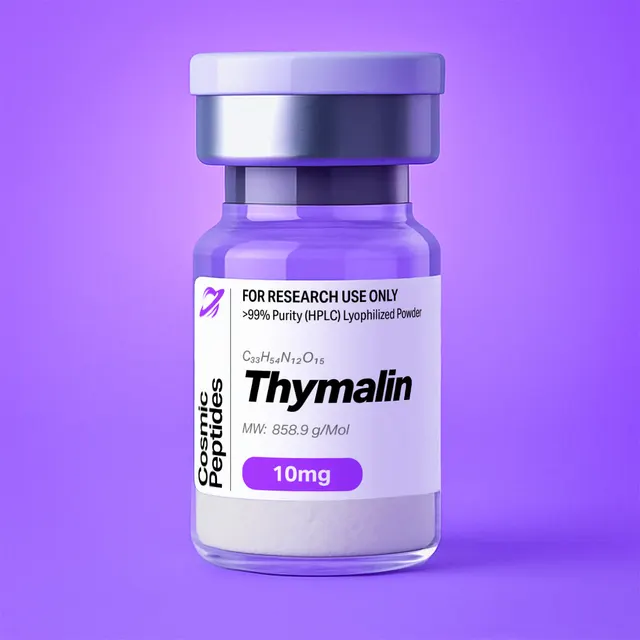
Thymalin
Commonly researched with these items
Patent-pending lot tracking: Complete transparency from production to vial.
Thymalin: Thymus-Derived Peptide Material and the Defined Nonapeptide pGlu-Ala-Lys-Ser-Gln-Gly-Gly-Ser-Asn
Disclaimer
Products described here are supplied for research use only and are not intended for diagnostic, therapeutic, or clinical application. All statements regarding biological activity reflect preclinical and in vitro findings exclusively and have not been evaluated by the U.S. Food and Drug Administration. These materials and related content are provided for educational and investigational purposes only and are intended solely for qualified researchers in laboratory and academic settings.
Key Facts
- Compound class
- Synthetic linear nonapeptide with an N-terminal pyroglutamyl residue and a free C-terminal acid; the sequence known in the literature as facteur thymique sérique (FTS), nonathymulin or, zinc-bound, thymulin. The name "thymalin" originally denotes a thymus-derived polypeptide mixture (see Molecular Identity).
- Sequence
Pyr-Ala-Lys-Ser-Gln-Gly-Gly-Ser-Asn-OH(Pyr = pGlu, L-pyroglutamyl; one-letter code pEAKSQGGSN), all chiral residues L- Molecular formula
- C33H54N12O15 (zinc-free peptide, no counterions)
- Molecular weight
- 858.9 g/mol (average, 858.86); monoisotopic mass 858.3832 Da
- CAS number
- 63958-90-7 for the defined nonapeptide (nonathymulin). The number 63340-72-7, also seen in catalogues, is registered to "thymic humoral factor", a different thymus-derived preparation (see Molecular Identity).
- PubChem CID
- 3085284
- Physical form as supplied
- Lyophilized powder
- Lot traceability
- Every unit is tied to a tracked lot; the lot number, recorded purity and verified quantity of the lot on sale are shown live on this page, with its certificate of analysis.
Molecular Identity and Classification
Several distinct materials share the vocabulary around thymalin, and only one of them is a single, chemically defined molecule.
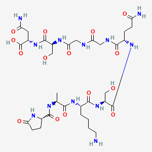
The defined nonapeptide. The material described by the formula, molecular weight and sequence on this page is pGlu-Ala-Lys-Ser-Gln-Gly-Gly-Ser-Asn-OH. Its PubChem IUPAC name, (2S)-4-amino-2-[[(2S)-2-[[2-[[2-[[(2S)-5-amino-2-[[(2S)-2-[[(2S)-6-amino-2-[[(2S)-2-[[(2S)-5-oxopyrrolidine-2-carbonyl]amino]propanoyl]amino]hexanoyl]amino]-3-hydroxypropanoyl]amino]-5-oxopentanoyl]amino]acetyl]amino]acetyl]amino]-3-hydroxypropanoyl]amino]-4-oxobutanoic acid, encodes seven stereocentres, all S. The formula is C33H54N12O15, the average molecular weight 858.86 g/mol and the monoisotopic mass 858.3832 Da. PubChem lists it as compound 3085284 under the names nonathymulin and thymic factor, CAS 63958-90-7, with "Thymalin" recorded as a synonym.
The sequence was established in 1977 for a peptide isolated from serum by Jean-François Bach, Mireille Dardenne and Jean-Marie Pléau in Paris, who named it facteur thymique sérique (FTS, serum thymic factor). Edman analysis of the intact peptide and of tryptic fragments, with the C-terminal residue identified by carboxypeptidase A digestion, gave the sequence, and synthesis confirmed it. In 1982 the same group showed that activity in an in vitro rosette assay depended on bound zinc and named the zinc complex thymulin (FTS-Zn), reserving FTS or nonathymulin for the zinc-free peptide.
The historical thymalin preparation. Thymalin in its original sense is a polypeptide complex obtained from calf thymus tissue by mild acid extraction, the preparation described under that name by Morozov and Khavinson. It is a mixture, not a molecule: no single formula, molecular weight or sequence describes it, and its composition depends on the extraction and fractionation used. Constituents have been resolved from it one at a time by chromatography — the dipeptide Glu-Trp was isolated from the preparation by reversed-phase HPLC, and a 2021 review places the dipeptide Lys-Glu (KE) in it as well — but a list of identified components is not a composition. Statements about the extract therefore do not transfer to the synthetic nonapeptide, or the reverse, and a methods section should say which material was used and how it was characterized.
Thymic humoral factor. The registry number 63340-72-7, sometimes printed beside thymalin, designates "thymic humoral factor" in the ChemIDplus records deposited in PubChem (substance SID 135324007): another thymus-derived preparation with no defined structure, not the nonapeptide.
One notational point follows from the chemistry. Because the pyroglutamyl residue leaves no free α-amine, the "H-" prefix that appears in some sequence listings for this peptide is an artefact of template notation: the correct form is Pyr-Ala-Lys-Ser-Gln-Gly-Gly-Ser-Asn-OH, or pGlu-… , with the free acid marked at the C-terminus only.
Structure and Chemical Features
The nine residues fall into three segments. pGlu-Ala-Lys carries the blocked N-terminus and the only basic side chain; Ser-Gln-Gly-Gly is polar and highly flexible; Ser-Asn-OH presents a hydroxyl, a side-chain amide and the free carboxylate — the cluster that forms the zinc site.
The pyroglutamyl N-terminus. Pyroglutamic acid (5-oxoproline) is a five-membered lactam formed when the side-chain carboxyl or amide of an N-terminal glutamic acid or glutamine condenses with its own α-amine. The peptide therefore has no free N-terminal amine: that end carries no positive charge, does not react with amine-reactive reagents such as ninhydrin or N-hydroxysuccinimide esters, resists most aminopeptidases, and blocks Edman degradation.
Charge and flexibility. With the N-terminus blocked, only two groups ionize in the usual pH range: the lysine ε-amine (pKa near 10.5) and the C-terminal acid (pKa near 3). Between pH 4 and pH 9 the molecule is a zwitterion of net charge close to zero, with a calculated isoelectric point near neutrality, roughly pH 6.5 to 7, so ion exchange separates it poorly and reversed-phase or hydrophilic-interaction chromatography is preferred. The chain is exceptionally mobile: Gly6-Gly7 leaves the backbone dihedrals unrestricted, the pyrrolidinone ring is the only rigid element, and PubChem computes 28 rotatable bonds across 60 heavy atoms.
The Metal-Binding Site
The most distinctive chemistry of this sequence is its coordination of divalent zinc. By gel filtration with 65Zn2+, Gastinel and colleagues found that the nonapeptide binds one zinc ion with an apparent dissociation constant of 5 ± 2 × 10⁻⁷ M at pH 7.5, with binding undetectable below pH 6.0 — consistent with deprotonation of the coordinating groups being required. Ga3+, Al3+, Mn2+ and Cu2+ compete for the same site.
The coordinating atoms were identified by NMR. Cung and colleagues followed 1H and 13C spectra in dimethyl sulfoxide-d6 across a range of metal-to-peptide ratios and found two species: a 1:2 complex in which one Zn(II) ion bridges two peptide molecules through the Ser4 hydroxyl oxygen and the Asn9 carboxylate, and a 1:1 complex in which the Ser8 hydroxyl joins as a third ligand. The 1:2 species predominates when zinc is substoichiometric, and exchange is fast on the NMR timescale, so spectra shift progressively rather than splitting. The [Ala4] and [Ala8] analogues, each missing one serine hydroxyl, confirmed the assignment, and parallel work with Al3+ showed that a harder trivalent ion reorganises the coordination sphere entirely.
Two practical points follow. The apo-peptide and the zinc complex are different chemical entities with different conformations, so a report must be read for which one it studied. And zinc is a ubiquitous laboratory contaminant — glassware, many buffer salts, water not purified for trace-metal content — so a nominally metal-free solution may partly complex adventitious zinc unless trace-metal-grade reagents or a chelating resin are used. The table below collects the differences that can actually be measured.
| Feature | Zinc-free peptide (FTS, nonathymulin) | Zinc complex (thymulin, FTS-Zn) | How to tell |
|---|---|---|---|
| Composition | C33H54N12O15 | Same chain plus one Zn2+, 1:1, with loss of protons from the ligating groups | ICP-MS or atomic absorption for zinc, the technique used in the 1982 work |
| Monoisotopic mass | 858.3832 Da | Adduct observed at m/z 921.304 for [M+Zn−H]+ | High-resolution MS; the zinc isotope envelope is unmistakable |
| Ligating atoms | None engaged | Ser4 Oγ and the Asn9 carboxylate, with Ser8 Oγ as a third ligand in the 1:1 species | 1H and 13C NMR titration; [Ala4] and [Ala8] analogues as controls |
| Stoichiometry in solution | Monomer | 1:2 Zn:peptide below equimolar metal, 1:1 above | Shift pattern across metal-to-peptide ratios; exchange is fast on the NMR timescale |
| pH window | Stable across the usual range | Formed above pH 6.0; Kd 5 ± 2 × 10⁻⁷ M at pH 7.5 | Gel filtration with 65Zn2+, or ITC at controlled pH |
| Conformation | Mobile ensemble in water | One defined conformation in the 1:1 species | Circular dichroism or NMR before and after metal addition |
| Antibody recognition | Not recognised by zinc-specific monoclonals | Recognised; the NMR work assigned this to the 1:1 complex only | Immunoassay with a zinc-specific monoclonal antibody |
Conformation in Solution
Laussac and colleagues characterized the free peptide by one- and two-dimensional 1H and 13C NMR, assigning spin systems by COSY and sequential connectivities by NOESY. In water the data fitted a mobile equilibrium among many conformations with no dominant fold, as a glycine-rich sequence predicts; in dimethyl sulfoxide-d6, where solvent does not compete for intramolecular hydrogen bonds, a more ordered structure with three internal hydrogen bonds appeared. Aqueous NMR of this peptide therefore reports an ensemble rather than a structure.
Synthesis and Manufacturing Chemistry
The defined nonapeptide is made by solid-phase peptide synthesis (SPPS), introduced by Merrifield in 1963, in which the chain is assembled C-terminus first on an insoluble support. Production uses the Fmoc/tBu strategy built on the base-labile 9-fluorenylmethoxycarbonyl group of Carpino and Han: Fmoc is removed with piperidine in dimethylformamide before each coupling, while side-chain protection survives to a single acid cleavage at the end.
A C-terminal asparagine. Synthesis starts by anchoring Fmoc-Asn(Trt)-OH to an acid-labile support such as Wang or 2-chlorotrityl chloride resin. Trityl protection of the side-chain amide is not optional in practice: unprotected Asn and Gln amides can be dehydrated to nitriles by carbodiimide activators, and the asparagine side-chain amide is the classic precursor of succinimide (cyclic-imide) formation, which a C-terminal position does nothing to prevent. A bulky 2-chlorotrityl linker also suppresses diketopiperazine formation at the dipeptide stage.
A glycine-glycine midsection. Gly6-Gly7 is the classic motif for on-resin aggregation: with no side chains to disrupt them, growing chains hydrogen-bond to one another, the resin shrinks and reagent access falls. Countermeasures are elevated-temperature or microwave-assisted coupling, solvents containing dimethyl sulfoxide or N-methylpyrrolidone, backbone-amide-linker chemistry where needed, and double coupling with colorimetric monitoring of residual free amine.
A pyroglutamyl cap. The final residue goes on as L-pyroglutamic acid. Its nitrogen is already in the lactam, so it needs no α-amino protection and it terminates the chain — nothing couples onto it and no Fmoc removal follows. Installing Glu or Gln and cyclising it in place is the alternative, but direct coupling leaves no doubt that the cap is complete. The capped N-terminus is also a purity handle: material with a free α-amine is a by-product.
Serine and lysine are protected as Ser(tBu) and Lys(Boc), glutamine as Gln(Trt). After the ninth coupling, trifluoroacetic acid with water and triisopropylsilane as scavengers releases the peptide and removes all side-chain protection at once; with no cysteine, methionine or tryptophan, the scavengers those residues need are unnecessary. The crude peptide is precipitated into cold diethyl ether, purified by preparative reversed-phase HPLC and lyophilized — normally as a trifluoroacetate salt, and zinc-free unless zinc is added deliberately.
The historical extract, by contrast, is made by aqueous extraction of thymus tissue and removal of high-molecular-weight material, typically by ultrafiltration and chromatographic fractionation. Its characterization is necessarily statistical — total peptide content, molecular-weight range, chromatographic fingerprint — rather than the single-molecule identity mass spectrometry gives a synthetic peptide.
Analytical Characterization
Chromatography. This peptide is extremely hydrophilic: PubChem computes an XLogP of −10.9 and a topological polar surface area of 452 Å2, and the sequence has no aromatic residue. On a conventional C18 column in water/acetonitrile with 0.1% trifluoroacetic acid it elutes at or near the void volume, unresolved from salts. Three fixes are routine: a polar-embedded or aqueous-compatible C18 phase at very low organic content; hydrophilic-interaction chromatography (HILIC) on bare silica or an amide phase, where the polarity that defeats C18 becomes the basis of retention; and ion-pairing with a longer-chain perfluorinated acid. Detection is at the amide band, 210 to 220 nm, since the absence of Trp, Tyr and Phe leaves no 280 nm signal — so A280 cannot give concentration, and quantitation relies on mass, amino-acid analysis or a 214 nm calibration curve.
Molecular mass. From the monoisotopic mass of 858.3832 Da, positive-mode electrospray gives [M+H]+ at m/z 859.390 and [M+2H]2+ at 430.199; with only the lysine side chain basic, the singly charged ion dominates. Sodium and potassium adducts appear at 881.372 and 897.346. Zinc, added or adventitious, gives [M+Zn−H]+ at 921.304 with the unmistakable zinc isotope pattern (64Zn, 66Zn, 67Zn, 68Zn at roughly 49, 28, 4 and 19%) — the quickest way to tell an apo-peptide from a complex.
Sequence Confirmation by Tandem MS
Collision-induced dissociation of the singly protonated molecule gives a b/y series from which the sequence can be read directly. The values below are monoisotopic and singly protonated, calculated from residue masses, and can be matched against a laboratory's own spectra.
| Position from N-terminus | Residue | b ion (m/z) | y ion (m/z) |
|---|---|---|---|
| 1 | Pyr (pGlu) | b1 112.039 | y9 = [M+H]+ 859.390 |
| 2 | Ala | b2 183.076 | y8 748.358 |
| 3 | Lys | b3 311.171 | y7 677.321 |
| 4 | Ser | b4 398.203 | y6 549.226 |
| 5 | Gln | b5 526.262 | y5 462.194 |
| 6 | Gly | b6 583.283 | y4 334.136 |
| 7 | Gly | b7 640.305 | y3 277.114 |
| 8 | Ser | b8 727.337 | y2 220.093 |
| 9 | Asn | b9 841.380 (rarely observed) | y1 133.061 |
Three details help in reading the spectrum. The b1 ion at m/z 112.039, normally absent for ordinary peptides, is observable for a lactam-capped N-terminus, so its presence is evidence of the cap. The glycine pair gives two consecutive 57.021 Da spacings that pin the Gly6-Gly7 segment. And b3 minus b2 is 128.095, the lysine residue mass, which is 0.036 Da below the 128.059 of glutamine — a distinction that needs resolving power of roughly 10,000 at m/z 300 and is the one place in this sequence where a low-resolution instrument can mis-assign a residue.
Amino-acid analysis. Hydrolysis in 6 M hydrochloric acid converts the side-chain amides to acids and opens the lactam, so the composition found is not that of the intact chain: Asn9 is recovered as aspartic acid and both pGlu1 and Gln5 as glutamic acid, giving Glu 2 : Ser 2 : Gly 2 : Ala 1 : Lys 1 : Asp 1, nine residues in all, with serine recovery below two because it is partly destroyed. Edman sequencing fails on the intact peptide; the classical workaround is unblocking with pyroglutamate aminopeptidase, described by Podell and Abraham in 1978, after which sequencing proceeds from Ala2.
Purity, Peptide Content and Counterions
HPLC purity and peptide content answer different questions, and conflating them is the commonest source of concentration error in peptide work. Purity is the fraction of UV-absorbing material that is the target; peptide content is the fraction of the weighed powder that is peptide at all, the rest being counterions, water and salts. The arithmetic here is instructive: one trifluoroacetate counterion (114.02 g/mol) on the single basic lysine gives a salt of 972.9 g/mol, so the theoretical maximum peptide content of a mono-trifluoroacetate salt is 858.86 / 972.9 = 88.3% by mass, rising to 93.5% for an acetate salt, before any residual water. Weighing the powder as though it were pure peptide therefore yields a solution 10 to 15% more dilute than intended.
Supporting assays complete the picture: ion chromatography or 19F NMR for trifluoroacetate, Karl Fischer titration for water, elemental analysis against the calculated C 46.15%, H 6.34%, N 19.57% and O 27.94%, chiral amino-acid analysis to exclude D-residues, and atomic absorption spectrometry or ICP-MS for zinc — the technique used in the 1982 work that defined thymulin.
Degradation markers. Two signatures matter in stored material: +0.984 Da, from deamidation of Asn9 or Gln5, and −18.011 Da, from cyclic-imide formation. Geiger and Clarke mapped the succinimide chemistry behind both in 1987, with rates depending sharply on pH and on the following residue. Deamidation products usually resolve from the parent peak by HILIC or a shallow reversed-phase gradient.
Physicochemical Properties
| Property | Value | Notes |
|---|---|---|
| Molecular formula | C33H54N12O15 | Apo-peptide, no counterion |
| Average molecular weight | 858.86 g/mol | Rounded to 858.9 in most catalogues |
| Monoisotopic mass | 858.3832 Da | For high-resolution MS |
| [M+H]+ / [M+2H]2+ | m/z 859.390 / 430.199 | Positive-mode ESI, calculated |
| [M+Na]+ / [M+K]+ | m/z 881.372 / 897.346 | Common alkali adducts |
| [M+Zn−H]+ | m/z 921.304 | Zinc complex; diagnostic Zn isotope pattern |
| Elemental composition | C 46.15, H 6.34, N 19.57, O 27.94% | Calculated from the formula |
| Ionizable groups | Lys ε-NH2 (pKa ≈ 10.5), C-terminal COOH (pKa ≈ 3) | N-terminus blocked as pyroglutamate |
| Net charge at pH 7 | Approximately zero (zwitterion) | One cation, one anion |
| Calculated isoelectric point | About 6.5–7 | Mid-point of the two pKa values |
| XLogP | −10.9 | Computed; exceptionally hydrophilic |
| Topological polar surface area | 452 Å2 | Computed |
| Hydrogen-bond donors / acceptors | 15 / 16 | Computed |
| Rotatable bonds / heavy atoms | 28 / 60 | Computed; highly flexible chain |
| Defined stereocentres | 7 | All S (L); Gly6 and Gly7 achiral |
| Zn2+ affinity | Kd 5 ± 2 × 10⁻⁷ M at pH 7.5 | Gel filtration with 65Zn; no binding below pH 6.0 |
| UV absorbance | Amide band only, about 214 nm | No Trp, Tyr or Phe; no 280 nm signal |
| Solubility | Freely soluble in water and aqueous buffers | Consistent with the computed logP and TPSA |
| Peptide content, mono-TFA salt | 88.3% maximum by mass | 858.86 / 972.9; acetate salt 93.5% |
The numbers describe a small, neutral, very polar, very flexible molecule. The XLogP of −10.9, the polar surface area of 452 Å2 and the 31 hydrogen-bonding atoms together mean it is solvated by water far more favourably than by any organic phase: octanol/water partition coefficients are too small to measure reliably, and the polarity rules out passive diffusion across lipid bilayers, so transport in any cell-based in vitro system must involve a carrier or endocytic route.
Two worked numbers: at 858.86 g/mol, 1 mg/mL is 1.164 mM, and 1 mM needs 0.859 mg/mL of pure peptide, or about 0.973 mg/mL of a mono-trifluoroacetate salt at 88.3% content. For metal stoichiometry, a 1 mM peptide solution requires 65.4 µg/mL of zinc for a 1:1 complex — far above the trace zinc of reagent water, yet within reach of some buffer salts, so apo- and holo-forms are not cleanly separable without deliberate control.
In the solid state the material is a hygroscopic amorphous powder. Serine- and amide-rich sequences are good glass formers, so there is no crystalline melting point: thermal analysis shows a glass transition and water loss. With no aromatic chromophore and no sulfur, it absorbs nothing above about 230 nm and so has no obvious route to photodegradation, but it is sensitive to moisture, which skews weighing and mobilises the hydrolytic chemistry above.
Research Context
The literature on this sequence is, at its core, about metal-dependent peptide recognition. The founding observation was chemical: material purified from serum, and synthetic peptide alike, lost activity in an in vitro rosette assay after exposure to the chelating resin Chelex 100, and activity returned on adding zinc salts, best at a 1:1 metal-to-peptide ratio, while other metals restored it less well. Atomic absorption spectrometry confirmed zinc in the synthetic material. That 1982 result from Dardenne and colleagues split one compound into two — an inactive apo-peptide and a zinc complex they named thymulin — and every study since has had to specify which form it used.
Binding and Conformational Measurements
The most quantitative in vitro work used tritiated synthetic peptide at 120 Ci/mmol on plasma-membrane preparations from the 1301 lymphoblastoid T-cell line. Gastinel and colleagues found binding that was temperature dependent, reversible, specific and saturable, at steady state within about 80 minutes at 37 °C with 1 nM labelled peptide. Scatchard analysis resolved at least two classes of site, with dissociation constants of 0.516 ± 0.2 nM and 110 ± 27.8 nM and densities of 0.186 ± 0.045 and 2.026 ± 0.367 pmol per milligram of membrane protein. Those numbers set the concentration window for any binding experiment, and two-site behaviour must be fitted explicitly. With no aromatic residue, fluorescent tracers need an external label, usually on the lysine ε-amine — next to the metal-binding region, so a labelled-peptide control matters.
Metal binding in cell-free systems. Contemporary equivalents of that gel-filtration measurement are isothermal titration calorimetry for binding enthalpy, far-UV circular dichroism for the conformational change on metal addition, and 1H NMR titration to locate the ligating atoms, with Ga3+, Al3+, Mn2+ or Cu2+ competition probing site specificity. The [Ala4] and [Ala8] analogues remain the cleanest controls.
Antibody recognition gives a second, independent read-out of conformation. Monoclonal antibodies raised against the zinc complex distinguish the two solution species — the NMR work concluded that only the 1:1 complex adopts the recognised conformation — so immunoassay with them is a conformational probe as much as a quantitative one, one reason immunoassay and mass-spectrometric quantitation of a sample can disagree.
In cultured cells, the classic in vitro experiment on peptide trafficking used primary thymic epithelial cells with colchicine, cytochalasin B and monensin. Both cytoskeletal inhibitors reduced the peptide reaching the supernatant in a concentration-dependent manner while raising its intracellular immunofluorescence signal, and monensin, which perturbs Golgi-derived vesicle traffic, behaved similarly. Savino and Dardenne read this as a conventional exocytic route: synthesis at the rough endoplasmic reticulum, passage through the Golgi, packaging into vesicles that acquire zinc, fusion with the plasma membrane.
Work on the extract-derived material has gone towards lineage markers in culture. In a 2020 study, thymalin applied to cultured hematopoietic stem cells lowered CD44 and CD117 immunofluorescence two- to three-fold and raised CD28 6.8-fold, read by the authors as CD117-positive cells progressing towards a CD28-positive T-lymphocyte phenotype. As cell-culture work on a mixture, it illustrates the standard read-outs: surface-marker immunofluorescence or flow cytometry, with marker ratios rather than absolute intensities as the endpoint.
Peptide–Nucleic Acid Interaction
A separate strand asks whether short, highly polar peptides bind DNA directly. The question predates both materials here: in 1975 Gianfranceschi and colleagues isolated a low-molecular-weight peptidic factor from aqueous calf thymus extracts and showed that it inhibited DNA-directed RNA polymerase in a cell-free system, attributing the effect to electrostatic peptide–DNA interaction rather than to nuclease or histone-fragment contamination. The modern version is computational: a 2021 systematic review from Khavinson and colleagues collects docking and molecular-dynamics studies of short peptides against double-stranded DNA, including the dipeptide Lys-Glu placed in the thymalin preparation. For the nonapeptide, one lysine is the only cationic anchor and the molecule is near-neutral at physiological pH, which makes it a stringent test case for electrostatics-based docking rather than an obvious DNA binder.
Names, Registry Numbers and What They Denote
| Name | What it denotes | Chemically defined? |
|---|---|---|
| FTS, facteur thymique sérique, serum thymic factor | The zinc-free nonapeptide pGlu-Ala-Lys-Ser-Gln-Gly-Gly-Ser-Asn-OH, as isolated and sequenced from serum | Yes |
| Nonathymulin | The same zinc-free nonapeptide; the name under which CAS 63958-90-7 and PubChem CID 3085284 are registered | Yes |
| Thymulin (FTS-Zn) | The 1:1 zinc complex of that nonapeptide, named in 1982 | Yes, as a complex of defined stoichiometry |
| Thymalin (historical sense) | A polypeptide complex extracted from thymus tissue; a mixture | No |
| Thymalin (research-chemical sense) | In catalogues, the synthetic nonapeptide; PubChem lists it as a synonym of CID 3085284 | Yes, when the formula and sequence are stated |
| Thymic humoral factor, CAS 63340-72-7 | A different thymus-derived preparation with no single structure | No |
| Thymic factor | Ambiguous: PubChem carries it as a synonym both of the defined nonapeptide (CID 3085284) and of the undefined preparation (SID 135324007), so on its own it identifies nothing | Not as written |
| Cytomedins | The collective term used by the originators of the extract for tissue-derived peptide fractions of this kind, not a structure | No |
The rule for a notebook entry: cite a formula, a monoisotopic mass and a sequence, not a trade name. Material for which those three cannot be stated is a mixture and should be described as one in a methods section. For this compound the minimum unambiguous identifier is the triple C33H54N12O15 / 858.3832 Da monoisotopic / Pyr-Ala-Lys-Ser-Gln-Gly-Gly-Ser-Asn-OH, with the metal state stated separately, because the sequence alone does not say whether zinc is present.
Related Research Compounds
Two other peptides in this catalogue invite direct comparison.
Thymosin Alpha-1 is the other well-known thymus-derived sequence but a different chemical proposition: a 28-residue, N-terminally acetylated, strongly acidic peptide of formula C129H215N33O55, with an acidic isoelectric point and no metal site of the kind described here. Side-by-side analysis shows how differently a nonapeptide and a 28-mer behave on reversed-phase columns and in electrospray charge-state distributions.
Among the short bioregulator peptides from the same research programme, Epitalon (Ala-Glu-Asp-Gly) and Pinealon (Glu-Asp-Arg) are the standard comparators and appear in the peptide–DNA docking literature cited above. Both have free N-termini and a clear net charge, which makes them useful controls when testing whether a behaviour depends on the blocked pyroglutamyl terminus or on charge.
Laboratory Handling and Storage
The solid. The lyophilized powder is kept tightly closed at −20 °C, dark and dry, or at −80 °C for long-term storage. It is hygroscopic, so a container from the freezer is allowed to reach room temperature in a desiccator before opening; opening it cold in humid air condenses water onto the powder, inflating the weighed mass and starting the chemistry that limits shelf life. Where accuracy matters, dissolving the entire contents and determining concentration analytically beats weighing a portion of a static-prone lyophilizate.
Solutions. The peptide dissolves readily in high-purity water and common aqueous buffers; no organic co-solvent is needed at a computed logP near −11. Stocks are typically 1 to 10 mM — 1 mM is 0.859 mg/mL of peptide — aliquoted for single use and frozen, avoiding repeated freeze–thaw. Where metal state must be defined, the choice has to be explicit: trace-metal-grade water and plasticware to hold the apo-form, or a known zinc concentration to drive the 1:1 complex. Buffer choice follows: phosphate precipitates zinc, Tris and histidine coordinate it, chelators strip it, so metal-controlled work runs in HEPES or MOPS.
pH and stability. The hydrolytic liabilities are deamidation of Asn9 and Gln5 and cyclic-imide formation at the C-terminal asparagine, both accelerated by alkaline pH and warmth. Solutions at pH 5 to 6 are the most stable, and material held at 37 °C for an assay should be checked afterwards by LC-MS for the 0.984 Da product rather than assumed intact. The pyroglutamyl cap helps: it resists the aminopeptidase activity of serum-containing media and removes the N-terminal Schiff-base chemistry that complicates peptides with free α-amines.
Surfaces and recovery. Extreme hydrophilicity makes adsorption to polypropylene less of a problem than for amphipathic peptides, but losses at low concentration still occur; low-binding plasticware and a carrier protein, where the design allows, improve recovery. Glassware is best avoided altogether when the metal state matters: glass surfaces both leach and adsorb divalent metal ions, and an acid-washed, trace-metal-grade plastic vessel is the more controllable container.
Re-verification. An LC-MS check of a stock — m/z 859.390 for [M+H]+, no peak 0.98 Da higher — confirms identity and integrity after storage in minutes, and ICP-MS or atomic absorption on the same stock quantifies zinc. Each lot's certificate of analysis, shown live on this page, records the results for that lot.
Controls specific to this material. Three are worth building into any analytical plan: a counterion-matched vehicle, since trifluoroacetate is not inert in cell culture, as Cornish and colleagues showed for osteoblast and chondrocyte proliferation; a zinc-only control at the metal concentration of the complex; and a chelator control, to show that an effect tracks the metal state of the peptide rather than the metal content of the medium.
Frequently Asked Research Questions
- What exactly is thymalin?
- Two things. Historically, a polypeptide complex obtained from calf thymus tissue by mild acid extraction — a mixture with no single formula or sequence, from which individual dipeptides such as Glu-Trp have been resolved chromatographically. In research-chemical catalogues, the defined synthetic nonapeptide pGlu-Ala-Lys-Ser-Gln-Gly-Gly-Ser-Asn-OH (C33H54N12O15, 858.86 g/mol), also called FTS or nonathymulin. Material supplied with a stated formula, molecular weight and sequence is the nonapeptide.
- What is the difference between this nonapeptide and thymulin?
- The same sequence in two states. The zinc-free peptide is FTS or nonathymulin; the 1:1 complex with Zn2+ was named thymulin in 1982, after chelation with Chelex 100 abolished activity in an in vitro rosette assay and added zinc returned it, most effectively at a 1:1 metal-to-peptide ratio.
- Which CAS number is correct?
- CAS 63958-90-7 is the defined nonapeptide (nonathymulin), PubChem CID 3085284. CAS 63340-72-7, also seen in listings, is registered in ChemIDplus records to "thymic humoral factor", a separate thymus-derived preparation.
- What masses should I expect by mass spectrometry?
- The monoisotopic mass is 858.3832 Da, so [M+H]+ falls at m/z 859.390 and [M+2H]2+ at 430.199, with sodium and potassium adducts at 881.372 and 897.346. A zinc complex gives [M+Zn−H]+ at 921.304 with the characteristic zinc isotope pattern.
- Why does this peptide show no absorbance at 280 nm?
- It contains no tryptophan, tyrosine or phenylalanine, the residues responsible for near-UV absorbance. Detection relies on the amide band near 214 nm, with quantitation by amino-acid analysis or by weight corrected for peptide content.
- Why does it elute so early on a C18 column?
- With a computed XLogP of −10.9 and a polar surface area of 452 Å2, it is far too polar for a conventional C18 phase to retain at usual organic content. HILIC, a polar-embedded reversed phase at very low acetonitrile, or ion pairing with a longer perfluorinated acid all give usable retention.
- Can I sequence it by Edman degradation?
- Not directly: the pyroglutamyl residue has no free α-amine for the phenyl isothiocyanate chemistry to attack. Enzymatic unblocking with pyroglutamate aminopeptidase, as described by Podell and Abraham, exposes Ala2; tandem mass spectrometry is the more direct modern route.
- Where does the zinc bind?
- NMR titrations in dimethyl sulfoxide-d6 placed the ligands at the Ser4 hydroxyl and the Asn9 carboxylate in the 1:2 metal-to-peptide complex, with the Ser8 hydroxyl joining as a third ligand in the 1:1 complex. Gel filtration gave an apparent Kd of 5 ± 2 × 10⁻⁷ M at pH 7.5, with no binding detectable below pH 6.0.
- What does amino-acid analysis report for this sequence?
- After 6 M hydrochloric acid hydrolysis the amides and the lactam are opened, giving Glu 2 (from pGlu1 and Gln5), Ser 2, Gly 2, Ala 1, Lys 1 and Asp 1 (from Asn9), nine residues in total. Serine recovery is characteristically low because it is partly destroyed under the hydrolysis conditions.
- How much of the weighed powder is actually peptide?
- At most 88.3% for a mono-trifluoroacetate salt (858.86 of 972.9 g/mol), about 93.5% for an acetate salt, before residual water. Molar concentrations follow from peptide content, not gross weight.
- What degradation should I look for in stored material?
- +0.984 Da indicates deamidation of Asn9 or Gln5; −18.011 Da indicates cyclic-imide formation. Both are favoured by alkaline pH and warmth, so single-use aliquots at pH 5 to 6, stored at −20 °C or below, are the countermeasure.
- Does this peptide cross cell membranes in culture?
- Not by passive diffusion: its polarity rules that out. Uptake in a cell-based in vitro system must involve a transporter or an endocytic route, and experiments should be interpreted accordingly.
Sources & Further Reading
- National Center for Biotechnology Information. PubChem Compound Summary for CID 3085284, Nonathymulin (pGlu-Ala-Lys-Ser-Gln-Gly-Gly-Ser-Asn-OH). PubChem CID 3085284.
- National Center for Biotechnology Information. PubChem Substance Record SID 135324007, Thymic humoral factor (CAS 63340-72-7), source ChemIDplus. PubChem SID 135324007.
- Morozov VG, Khavinson VK. Natural and synthetic thymic peptides as therapeutics for immune dysfunction. International Journal of Immunopharmacology 1997;19(9–10):501–505. doi:10.1016/S0192-0561(97)00058-1
- Bach JF, Dardenne M, Pleau JM, Rosa J. Biochemical characterisation of a serum thymic factor. Nature 1977;266(5597):55–57. doi:10.1038/266055a0
- Pleau JM, Dardenne M, Blouquit Y, Bach JF. Structural study of circulating thymic factor: a peptide isolated from pig serum. II. Amino acid sequence. Journal of Biological Chemistry 1977;252(22):8045–8047. doi:10.1016/S0021-9258(17)40933-1
- Dardenne M, Pléau JM, Nabarra B, Lefrancier P, Derrien M, Choay J, Bach JF. Contribution of zinc and other metals to the biological activity of the serum thymic factor. Proceedings of the National Academy of Sciences 1982;79(17):5370–5373. doi:10.1073/pnas.79.17.5370
- Gastinel LN, Dardenne M, Pleau JM, Bach JF. Studies on the zinc binding site to the serum thymic factor. Biochimica et Biophysica Acta (General Subjects) 1984;797(2):147–155. doi:10.1016/0304-4165(84)90116-8
- Laussac JP, Cung MT, Pasdeloup M, Haran R, Marraud M, Lefrancier P, Dardenne M, Bach JF. NMR study of thymulin, a lymphocyte differentiating thymic nonapeptide. Conformational states of free peptide in solution. Journal of Biological Chemistry 1986;261(17):7784–7790. doi:10.1016/S0021-9258(19)57469-5
- Cung MT, Marraud M, Lefrancier P, Dardenne M, Bach JF, Laussac JP. NMR study of a lymphocyte differentiating thymic factor. An investigation of the Zn(II)-nonapeptide complexes (thymulin). Journal of Biological Chemistry 1988;263(12):5574–5580. doi:10.1016/S0021-9258(18)60603-9
- Laussac JP, Lefrancier P, Dardenne M, Bach JF, Marraud M, Cung MT. Structural and conformational study of the aluminum-thymulin complex using 1-D and 2-D NMR techniques. Inorganic Chemistry 1988;27(23):4094–4099. doi:10.1021/ic00296a005
- Gastinel LN, Pleau JM, Dardenne M, Sasaki A, Bricas E, Morgat JL, Bach JF. High affinity binding sites on plasma membrane obtained from the lymphoblastoid cultured 1301 cell line for highly radioactive serum thymic factor. Biochimica et Biophysica Acta (Biomembranes) 1982;684(1):117–126. doi:10.1016/0005-2736(82)90055-4
- Savino W, Dardenne M. Thymic hormone-containing cells. VIII. Effects of colchicine, cytochalasin B, and monensin on secretion of thymulin by cultured human thymic epithelial cells. Journal of Histochemistry & Cytochemistry 1986;34(12):1719–1723. doi:10.1177/34.12.3782779
- Khavinson VK, Linkova NS, Kvetnoy IM, Polyakova VO, Drobintseva AO, Kvetnaia TV, Ivko OM. Thymalin: activation of differentiation of human hematopoietic stem cells. Bulletin of Experimental Biology and Medicine 2020;170(1):118–122. doi:10.1007/s10517-020-05016-z
- Khavinson VK, Popovich IG, Linkova NS, Mironova ES, Ilina AR. Peptide regulation of gene expression: a systematic review. Molecules 2021;26(22):7053. doi:10.3390/molecules26227053
- Gianfranceschi GL, Amici D, Guglielmi L. Evidence for the presence in calf thymus of a peptidic factor controlling DNA transcription in vitro. Biochimica et Biophysica Acta (Nucleic Acids and Protein Synthesis) 1975;414(1):9–19. doi:10.1016/0005-2787(75)90121-5
- Geiger T, Clarke S. Deamidation, isomerization, and racemization at asparaginyl and aspartyl residues in peptides. Succinimide-linked reactions that contribute to protein degradation. Journal of Biological Chemistry 1987;262(2):785–794. doi:10.1016/S0021-9258(19)75855-4
- Podell DN, Abraham GN. A technique for the removal of pyroglutamic acid from the amino terminus of proteins using calf liver pyroglutamate amino peptidase. Biochemical and Biophysical Research Communications 1978;81(1):176–185. doi:10.1016/0006-291X(78)91646-7
- Merrifield RB. Solid phase peptide synthesis. I. The synthesis of a tetrapeptide. Journal of the American Chemical Society 1963;85(14):2149–2154. doi:10.1021/ja00897a025
- Carpino LA, Han GY. 9-Fluorenylmethoxycarbonyl amino-protecting group. The Journal of Organic Chemistry 1972;37(22):3404–3409. doi:10.1021/jo00795a005
- Cornish J, Callon KE, Lin CQ, Xiao CL, Mulvey TB, Cooper GJS, Reid IR. Trifluoroacetate, a contaminant in purified proteins, inhibits proliferation of osteoblasts and chondrocytes. American Journal of Physiology-Endocrinology and Metabolism 1999;277(5):E779–E783. doi:10.1152/ajpendo.1999.277.5.E779
Last reviewed:
| CAS Number | 63340-72-7 |
|---|---|
| Molecular Formula | C33H54N12O15 |
| Molecular Weight | 858.9 g/Mol |
| Purity | 99.8% |
| Lot Number | 26075 |
| Quantity | 10.11mg |
| Sequence | H-Pyr-Ala-Lys-Ser-Gln-Gly-Gly-Ser-Asn-OH |
Cosmic Peptides Certificate of Analysis
Unable to load certificate. Download COA