Thymosin Alpha-1
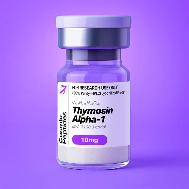
Thymosin Alpha-1
Commonly researched with these items
Patent-pending lot tracking: Complete transparency from production to vial.
Thymosin Alpha-1: The N-Acetylated 28-Residue Acidic Fragment of Prothymosin Alpha
Disclaimer
Products described here are supplied for research use only and are not intended for diagnostic, therapeutic, or clinical application. All statements regarding biological activity reflect preclinical and in vitro findings exclusively and have not been evaluated by the U.S. Food and Drug Administration. These materials and related content are provided for educational and investigational purposes only and are intended solely for qualified researchers in laboratory and academic settings.
Key Facts
- Compound class
- Linear 28-residue polypeptide; N-terminally acetylated acidic peptide corresponding to residues 2–29 of prothymosin alpha
- Sequence
Ac-Ser-Asp-Ala-Ala-Val-Asp-Thr-Ser-Ser-Glu-Ile-Thr-Thr-Lys-Asp-Leu-Lys-Glu-Lys-Lys-Glu-Val-Val-Glu-Glu-Ala-Glu-Asn-OH(one-letter code, Ac-SDAAVDTSSEITTKDLKEKKEVVEEAEN-OH), all chiral residues in the L-configuration- Molecular formula
- C129H215N33O55
- Molecular weight
- 3108.3 g/mol (average); monoisotopic mass 3106.504 Da
- CAS number
- 62304-98-7
- PubChem CID
- 16130571
- Defining modification
- Nα-acetyl serine at position 1; the free C-terminus is an unmodified asparagine carboxylic acid
- Net charge and isoelectric point
- Approximately −6 at pH 7.0; calculated pI about 3.8–4.1 depending on the pKa set (a strongly acidic peptide)
- Physical form as supplied
- Lyophilized powder
- Lot traceability
- Every unit is tied to a tracked lot; the lot number, recorded purity and verified quantity of the lot on sale are shown live on this page, with its certificate of analysis.
Molecular Identity and Classification
Thymosin alpha-1 (commonly abbreviated Tα1) is a single-chain polypeptide of 28 amino acid residues whose N-terminal serine carries an acetyl group on its α-amino nitrogen. Its full sequence is Ac-Ser-Asp-Ala-Ala-Val-Asp-Thr-Ser-Ser-Glu-Ile-Thr-Thr-Lys-Asp-Leu-Lys-Glu-Lys-Lys-Glu-Val-Val-Glu-Glu-Ala-Glu-Asn-OH, written in one-letter code as Ac-SDAAVDTSSEITTKDLKEKKEVVEEAEN-OH. The molecular formula of the free peptide acid is C129H215N33O55, corresponding to an average molecular weight of 3108.3 g/mol and a monoisotopic mass of 3106.504 Da. It is registered under CAS number 62304-98-7 and catalogued as PubChem compound 16130571.
The molecule is not an independent gene product. It corresponds to residues 2–29 of prothymosin alpha, a 111-residue intracellular acidic protein encoded by the PTMA gene (UniProt P06454). The initiator methionine is removed and the exposed serine is Nα-acetylated; counting from that serine (the convention of the processing literature, one less than the UniProt numbering), the peptide ends at Asn28. Sarandeses and colleagues showed in 2003 that a lysosomal asparaginyl endopeptidase, identified as mammalian legumain, cleaves the Asn28–Gly29 and Asn35–Gly36 bonds of prothymosin alpha with similar efficiency in vitro, generating the 28- and 35-residue fragments thymosin alpha-1 and thymosin alpha-11. Numbering, acetylation state and the free C-terminal Asn all follow from the precursor.
A frequent source of confusion is the relationship between thymosin alpha-1 and the beta-thymosins. Despite the shared historical name, the two families are chemically and genetically unrelated: the beta-thymosins, such as thymosin beta-4, are 40–44-residue actin-sequestering peptides from a distinct gene family, whereas the alpha-thymosin nomenclature groups fragments of prothymosin alpha. (TB-500, the acetylated heptapeptide Ac-LKKTETQ-OH, is a short synthetic sequence taken from the actin-binding region of thymosin beta-4, and is unrelated in sequence to thymosin alpha-1.) The common name arose because both families were first isolated from the same partially purified calf thymus extract, "thymosin fraction 5", for which Goldstein and colleagues proposed in 1977 a nomenclature based on isoelectric focusing: the alpha designation went to the most acidic components. Thymosin alpha-1 is, by composition, exactly that: six glutamate and three aspartate residues against four lysines and no arginine or histidine.
The compound should also be distinguished from Thymalin, which is catalogued as the unrelated nonapeptide pGlu-Ala-Lys-Ser-Gln-Gly-Gly-Ser-Asn, and from the unprocessed precursor protein, which behaves very differently in solution (see Structure and Chemical Features).
Structure and Chemical Features
The amino-acid composition explains almost all of the physical behaviour. The 28 residues comprise Glu ×6, Lys ×4, three each of Ser, Asp, Ala, Val and Thr, and single Ile, Leu and Asn residues. There is no Trp, Tyr, Phe, Cys, Met, Pro, Gly, Arg or His. Hence essentially no absorbance at 280 nm, no oxidation-prone or disulfide-forming side chain, and no proline to impose local rigidity or cis–trans isomerism.
Charge is dominated by the carboxylates. Nine side-chain acids plus the C-terminal carboxyl oppose only four lysine ε-amines, and the acetylated N-terminus contributes no positive charge. Summing side-chain ionisation gives a net charge of approximately −6 at pH 7.0 (−5.98 to −5.99 across three common pKa sets) and a calculated isoelectric point between 3.8 and 4.1. This is the most practically important property of the molecule: at neutral pH it is a polyanion that binds anion-exchange resins strongly, is retained weakly on reversed-phase media unless the mobile phase is acidified, and ionises well in negative-mode electrospray.
The N-terminal acetyl group converts the α-amine into a neutral amide, removing one positive charge, eliminating the free amine needed for Edman sequencing or amine-reactive labelling, and blocking aminopeptidase attack at that end. It adds exactly 42.0106 Da (C2H2O) to the monoisotopic mass, so a des-acetyl impurity appears 42.011 Da lighter, a difference that is unambiguous at any useful resolution.
Conformation in Solution and at Interfaces
Thymosin alpha-1 is an intrinsically disordered peptide in water. Grottesi and colleagues (1998) examined it by circular dichroism and two-dimensional NMR and reported no preferred conformation in aqueous solution, with partial structure appearing in the presence of small unilamellar vesicles of dimyristoylphosphatidylcholine and dimyristoylphosphatidic acid (10:1), in sodium dodecyl sulfate micelles, or on addition of zinc ions. In water–trifluoroethanol mixtures the peptide became appreciably structured, with a β-turn between residues 5 and 8 and an α-helix between residues 17 and 24.
A higher-field structure determination followed. Elizondo-Riojas and colleagues (2011) solved the conformation at 800 MHz in 40% trifluoroethanol / 60% water and found two stable regions: an α-helix spanning residues 14 to 26 and two double β-turns in the N-terminal twelve residues forming a distorted helical arrangement. The coordinates are deposited in the Protein Data Bank as entry 2L9I. The two studies agree on the essentials (a helical C-terminal half and turn structure near the N-terminus) and differ on the helix boundaries, which is expected given the different trifluoroethanol content and field strength.
| Region | Charged residues | Net charge at pH 7 | Structure and chemistry |
|---|---|---|---|
| 1–12, Ac-SDAAVDTSSEIT | Asp2, Asp6, Glu10; no Lys | −3 | Two double β-turns; acetylated terminus; both aspartimide-prone motifs (Asp-Ala, Asp-Thr) |
| 13–26, TKDLKEKKEVVEEA | Lys14,17,19,20; Asp15, Glu18,21,24,25 | −1 | α-Helix 14–26; carries all four lysines, with Leu16 and Val22-Val23 |
| 27–28, EN-OH | Glu27, C-terminal carboxylate | −2 | Free Asn28 acid left by Asn–Gly cleavage; C-terminal region implicated in albumin contact |
The lack of a stable fold is inherited from the precursor. Gast and colleagues (1995) showed by small-angle X-ray scattering, dynamic light scattering and circular dichroism that prothymosin alpha is a monomer with a random-coil-like conformation, a Stokes radius of 3.07 nm and a radius of gyration of 4.76 nm, 1.77 and 3.42 times the values expected for a compactly folded protein of that length.
Synthesis and Manufacturing Chemistry
Research material of this kind is typically made by solid-phase peptide synthesis (SPPS), introduced by Merrifield in 1963, in the Fmoc/tBu mode built on the 9-fluorenylmethoxycarbonyl group of Carpino and Han (1972). For the free C-terminal acid the chain is anchored through Asn to an acid-labile Wang or 2-chlorotrityl resin and assembled toward the N-terminus, each cycle removing Fmoc with 20% piperidine in dimethylformamide and coupling the next residue with an activator such as DIC/Oxyma, HBTU or HATU.
Three features of this particular sequence dominate the chemistry. First, side-chain protection is unusually uniform: Glu and Asp are used as their tert-butyl esters, Ser and Thr as tert-butyl ethers, Lys as the Boc derivative and Asn as the trityl amide, all removable in a single trifluoroacetic acid (TFA) cleavage. There is no cysteine to oxidise, no methionine to protect against alkylation and no arginine with its slow-to-remove Pbf group.
Second, the aspartate content creates a well-documented side-reaction risk. Under repeated piperidine exposure an aspartyl β-ester can cyclise onto the backbone nitrogen of the following residue to give a five-membered aspartimide, which then opens to the α- or β-aspartyl (iso-aspartyl) peptide, epimerises, or is captured by piperidine. The risk is highest when the following residue is small or polar (Gly, Asn, Ser, Thr, Ala) and lower for bulky residues; this sequence contains Asp-Ala at positions 2–3, Asp-Thr at 6–7 and Asp-Leu at 15–16, so the first two are the sites to watch. Mergler and colleagues (2003) examined the problem systematically in Fmoc SPPS; the usual countermeasures are bulkier β-esters such as OMpe in place of OtBu, shorter deprotection times, and an acidic additive such as HOBt in the piperidine solution. The by-products have characteristic masses: the aspartimide itself is 18.011 Da lighter than the target, the piperidide adducts 67.079 Da heavier, while the β-aspartyl and D-aspartyl products are exactly isobaric — which is why reversed-phase separation, and not mass alone, is the decisive purity criterion for this peptide.
Third, the N-terminal acetyl group must be installed deliberately: after the final Fmoc removal the resin-bound peptide is capped with acetic anhydride and a hindered base. Incomplete capping leaves the des-acetyl peptide (−42.011 Da, one extra positive charge), a standard specified impurity. Recombinant routes exist as well: Fang and colleagues (2009) showed that the prokaryotic Nα-acetyltransferase RimJ is responsible for Nα-acetylation of thymosin alpha-1 expressed as a fusion protein in Escherichia coli, and that fully acetylated peptide can be obtained by co-expression with RimJ.
After TFA cleavage with water and triisopropylsilane as scavengers (sufficient for a composition without Cys, Met, Trp or Arg), the crude peptide is precipitated in cold ether, purified by preparative reversed-phase HPLC and lyophilised. The counterion depends on the last step: a TFA-containing mobile phase leaves trifluoroacetate paired with the four lysines, whereas an acetate buffer or a salt exchange gives an acetate or sodium salt.
Analytical Characterization
Reversed-phase HPLC. Purity is reported as percentage peak area from a gradient separation with UV detection at 210–220 nm. With no Trp, Tyr or Phe there is no usable signal at 280 nm, so quantification must use peptide-bond absorbance, a colorimetric assay or amino-acid analysis. A C18 column with 0.1% TFA in water and acetonitrile is typical; the acid matters here, because at neutral pH the polyanion is poorly retained. Ammonium formate at pH 2.5–3.5 is a volatile alternative when the eluate goes to the mass spectrometer.
Mass spectrometry. The monoisotopic mass of the neutral peptide is 3106.504 Da and the average mass 3108.3 Da. In positive-mode electrospray the four lysines readily support multiple charging, giving [M+3H]3+ at m/z 1036.51 and [M+4H]4+ at m/z 777.63 (monoisotopic), with [M+2H]2+ at m/z 1554.26. With ten carboxyl groups, negative mode is a useful orthogonal check: [M−3H]3− appears at m/z 1034.49 and [M−2H]2− at m/z 1552.24.
| Species | Calculated m/z or Δmass | Analytical meaning |
|---|---|---|
| Neutral monoisotopic M | 3106.504 Da | Reference mass, C129H215N33O55 |
| [M+2H]2+ | 1554.26 | Positive ESI, low charge state |
| [M+3H]3+ | 1036.51 | Positive ESI, typically among the most abundant ions |
| [M+4H]4+ | 777.63 | Four lysines are the only basic sites (N-terminus blocked) |
| [M−2H]2− | 1552.24 | Negative ESI; favoured by the acidic composition |
| [M−3H]3− | 1034.49 | Negative ESI; higher charge states also possible |
| Des-acetyl peptide | −42.011 Da (M = 3064.494) | Incomplete N-terminal capping |
| Deamidated peptide | +0.984 Da | Asn28 side-chain amide hydrolysed to Asp; one extra negative charge |
| Aspartimide intermediate | −18.011 Da (M = 3088.494) | Cyclised Asp2 or Asp6 (SPPS by-product) |
| Piperidide adduct | +67.079 Da (M = 3173.583) | Aspartimide opened by piperidine |
| tert-Butyl adduct | +56.063 Da | Incomplete side-chain deprotection |
| Iso-aspartyl rearrangement product | 0.000 Da (isobaric) | Aspartimide pathway; resolvable only chromatographically |
| Trifluoroacetate adduct | +113.993 Da per TFA | Counterion carry-over, not a covalent impurity |
Tandem MS and sequence confirmation. Collision-induced dissociation yields b and y ions that read the sequence from both ends; useful low-mass anchors are the y1 ion of Asn at m/z 133.06, y2 (Glu-Asn) at 262.10 and y3 (Ala-Glu-Asn) at 333.14, while the acetyl-bearing N-terminal series starts at b2 (Ac-Ser-Asp) at m/z 245.08 and b3 (Ac-Ser-Asp-Ala) at 316.11. Because the N-terminus is blocked, Edman sequencing fails outright on intact material, so sequence verification is done by MS/MS.
Amino-acid analysis. Acid hydrolysis followed by amino-acid analysis should return Glx 6 (all Glu; there is no Gln), Asx 4 (three Asp plus the Asn, which hydrolyses to Asp), Lys 4, Ser 3, Ala 3, Val 3, Thr 3, Ile 1 and Leu 1 (Ser and Thr partly destroyed and Val–Val and Ile bonds slow to cleave under standard 6 M HCl conditions, so those values are usually corrected by time-course extrapolation), with no detectable Arg, His, Phe, Tyr, Gly, Pro, Met or Cys. The absence of those eight residues makes the composition profile unusually diagnostic: an extraneous Gly or Arg signal points to a different peptide or to carry-over.
Purity versus peptide content. HPLC purity is the fraction of UV-absorbing material that is the target; peptide content is the fraction of the weighed powder that is peptide at all, the rest being counterions, water and salts. For this sequence the counterion term is modest: four trifluoroacetate ions add about 456 g/mol to a 3108 g/mol peptide, about 13% of the mass of the tetra-TFA salt. Karl Fischer titration quantifies water, ion chromatography or 19F NMR quantifies trifluoroacetate, and quantitative amino-acid analysis or 1H qNMR against a certified standard gives peptide content directly.
Orthogonal identity methods. Capillary electrophoresis and anion-exchange chromatography both exploit the −6 net charge and separate charge variants (deamidated and des-acetyl forms) that reversed-phase HPLC may resolve poorly. Circular dichroism in water should show a spectrum characteristic of a disordered chain, with a strong negative band near 200 nm and little signal at 222 nm; the appearance of substantial helical signature in plain aqueous buffer would indicate a formulation artefact or a different material.
Physicochemical Properties
| Property | Value | Notes |
|---|---|---|
| Molecular formula | C129H215N33O55 | Free acid, no counterions |
| Average molecular weight | 3108.3 g/mol | PubChem CID 16130571 |
| Monoisotopic mass | 3106.504 Da | For high-resolution MS |
| Residue count | 28 | Residues 2–29 of prothymosin alpha |
| Acidic / basic side chains | 9 / 4 | 6 Glu + 3 Asp against 4 Lys |
| Net charge at pH 7.0 | approximately −6 | Calculated; N-terminus blocked by acetylation |
| Isoelectric point | about 3.8–4.1 (calculated) | Range reflects the pKa set used |
| XLogP3 | −24 | Computed; extremely hydrophilic |
| Topological polar surface area | 1460 Å2 | Computed |
| Hydrogen-bond donors / acceptors | 49 / 59 | Computed |
| Rotatable bonds | 111 | Computed; consistent with a disordered chain |
| Grand average of hydropathicity (GRAVY) | −1.03 | Kyte–Doolittle, calculated from the sequence |
| UV absorbance | Peptide bond only (about 214 nm) | No Trp, Tyr or Phe; no 280 nm band |
| Secondary structure in water | Disordered (random-coil-like) | Circular dichroism and NMR; partly helical in trifluoroethanol or micelles |
| Oxidation-sensitive residues | None | No Met, Cys or Trp |
| Solubility | Freely soluble in water and in neutral aqueous buffers | Follows from high charge density and GRAVY |
Every entry in the table points the same way: a chain with no aromatic or sulfur-containing residues, 111 rotatable bonds and a computed XLogP of −24 is a hydrophilic, conformationally mobile polyanion. It dissolves readily in water, aggregates far less than amphipathic peptides of similar length, and does not partition into lipid by passive means; the membrane association described under Research Context depends on negatively charged surfaces.
The chemical liabilities are correspondingly narrow. The single amide side chain, Asn28, is the C-terminal residue; with no residue after it, the fast succinimide route that makes internal Asn-Gly and Asn-Ser sites labile is not available, and its deamidation (+0.984 Da, one extra negative charge) is comparatively slow, favoured by pH extremes and heat. The Asp-Ala and Asp-Thr motifs at positions 2–3 and 6–7 are the sites to watch for slow iso-aspartate formation in solution. The lyophilised solid is hygroscopic. There is no methionine sulfoxide formation, disulfide scrambling or tryptophan photodegradation to guard against.
Worked example. 1.00 mL of a 1.00 mM solution requires 3.108 mg of free peptide acid. If a lot's certificate of analysis reports a peptide content of 85% by weight, weigh 3.108 / 0.85 = 3.66 mg. Ignoring that correction overstates the concentration by about 18%, a common reason why nominally identical concentration–response curves from two laboratories do not superimpose.
Research Context
In vitro research on thymosin alpha-1 asks two distinct questions: how a disordered, highly charged peptide engages membranes and carrier proteins, and which receptor-proximal signalling events can be detected in cultured immune cells.
Membrane and Carrier-Protein Interaction
Because the peptide is unstructured in bulk water and acquires helical character only in low-polarity or micellar environments, any model of its molecular recognition must include folding at the interface as part of the binding event. Mandaliti and colleagues (2016) used 1H and natural-abundance 15N NMR to show that association with mixed phosphatidylcholine/phosphatidylserine vesicles occurs only when negatively charged phosphatidylserine is exposed, and observed a direct interaction with K562 cells in which phosphatidylserine had been externalised by resveratrol-induced apoptosis; they proposed that the N-terminal segment inserts into such negatively charged membrane regions. The same work used saturation-transfer difference, transferred-NOE and diffusion-ordered spectroscopy to show that the C-terminal region contacts human serum albumin, so in an albumin-containing medium the free peptide concentration is lower than the nominal one. Comparing serum-free, defined-albumin and serum-containing media is therefore informative, and surface plasmon resonance, isothermal titration calorimetry or fluorescence anisotropy are natural quantitative complements to the NMR work.
Cellular Signalling Read-Outs
The cell-based literature centres on pattern-recognition receptor signalling in cultured dendritic cells. Romani and colleagues (2004) reported that the peptide induced functional maturation and interleukin-12 production in fungus-pulsed dendritic cells through a p38 mitogen-activated protein kinase / nuclear factor-κB-dependent route, with signalling dependent on the adaptor myeloid differentiation factor 88 and involving distinct Toll-like receptors. A second study from the same group (2006) reported that the peptide induced expression and activity of the tryptophan-catabolising enzyme indoleamine 2,3-dioxygenase in dendritic cells, that this induction required Toll-like receptor 9 and type I interferon receptor signalling, and that it was associated with interleukin-10 production and the generation of regulatory T cells. These two papers define the standard in vitro read-outs that later work uses: surface maturation markers by flow cytometry, cytokine output by ELISA or multiplex bead assay, phosphorylation of p38 and of the NF-κB pathway by immunoblot, kynurenine accumulation as a measure of indoleamine 2,3-dioxygenase activity, and receptor- or adaptor-deficient cells as the specificity control.
An earlier cell-culture observation concerns the interleukin-2 receptor. Leichtling, Serrate and Sztein (1990) reported that the peptide increased the number of high-affinity interleukin-2 receptors on peripheral blood lymphocytes stimulated in vitro with phytohaemagglutinin, without altering receptor affinity, and had no effect without mitogenic stimulation. The concentration–response was bimodal, with peaks in most experiments near 10−8 M and 10−12 M and positions that varied between lymphocyte preparations; the increase tracked Tac antigen (CD25) expression by flow cytometry and higher interleukin-2 production. Two lessons remain relevant: a monotonic concentration–response should not be assumed, and the co-stimulation state of the culture is part of the experimental variable.
Typical designs therefore span wide logarithmic concentration ranges, include stimulated and unstimulated arms, and control for counterion and albumin content. Two further controls are informative: the des-acetyl peptide, which isolates the contribution of the blocked terminus, and a scrambled peptide of identical composition, which separates sequence-specific effects from the generic behaviour of a highly acidic polypeptide.
Discovery as a Problem in Protein Chemistry
| Year | Advance | Chemical significance |
|---|---|---|
| 1966 | Partial purification of a thymic factor named thymosin (Goldstein, Slater, White) | Origin of the crude calf thymus extracts later standardised as fraction 5 |
| 1977 | Isolation and sequence of thymosin alpha-1 from fraction 5; nomenclature proposed (Goldstein, Low and co-workers) | 28-residue, heat-stable, highly acidic chain; alpha/beta/gamma names assigned by isoelectric point |
| 1979 | Full isolation and sequence papers in the Journal of Biological Chemistry (Low, Goldstein and co-workers) | Companion papers I (isolation) and II (sequence analysis) |
| 1979 | Independent chemical syntheses (Wang, Kulesha, Winter; Birr, Stollenwerk) | Structure confirmed by identity of natural and synthetic material |
| 1984–1985 | Prothymosin alpha identified and sequenced (Haritos, Horecker and co-workers) | The peptide recognised as the N-terminal fragment of a larger acidic protein |
| 1995 | Random-coil conformation of the precursor (Gast and co-workers) | One of the early quantitative descriptions of an intrinsically disordered protein |
| 2003 | Legumain identified as a processing enzyme (Sarandeses and co-workers) | Asn–Gly cleavage explains the free C-terminal Asn |
| 2011 | 800 MHz NMR structure in 40% trifluoroethanol, PDB 2L9I (Elizondo-Riojas and co-workers) | Helix 14–26 and N-terminal double β-turns defined |
A blocked N-terminus makes direct Edman degradation of the intact chain impossible, so sequence work on a peptide of this kind depends on fragments generated by enzymatic or chemical cleavage, and the two independent 1979 syntheses served as proof of structure in the classical sense. When the 1984 precursor work showed that most immunoreactive material in thymic tissue was the larger protein, the question changed from "what is this peptide?" to "how is it cut out?", which the 2003 identification of an asparaginyl endopeptidase acting at Asn–Gly bonds answered in chemical terms.
Laboratory Handling and Storage
The lyophilised solid. Store tightly closed and desiccated at −20 °C or below. The powder takes up atmospheric moisture quickly, so let a vial reach room temperature in a desiccator before opening; otherwise condensation adds to the weighed mass. Anti-static weighing tools avoid losses from a few-milligram lyophilisate.
Preparing solutions. The peptide dissolves readily in high-purity water and in neutral buffers such as phosphate-buffered saline; no organic co-solvent is needed, and trifluoroethanol-type media should be avoided unless a change of conformation is intended. Stocks of 0.1–1 mM are convenient; correct the weighed mass for peptide content as in the worked example. An acidic peptide salt can shift the pH of a small, weakly buffered volume, so check the pH of a concentrated stock.
Storage of solutions. Divide stocks into single-use aliquots in low-binding polypropylene tubes at −80 °C (long term) or −20 °C (short term) and avoid repeated freeze–thaw cycles. The slow changes possible in solution (iso-aspartate formation at Asp-Ala and Asp-Thr, deamidation of the C-terminal Asn) are minimised by keeping stocks near neutral pH and cold. For cell culture, filter a concentrated stock rather than a dilute working solution through a 0.22 µm low-protein-binding membrane, so any adsorptive loss stays proportionally negligible.
Working at low concentrations. Peptides adsorb to surfaces at sub-micromolar concentrations; low-binding consumables help, and a carrier protein can too, but the albumin-binding results above make a carrier protein an experimental variable in its own right for this compound.
Re-verification. After long storage, a reversed-phase LC-MS check confirms identity and integrity: look for [M+3H]3+ at m/z 1036.5 (or [M−3H]3− at 1034.5), and for neighbouring peaks at −42.011 Da (des-acetyl), +0.984 Da (deamidated) or with the target mass but a shifted retention time (iso-aspartyl). Each lot's certificate of analysis, shown live on this page, records the results obtained for that lot.
Frequently Asked Research Questions
- What is thymosin alpha-1?
- Thymosin alpha-1 is a 28-residue polypeptide with an Nα-acetylated serine at position 1, corresponding to residues 2–29 of the 111-residue intracellular protein prothymosin alpha. Its molecular formula is C129H215N33O55, its average molecular weight 3108.3 g/mol and its monoisotopic mass 3106.504 Da.
- What is the sequence of thymosin alpha-1?
Ac-Ser-Asp-Ala-Ala-Val-Asp-Thr-Ser-Ser-Glu-Ile-Thr-Thr-Lys-Asp-Leu-Lys-Glu-Lys-Lys-Glu-Val-Val-Glu-Glu-Ala-Glu-Asn-OH, written in one-letter code as Ac-SDAAVDTSSEITTKDLKEKKEVVEEAEN-OH. It contains no aromatic residue, no cysteine, no methionine, no proline and no glycine.- What is the CAS number of thymosin alpha-1?
- 62304-98-7. The compound is catalogued in PubChem as CID 16130571, and its parent protein is UniProt entry P06454.
- Is thymosin alpha-1 acidic or basic?
- Strongly acidic. Nine carboxylate side chains (six Glu, three Asp) plus the C-terminal acid oppose only four lysines, and the N-terminal amine is blocked by acetylation, giving a net charge of about −6 at pH 7.0 and a calculated isoelectric point of about 3.8–4.1.
- What masses and charge states should I expect by LC-MS?
- In positive electrospray, monoisotopic [M+3H]3+ at m/z 1036.51, [M+4H]4+ at 777.63 and [M+2H]2+ at 1554.26. In negative mode, [M−3H]3− at 1034.49 and [M−2H]2− at 1552.24. Negative mode gives an orthogonal check well suited to an acidic sequence.
- Why is there no absorbance at 280 nm?
- The sequence contains no tryptophan, tyrosine or phenylalanine, the residues responsible for near-UV absorbance. Detection and quantification must rely on peptide-bond absorbance near 214 nm, a colorimetric assay, or amino-acid analysis.
- Why does Edman sequencing fail on this peptide?
- Edman chemistry requires a free α-amino group. The N-terminal serine is acetylated, so the intact peptide is blocked; sequence confirmation is done by tandem mass spectrometry, and any Edman signal from intact material points to a des-acetyl impurity.
- What impurities are specific to this sequence?
- The des-acetyl peptide (−42.011 Da, from incomplete N-terminal capping), the deamidated form (+0.984 Da at the C-terminal Asn), aspartimide-derived species from SPPS at the Asp-Ala and Asp-Thr motifs (the imide at −18.011 Da, piperidides at +67.079 Da, and isobaric iso-aspartyl peptides resolvable only chromatographically), and residual tert-butyl adducts (+56.063 Da).
- What secondary structure does thymosin alpha-1 have?
- None stable in water: circular dichroism and NMR show a disordered chain. In 40% trifluoroethanol an α-helix spanning residues 14–26 and two double β-turns in the N-terminal twelve residues were determined at 800 MHz by Elizondo-Riojas and colleagues (2011); the coordinates are PDB entry 2L9I. Partial structure also appears in micelles, in negatively charged vesicles and in the presence of zinc ions.
- How is thymosin alpha-1 related to thymosin beta-4?
- Only historically. Both were first named from the same partially purified calf thymus extract, fraction 5, with the alpha label marking the most acidic components. Thymosin alpha-1 is a fragment of prothymosin alpha; the beta-thymosins are 40–44-residue actin-binding peptides from an unrelated gene family.
- How should molar concentrations be calculated?
- From the lot's peptide content, not from the weighed mass alone. 1.00 mL of a 1.00 mM solution requires 3.108 mg of free peptide acid; at 85% peptide content, weigh 3.66 mg. Counterions (four trifluoroacetate ions add about 456 g/mol) and water make up the balance.
- Where can I see the purity of the material I am buying?
- This page shows the lot number, recorded purity and verified quantity of the lot currently on sale, taken live from the shop's lot-tracking database, together with that lot's certificate of analysis.
Sources & Further Reading
- National Center for Biotechnology Information. PubChem Compound Summary for CID 16130571, Thymalfasin. PubChem CID 16130571.
- UniProt Consortium. Prothymosin alpha (PTMA), Homo sapiens. UniProtKB entry P06454.
- Goldstein AL, Slater FD, White A. Preparation, assay, and partial purification of a thymic lymphocytopoietic factor (thymosin). Proceedings of the National Academy of Sciences 1966;56(3):1010–1017. doi:10.1073/pnas.56.3.1010
- Goldstein AL, Low TLK, McAdoo M, McClure J, Thurman GB, Rossio J, Lai CY, Chang D, Wang SS, Harvey C, Ramel AH, Meienhofer J. Thymosin alpha1: isolation and sequence analysis of an immunologically active thymic polypeptide. Proceedings of the National Academy of Sciences 1977;74(2):725–729. doi:10.1073/pnas.74.2.725
- Low TLK, Thurman GB, McAdoo M, McClure J, Rossio JL, Naylor PH, Goldstein AL. The chemistry and biology of thymosin. I. Isolation, characterization, and biological activities of thymosin alpha1 and polypeptide beta1 from calf thymus. Journal of Biological Chemistry 1979;254(3):981–986. doi:10.1016/s0021-9258(17)37900-0
- Low TLK, Goldstein AL. The chemistry and biology of thymosin. II. Amino acid sequence analysis of thymosin alpha1 and polypeptide beta1. Journal of Biological Chemistry 1979;254(3):987–995. doi:10.1016/s0021-9258(17)37901-2
- Wang SS, Kulesha ID, Winter DP. Synthesis of thymosin alpha-1. Journal of the American Chemical Society 1979;101(1):253–254. doi:10.1021/ja00495a060
- Birr C, Stollenwerk U. Synthesis of thymosin alpha-1, a polypeptide of the thymus. Angewandte Chemie International Edition in English 1979;18(5):394–395. doi:10.1002/anie.197903941
- Haritos AA, Goodall GJ, Horecker BL. Prothymosin alpha: isolation and properties of the major immunoreactive form of thymosin alpha 1 in rat thymus. Proceedings of the National Academy of Sciences 1984;81(4):1008–1011. doi:10.1073/pnas.81.4.1008
- Haritos AA, Blacher R, Stein S, Caldarella J, Horecker BL. Primary structure of rat thymus prothymosin alpha. Proceedings of the National Academy of Sciences 1985;82(2):343–346. doi:10.1073/pnas.82.2.343
- Gast K, Damaschun H, Eckert K, Schulze-Forster K, Maurer HR, Müller-Frohne M, Zirwer D, Czarnecki J, Damaschun G. Prothymosin alpha: a biologically active protein with random coil conformation. Biochemistry 1995;34(40):13211–13218. doi:10.1021/bi00040a037
- Sarandeses CS, Covelo G, Díaz-Jullien C, Freire M. Prothymosin alpha is processed to thymosin alpha 1 and thymosin alpha 11 by a lysosomal asparaginyl endopeptidase. Journal of Biological Chemistry 2003;278(15):13286–13293. doi:10.1074/jbc.M213005200
- Grottesi A, Sette M, Palamara T, Rotilio G, Garaci E, Paci M. The conformation of peptide thymosin alpha 1 in solution and in a membrane-like environment by circular dichroism and NMR spectroscopy. A possible model for its interaction with the lymphocyte membrane. Peptides 1998;19(10):1731–1738. doi:10.1016/s0196-9781(98)00132-6
- Elizondo-Riojas MA, Chamow SM, Tuthill CW, Gorenstein DG, Volk DE. NMR structure of human thymosin alpha-1. Biochemical and Biophysical Research Communications 2011;416(3–4):356–361. doi:10.1016/j.bbrc.2011.11.041
- Mandaliti W, Nepravishta R, Sinibaldi Vallebona P, Pica F, Garaci E, Paci M. Thymosin alpha1 interacts with exposed phosphatidylserine in membrane models and in cells and uses serum albumin as a carrier. Biochemistry 2016;55(10):1462–1472. doi:10.1021/acs.biochem.5b01345
- Romani L, Bistoni F, Gaziano R, Bozza S, Montagnoli C, Perruccio K, et al. Thymosin alpha 1 activates dendritic cells for antifungal Th1 resistance through Toll-like receptor signaling. Blood 2004;103(11):4232–4239. doi:10.1182/blood-2003-11-4036
- Romani L, Bistoni F, Perruccio K, Montagnoli C, Gaziano R, Bozza S, et al. Thymosin alpha1 activates dendritic cell tryptophan catabolism and establishes a regulatory environment for balance of inflammation and tolerance. Blood 2006;108(7):2265–2274. doi:10.1182/blood-2006-02-004762
- Leichtling KD, Serrate SA, Sztein MB. Thymosin alpha 1 modulates the expression of high affinity interleukin-2 receptors on normal human lymphocytes. International Journal of Immunopharmacology 1990;12(1):19–29. doi:10.1016/0192-0561(90)90064-t
- Fang H, Zhang X, Shen L, Si X, Ren Y, Dai H, et al. RimJ is responsible for N-alpha-acetylation of thymosin alpha1 in Escherichia coli. Applied Microbiology and Biotechnology 2009;84(1):99–104. doi:10.1007/s00253-009-1994-8
- Mergler M, Dick F, Sax B, Weiler P, Vorherr T. The aspartimide problem in Fmoc-based SPPS. Part I. Journal of Peptide Science 2003;9(1):36–46. doi:10.1002/psc.430
Last reviewed:
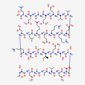
| CAS Number | 62304-98-7 |
|---|---|
| Molecular Formula | C129H215N33O55 |
| Molecular Weight | 3108.3 g/Mol |
| Purity | 99.7% |
| Lot Number | 26088 |
| Quantity | 5.48mg |
| Sequence | Ac-Ser-Asp-Ala-Ala-Val-Asp-Thr-Ser-Ser-Glu-Ile-Thr-Thr-Lys-Asp-Leu-Lys-Glu-Lys-Lys-Glu-Val-Val-Glu-Glu-Ala-Glu-Asn-OH |
Cosmic Peptides Certificate of Analysis
Unable to load certificate. Download COA