VIP
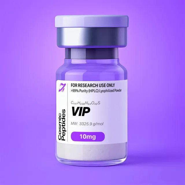
VIP
Commonly researched with these items
Patent-pending lot tracking: Complete transparency from production to vial.
VIP (Vasoactive Intestinal Peptide): The 28-Residue Amphipathic Neuropeptide and Prototype VPAC Receptor Ligand
Disclaimer
Products described here are supplied for research use only and are not intended for diagnostic, therapeutic, or clinical application. All statements regarding biological activity reflect preclinical and in vitro findings exclusively and have not been evaluated by the U.S. Food and Drug Administration. These materials and related content are provided for educational and investigational purposes only and are intended solely for qualified researchers in laboratory and academic settings.
Key Facts
- Compound class
- Linear 28-residue neuropeptide of the secretin/PACAP peptide family; endogenous agonist of the class B1 G protein-coupled receptors VPAC1 and VPAC2
- Sequence / structure
H-His-Ser-Asp-Ala-Val-Phe-Thr-Asp-Asn-Tyr-Thr-Arg-Leu-Arg-Lys-Gln-Met-Ala-Val-Lys-Lys-Tyr-Leu-Asn-Ser-Ile-Leu-Asn(one-letter code: HSDAVFTDNYTRLRKQMAVKKYLNSILN); the native peptide ends in a C-terminal asparagine amide (Asn-NH2)- Molecular formula
- C147H237N43O43S as catalogued (the formula of the free C-terminal acid, as depicted in PubChem); the C-terminally amidated native form is C147H238N44O42S
- Molecular weight
- 3326.8 g/mol as catalogued (free acid; monoisotopic mass 3324.7401 Da); 3325.8 g/mol for the amide (monoisotopic mass 3323.7561 Da)
- CAS number
- 37221-79-7 (the registry number 40077-57-4 is also used for the same sequence)
- PubChem CID
- 53314964 (CAS 37221-79-7); 16132300 (CAS 40077-57-4)
- Physical form as supplied
- Lyophilized powder
- Lot traceability
- Every unit is tied to a tracked lot; the lot number, recorded purity and verified quantity of the lot on sale are shown live on this page, with its certificate of analysis.
Molecular Identity and Classification
Vasoactive intestinal peptide (VIP) is a 28-amino-acid linear peptide with the sequence His-Ser-Asp-Ala-Val-Phe-Thr-Asp-Asn-Tyr-Thr-Arg-Leu-Arg-Lys-Gln-Met-Ala-Val-Lys-Lys-Tyr-Leu-Asn-Ser-Ile-Leu-Asn. It was isolated by Sami Said and Viktor Mutt in 1970 from extracts of small intestine, purified by following its activity in the bioassays of the time, and its complete primary structure was reported by the same authors in 1974. The same 28-residue sequence is shared by many mammalian orthologues, which is why catalogues and chemical registries list it as a single compound.
VIP belongs to the secretin/PACAP family of peptides, a group of structurally related regulatory peptides that includes pituitary adenylate cyclase-activating polypeptide (PACAP), peptide histidine methionine (PHM), secretin and growth hormone-releasing hormone (GHRH). The family is defined by a shared architecture: an N-terminal segment beginning with histidine (or a closely related residue) that is needed for receptor activation, followed by a long stretch with a strong tendency to form an amphipathic α-helix that provides most of the binding energy. The receptors of the family are class B1 (secretin-like) G protein-coupled receptors, and VIP is the prototypical ligand of two of them, VPAC1 and VPAC2.
In the cell, VIP is cut from a larger precursor. Cloning of the precursor cDNA by Itoh and colleagues in 1983 showed that prepro-VIP encodes two bioactive peptides, VIP itself and PHM-27, separated by short spacer sequences and flanked by basic cleavage sites. VIP is released from the precursor as a glycine-extended intermediate, VIP-Gly, and the glycine is then converted into the C-terminal amide of Asn28 by peptidylglycine α-amidating monooxygenase. The native peptide therefore ends in …Ile-Leu-Asn-NH2, a modification reviewed by Bradbury and Smyth (1991).
That terminal amide is the single most important point of chemical identity for this product, because chemical registries do not represent it consistently. PubChem's records for both registry numbers in common use, CAS 37221-79-7 (CID 53314964) and CAS 40077-57-4 (CID 16132300), depict the 28-residue chain with a free C-terminal carboxylic acid and give the formula C147H237N43O43S and molecular weight 3326.8 g/mol, the values listed for this product. The amidated native peptide, the form most suppliers and the research literature refer to as VIP, has the formula C147H238N44O42S and a molecular weight of 3325.8 g/mol. The two forms differ by exactly one NH-for-O exchange, 0.984 Da, and this page gives analytical values for both so that a laboratory can confirm which form a given lot represents from its certificate of analysis or by its own mass spectrometry.
Structure and Chemical Features
The 28 residues of VIP comprise 15 different amino acids: three each of asparagine, leucine and lysine; two each of serine, aspartic acid, alanine, valine, threonine, tyrosine and arginine; and one each of histidine, phenylalanine, glutamine, methionine and isoleucine. There is no cysteine, proline, glycine, tryptophan or glutamic acid. The absence of cysteine means VIP has no disulfide bonds and no covalent constraint on its fold; the absence of proline and glycine, the two strongest helix breakers, is consistent with the long helical segment the peptide adopts in membrane-like environments.
Charge is distributed unevenly along the chain. The basic residues (Arg12, Arg14, Lys15, Lys20 and Lys21) cluster in the central and C-terminal half, whereas the two acidic residues, Asp3 and Asp8, lie in the N-terminal third alongside His1. With a free N-terminal amine, two arginines and three lysines and only two carboxylate side chains, the amidated peptide carries a net charge of about +4 at pH 7 (about +3 for the free acid) and has a calculated isoelectric point of roughly 10.2 to 10.5, depending on the pKa scale used. The calculated grand average of hydropathy (GRAVY, Kyte-Doolittle) is −0.64, a moderately hydrophilic value that conceals a strongly amphipathic distribution: when residues 10 to 28 are drawn on a helical wheel (100° per residue), Leu13, Val19, Leu23, Ile26 and Leu27 cluster within an arc of about 140°, Arg14, Lys15 and Lys21 lie on the opposite face, and Arg12 and Lys20 sit at the edges of the hydrophobic arc, where their long side chains can reach the polar interface of a membrane or micelle.
Conformation in Solution and in Membrane Mimetics
In dilute aqueous buffer VIP is largely disordered, with circular dichroism spectra dominated by random-coil character. In helix-promoting solvents such as methanol or trifluoroethanol, and in the presence of detergent micelles or phospholipid vesicles, it folds into a long α-helix. Umetsu and colleagues (2011) compared the NMR structures of uniformly 13C/15N-labelled VIP in methanol and in dodecylphosphocholine (DPC) micelles (PDB entries 2RRH and 2RRI). The labelled peptide used in that work was the glycine-extended form, VIP-Gly, and the deposited coordinate files annotate a continuous helix from Asp3 to the C-terminus in methanol and from Val5 to Leu27 in DPC micelles, with the authors describing measurable differences in helix extent and orientation between the two environments.
The C-terminal amide also has structural consequences. Replacing the terminal carboxylate with a neutral amide removes an unfavourable interaction between a negative charge and the negative pole of the helix dipole at the C-terminus, and changes the peptide's net charge by one unit, which in turn affects retention in reversed-phase and ion-exchange chromatography. In the receptor-bound state, the C-terminal helix is the part of the peptide that is held in the groove of the receptor's N-terminal extracellular domain, so the terminal group sits in a region that contributes to binding affinity.
Chemical Liabilities Built Into the Sequence
Several residues give VIP well-defined degradation pathways. Met17 is the only sulfur-containing residue and is readily oxidized to methionine sulfoxide (+15.995 Da), during synthesis, cleavage, storage in solution or exposure to peroxide-containing solvents; replacement of Met17 by the isosteric, non-oxidizable norleucine ([Nle17]VIP) is a classic analogue design. Asn24-Ser25 is an Asn-Ser motif, among the sequence contexts most prone to deamidation through a succinimide intermediate, giving Asp and isoAsp variants (+0.984 Da). Asp3-Ala4 and Asp8-Asn9 can form aspartimide during synthesis and slowly isomerize in solution. Finally, the Arg and Lys residues at positions 12, 14, 15, 20 and 21 are trypsin-like cleavage sites, which makes VIP susceptible to serine proteases present in serum-containing culture media and in cell extracts.
Synthesis and Manufacturing Chemistry
The first chemical synthesis of the full VIP sequence was reported by Bodanszky, Klausner and Said in 1973, using classical solution-phase fragment chemistry, and the synthetic peptide was compared directly with the natural material. Today VIP is made by solid-phase peptide synthesis (SPPS), the approach introduced by Merrifield in 1963, using Fmoc/tert-butyl chemistry.
The choice of resin decides the C-terminal group, which is why it matters for this product. Assembly on an amide-generating resin, such as Rink amide or Sieber amide, releases the peptide as the C-terminal amide (the native form). Assembly on Wang or 2-chlorotrityl resin releases the free acid. Because both forms are offered as "VIP" in the research market and the registry depictions differ, the certificate of analysis and the observed mass are the authoritative description of a given lot.
Side-chain protection follows standard practice for a 28-mer: His(Trt), Ser(tBu), Thr(tBu), Tyr(tBu), Asp(OtBu), Asn(Trt), Gln(Trt), Arg(Pbf) and Lys(Boc), with methionine and the hydrophobic residues unprotected. Several sequence-specific issues affect crude purity:
- Aspartimide formation at Asp3-Ala4 and Asp8-Asn9 during repeated piperidine deprotection cycles, giving α- and β-linked by-products and piperidides; additives such as Oxyma or HOBt in the deprotection solution, or bulkier Asp side-chain esters, limit it.
- Histidine racemization at the N-terminal coupling step, which requires careful choice of activator and short pre-activation times.
- Methionine oxidation during the final trifluoroacetic acid (TFA) cleavage, controlled with thiol and thioether scavengers (for example 1,2-ethanedithiol and thioanisole) in the cleavage mixture.
- Chain aggregation in the hydrophobic C-terminal stretch (Tyr-Leu-Asn-Ser-Ile-Leu-Asn), the first residues assembled, which can slow couplings and is addressed with low-loading resins, elevated coupling temperature and double couplings.
After cleavage, the crude peptide is precipitated in cold ether, purified by preparative reversed-phase HPLC with water/acetonitrile gradients containing 0.1% TFA, and lyophilized. The product is therefore usually a trifluoroacetate salt unless a counterion exchange to acetate or chloride has been performed.
Analytical Characterization
Reversed-phase HPLC. VIP is well retained on C18 and C8 columns and, with 0.1% TFA, elutes in the mid-range of a typical 5–65% acetonitrile gradient. UV detection at 214–220 nm follows the 27 peptide bonds; because the sequence contains two tyrosines and one phenylalanine but no tryptophan, a 280 nm signal is present but weak, and is useful mainly as a secondary identity check. Methionine sulfoxide variants elute measurably earlier than the parent peptide on reversed-phase columns, and deamidated and isoAsp variants often appear as closely eluting shoulders, so shallow gradients (for example 0.5% acetonitrile per minute across the elution window) are recommended when degradation products are of interest.
Intact mass spectrometry. In positive-mode electrospray ionization a peptide of this size and basicity gives a charge-state envelope, typically dominated by the 3+, 4+ and 5+ ions under acidic conditions. The table below lists the monoisotopic m/z values calculated for both C-terminal forms. Note that for a molecule containing 147 carbon atoms the monoisotopic peak is not the most intense peak of the isotope cluster: the expected 13C contribution alone (147 × 1.07%, about 1.6 relative to the monoisotopic peak) makes the second isotopic peak the tallest, so instruments that report the most abundant or average mass will give values about 1–2 Da above the monoisotopic figure. PubChem's "exact mass" of 3325.7435 Da for the free-acid record is such a most-abundant-isotopologue value; the monoisotopic mass is 3324.7401 Da.
| Ion | C-terminal amide (native) | C-terminal free acid (as catalogued) |
|---|---|---|
| Neutral monoisotopic mass | 3323.7561 Da | 3324.7401 Da |
| [M+2H]2+ | 1662.885 | 1663.377 |
| [M+3H]3+ | 1108.926 | 1109.254 |
| [M+4H]4+ | 831.946 | 832.192 |
| [M+5H]5+ | 665.759 | 665.955 |
| [M+6H]6+ | 554.967 | 555.131 |
| Average-mass deconvolution | 3325.85 Da | 3326.83 Da |
The 0.984 Da problem. The difference between the amide and the free acid (replacement of NH by O, +0.98402 Da) is exactly the same mass change as the deamidation of one asparagine or glutamine. Intact mass alone therefore cannot distinguish a free-acid peptide from an amidated peptide carrying one deamidated residue, and VIP contains three asparagines and one glutamine. The two are separated by localizing the change: trypsin digestion followed by LC-MS/MS of the C-terminal fragment settles the question directly.
Peptide mapping. Trypsin cleaves after Arg12, Arg14, Lys15, Lys20 and Lys21. The C-terminal fragment YLNSILN is diagnostic for the C-terminal form, and its y-ion series distinguishes a terminal amide from deamidation at Asn24. Missed cleavages at the adjacent basic pairs (LRK, QMAVKK) are common.
| Residues | Fragment | [M+H]+ | Diagnostic use |
|---|---|---|---|
| 1–12 | HSDAVFTDNYTR | 1425.639 (2+: 713.323) | N-terminus; Asp3 and Asp8 isomerization, Asn9 deamidation |
| 13–14 | LR | 288.203 | Elutes early; often lost in the void |
| 15, 21 | K | 147.113 | Free lysine, rarely monitored |
| 16–20 | QMAVK | 576.317 | +15.995 Da locates Met17 oxidation |
| 22–28 | YLNSILN | 835.467 (amide); 836.451 (acid) | C-terminal form; Asn24 deamidation |
Amino-acid analysis and peptide content. After acid hydrolysis the expected ratios are Asx 5 (two Asp plus three Asn, converted to Asp), Leu 3, Lys 3, Ser 2, Ala 2, Val 2, Thr 2, Tyr 2, Arg 2, and Glx, His, Phe, Met and Ile 1 each. Amino-acid analysis, together with ion chromatography for the counterion and Karl Fischer titration for water, determines peptide content, which for a basic 28-mer is substantially below 100% by weight. The peptide has seven protonatable groups (the N-terminal amine, His1, two Arg and three Lys); as a fully protonated trifluoroacetate salt with seven TFA counterions (114.02 g/mol each), the amidated peptide would have a formula weight of about 4124 g/mol, a peptide content of about 80.6%. The corresponding acetate salt (60.05 g/mol per counterion) would be about 3746 g/mol, or 88.8% peptide. Real lots usually lie in between.
Physicochemical Properties
| Property | Value | Notes |
|---|---|---|
| Chain length | 28 residues | No Cys, Pro, Gly, Trp or Glu |
| Molecular formula (free acid) | C147H237N43O43S | PubChem CID 53314964 and 16132300; catalogued value |
| Molecular formula (amide) | C147H238N44O42S | Native C-terminal Asn-NH2; calculated |
| Average molecular weight | 3326.8 g/mol (acid); 3325.8 g/mol (amide) | Neutral peptide, without counterions |
| Monoisotopic mass | 3324.7401 Da (acid); 3323.7561 Da (amide) | Calculated; acid value matches PubChem |
| Heavy atoms | 234 | PubChem, free acid |
| Hydrogen-bond donors / acceptors | 51 / 51 | PubChem computed, free acid |
| Rotatable bonds | 115 | PubChem computed |
| Topological polar surface area | 1470 Å2 | PubChem computed |
| XLogP3 | −15.9 | PubChem computed; strongly hydrophilic overall |
| Net charge at pH 7 | about +4 (amide); about +3 (acid) | Calculated from side-chain pKa values |
| Isoelectric point | about 10.2–10.5 (calculated) | Basic, dominated by Arg and Lys |
| GRAVY (Kyte-Doolittle) | −0.64 | Calculated; amphipathic helix in residues 10–28 |
| Molar absorption coefficient at 280 nm | 2980 M−1 cm−1 | 2 Tyr × 1490 M−1 cm−1 (Pace and colleagues, 1995); A280 of a 1 mg/mL solution about 0.90 |
| Molar quantity | about 300.7 nmol per mg | Amide, as pure peptide; correct for peptide content |
The very negative XLogP and large polar surface area describe a freely water-soluble molecule that cannot cross lipid membranes by diffusion, yet it carries a hydrophobic helical face that lets it partition onto membrane surfaces, into micelles and onto the walls of plastic and glass containers. The high positive charge reinforces adsorption to negatively charged surfaces such as untreated glass and silica. In practice this means VIP solutions at nanomolar concentrations can lose a large fraction of their peptide to the container unless a carrier protein or low-binding plastic is used.
Worked calculation: concentration from absorbance. Because VIP contains two tyrosines and no tryptophan, its absorbance at 280 nm is modest. A 100 µM solution of the amide has an expected A280 of about 0.30 in a 1 cm cell (2980 M−1 cm−1 × 1.0 × 10−4 M), which is measurable on a standard spectrophotometer, while a 1 µM working solution (A280 about 0.003) is not. For dilute solutions, quantitation by amino-acid analysis, by absorbance at 205–214 nm against a standard, or by HPLC against a reference preparation is more reliable.
Research Context
VIP is one of the most extensively characterized class B1 receptor ligands and a reference agonist in receptor pharmacology, structural biology and cell signalling. The nomenclature and pharmacology of its receptors were set out by the IUPHAR committee in the review by Harmar and colleagues (2012), which remains the standard reference for the field.
Receptors: VPAC1, VPAC2 and PAC1
Three receptors recognize VIP and PACAP. VPAC1 (gene VIPR1), first cloned by Ishihara and colleagues in 1992, and VPAC2 (gene VIPR2), first cloned by Lutz and colleagues in 1993, bind VIP and PACAP with similar, nanomolar affinity. The third receptor, PAC1 (gene ADCYAP1R1), cloned by Hashimoto and colleagues in 1993, binds PACAP with an affinity two to three orders of magnitude higher than VIP. All three couple predominantly to Gs and adenylyl cyclase, raising intracellular cyclic AMP, and additional coupling to Gq-dependent calcium mobilization and to other pathways has been reported in some cell systems. Couvineau and Laburthe (2012) reviewed the molecular pharmacology of the VPAC receptors, including their interaction with accessory proteins such as receptor activity-modifying proteins (RAMPs), which can modify signalling in recombinant expression systems.
The selectivity of VIP itself is therefore modest: it activates VPAC1 and VPAC2 about equally and PAC1 weakly. To separate the two VPAC receptors, researchers use engineered analogues. Nicole and colleagues (2000) carried out an alanine scan of VIP at the human receptors VPAC1 and VPAC2, identifying residues in both the N-terminal segment and the helical region that are required for high-affinity binding, and used the results to design a VPAC1-selective agonist. Gourlet and colleagues (1997) showed that the cyclic, lactam-bridged analogue Ro 25-1553 is highly selective for VPAC2. Such selective ligands, with VIP and PACAP, are the standard toolkit for assigning a response to a receptor subtype.
The Two-Domain Binding Mechanism
Class B1 receptors bind their peptide ligands in two steps, a model that VIP research helped establish. The receptor's large N-terminal extracellular domain (ECD) first captures the C-terminal helical portion of the peptide, providing affinity and selectivity; the free N-terminal segment of the peptide then inserts into a pocket within the seven-transmembrane bundle and triggers the conformational change that activates the G protein. Laburthe, Couvineau and Tan (2007) summarized the photoaffinity labelling, mutagenesis and modelling data supporting this mechanism for VPAC receptors. It explains why N-terminally modified VIP analogues tend to retain binding but lose efficacy, a classic route to antagonists.
High-resolution structures confirmed the model. Duan and colleagues (2020) reported a 3.2 Å cryo-electron microscopy structure of the human receptor VPAC1 in complex with PACAP-27 and the heterotrimeric Gs protein (PDB 6VN7), stabilized with a NanoBiT tethering strategy. The structure shows the peptide's N-terminus deep in the transmembrane ligand-binding pocket and its C-terminal helix held by the ECD. Because the first 27 residues of PACAP share 19 identities with VIP, including the N-terminal His-Ser-Asp and the C-terminal helical motif Gln-Met-Ala-Val-Lys-Lys-Tyr-Leu, the VPAC1-PACAP-27 complex is widely used as a template for modelling VIP binding.
In Vitro Assay Systems and Read-Outs
Typical VIP experiments use recombinant cell lines such as CHO cells or human embryonic kidney (HEK293) cells stably expressing the human receptors VPAC1, VPAC2 or PAC1, or cell lines that express the receptors endogenously, such as the colon-derived epithelial cell line HT-29, a long-standing VPAC1 system. Common read-outs include:
- Radioligand binding with 125I-labelled VIP on cell membranes, giving IC50 and Ki values for unlabelled ligands in competition experiments.
- Cyclic AMP accumulation by homogeneous time-resolved fluorescence (HTRF), enzyme immunoassay or luciferase-based biosensors, usually in the presence of a phosphodiesterase inhibitor, to obtain EC50 and maximal response.
- Adenylyl cyclase activity in membrane preparations, the classical assay that gave PACAP its name.
- β-arrestin recruitment and receptor internalization assays, used to study biased signalling and receptor trafficking.
Concentration-response series for VIP usually span about 1 pM to 1 µM, with half-maximal cAMP responses at recombinant VPAC receptors typically in the sub-nanomolar to low-nanomolar range. At these concentrations, surface losses and proteolysis are the main sources of error: binding and functional buffers normally contain bovine serum albumin (often 0.1–1%) and, where serum or cell extracts are present, protease inhibitors such as bacitracin or aprotinin. A matched counterion vehicle control is advisable, since Cornish and colleagues showed that trifluoroacetate alone can affect cell proliferation in culture.
VIP Compared With Related Family Peptides
Placing VIP next to its closest relatives shows which positions are conserved for receptor activation and which create selectivity. The alignment below compares VIP with PACAP-27, the N-terminal 27 residues of the 38-residue PACAP isolated by Miyata and colleagues in 1989.
| Feature | VIP | PACAP-27 |
|---|---|---|
| Sequence | HSDAVFTDNYTRLRKQMAVKKYLNSILN-NH2 | HSDGIFTDSYSRYRKQMAVKKYLAAVL-NH2 |
| Length | 28 residues | 27 residues (38 in PACAP-38) |
| Identical positions | 19 of 27 (about 70%) | Differences at 4, 5, 9, 11, 13, 24, 25 and 26 |
| C-terminus | Asn-NH2 | Leu-NH2 |
| Receptor preference | VPAC1 ≈ VPAC2 ≫ PAC1 | PAC1 ≈ VPAC1 ≈ VPAC2 |
| Methionine | Met17 | Met17 |
The shared N-terminal His-Ser-Asp, the Phe6-Thr7-Asp8 motif and the long identical stretch from Arg14 to Leu23 account for the equal potency of the two peptides at the VPAC receptors. The residues that let PAC1 discriminate against VIP are scattered across the sequence.
Other members of the family are offered in this catalogue as research compounds. Sermorelin is GHRH(1–29) amide and Tesamorelin is an N-terminally modified analogue of the full 44-residue GHRH; both share the class B1 receptor logic and the C-terminal amide of VIP, and they make useful comparison compounds for analytical method development on secretin-family peptides of similar size. C-terminal amidation by the same enzymatic pathway is also a defining feature of Oxytocin and Kisspeptin-10.
Laboratory Handling and Storage
Storage of the solid. Lyophilized VIP is best stored tightly closed at −20 °C or below, protected from light and moisture. The powder is hygroscopic; allow the closed vial to reach room temperature, preferably in a desiccator, before opening it, so that atmospheric moisture does not condense on the cold solid. Limiting air exposure protects Met17.
Preparing stock solutions. VIP dissolves readily in high-purity water, and its basic character means that dilute acetic acid (for example 0.1–1%) is a good first solvent if a lot dissolves slowly. A 1 mM stock of the amide corresponds to about 3.33 mg of pure peptide per mL; correct the weighed mass for peptide content as described above. Stocks are often made at 0.1–1 mM in water or dilute acid, divided into single-use aliquots in low-binding polypropylene tubes and frozen at −20 °C or −80 °C. Avoid repeated freeze-thaw cycles. Neutral and alkaline buffers accelerate deamidation at Asn24-Ser25, so avoid long-term storage at pH 7.4.
Working solutions. Prepare working dilutions immediately before use in buffer containing a carrier protein such as 0.1% bovine serum albumin, use low-binding plastics, and avoid untreated glass. Solutions intended for cell culture are usually passed through a 0.22 µm low-protein-binding membrane.
Oxidation control. Use peroxide-free solvents and avoid aged polysorbate-containing buffers, which accumulate peroxides.
Verification. An LC-MS check of a stock after long storage should show the 4+ and 5+ ions near m/z 832 and 666 (see the table above for exact values of each C-terminal form), with a +4.0 m/z satellite on the 4+ ion indicating methionine sulfoxide. Each lot's certificate of analysis, linked on this page, records the analytical results for that lot.
Frequently Asked Research Questions
- What is vasoactive intestinal peptide?
- Vasoactive intestinal peptide (VIP) is a 28-residue linear neuropeptide, HSDAVFTDNYTRLRKQMAVKKYLNSILN, of the secretin/PACAP family. It was isolated by Said and Mutt in 1970, sequenced in 1974, and is the endogenous agonist of the class B1 G protein-coupled receptors VPAC1 and VPAC2.
- Is VIP C-terminally amidated?
- Native VIP ends in asparagine amide (Asn-NH2), formed from a glycine-extended precursor. Synthetic VIP can be made as either the amide or the free acid depending on the resin used; PubChem's records depict the free acid. The certificate of analysis and the observed mass identify the form of a given lot.
- What are the molecular formula and molecular weight of VIP?
- The free acid is C147H237N43O43S, 3326.8 g/mol (monoisotopic mass 3324.7401 Da), the values in PubChem and in this product's catalogue entry. The native amide is C147H238N44O42S, 3325.8 g/mol (monoisotopic mass 3323.7561 Da).
- What is the CAS number of VIP?
- VIP is listed under CAS 37221-79-7 (PubChem CID 53314964). The registry number 40077-57-4 (PubChem CID 16132300) is also used for the same 28-residue sequence.
- Which ions should I see for VIP by electrospray mass spectrometry?
- A charge-state envelope dominated by the 3+, 4+ and 5+ ions. For the amide the monoisotopic values are m/z 1108.926, 831.946 and 665.759; for the free acid they are 1109.254, 832.192 and 665.955. The most intense isotope peak lies about 1 Da above the monoisotopic peak.
- How can I tell the free acid from a deamidated amide?
- Both differ from the native amide by +0.984 Da, so intact mass cannot separate them. Digest with trypsin and analyse the C-terminal fragment YLNSILN by LC-MS/MS: [M+H]+ m/z 835.467 for the amide and 836.451 for the free acid, with the y-ion series locating any deamidation.
- Which receptors does VIP activate?
- VIP binds VPAC1 and VPAC2 with similar nanomolar affinity and binds PAC1 two to three orders of magnitude more weakly than PACAP does. All three receptors couple mainly to Gs and cyclic AMP production.
- How is VIP related to PACAP?
- PACAP-27 shares 19 of its 27 residues with VIP, including the N-terminal His-Ser-Asp and the helical segment from Arg14 to Leu23. Both are amidated members of the secretin/PACAP family, and the cryo-EM structure of VPAC1 with PACAP-27 (PDB 6VN7) is a common template for VIP binding models.
- Does VIP have a defined three-dimensional structure?
- In water it is largely disordered. In methanol, trifluoroethanol or detergent micelles it forms a long α-helix covering most of the sequence; NMR structures in methanol and DPC micelles are deposited as PDB 2RRH and 2RRI (Umetsu and colleagues, 2011).
- What are the main degradation pathways of VIP?
- Oxidation of Met17 to the sulfoxide (+15.995 Da), deamidation of asparagine, especially at the Asn24-Ser25 motif (+0.984 Da), aspartate isomerization, and cleavage by trypsin-like proteases after its arginine and lysine residues.
- Why do my VIP concentrations fall at low nanomolar levels?
- VIP's amphipathic helix and high positive charge make it adsorb to plastic and glass surfaces. Use low-binding plastics, a carrier protein such as 0.1% bovine serum albumin, and fresh working dilutions.
- Where can I see the purity of the VIP I am buying?
- This page shows the lot number, recorded purity and verified quantity of the lot currently on sale, taken live from the shop's lot-tracking database, together with that lot's certificate of analysis.
Sources & Further Reading
- National Center for Biotechnology Information. PubChem Compound Summary for CID 53314964 (CAS 37221-79-7) and CID 16132300 (CAS 40077-57-4), vasoactive intestinal peptide. PubChem CID 53314964; PubChem CID 16132300.
- Said SI, Mutt V. Polypeptide with broad biological activity: isolation from small intestine. Science 1970;169(3951):1217–1218. doi:10.1126/science.169.3951.1217
- Mutt V, Said SI. Structure of the porcine vasoactive intestinal octacosapeptide. European Journal of Biochemistry 1974;42(2):581–589. doi:10.1111/j.1432-1033.1974.tb03373.x
- Bodanszky M, Klausner YS, Said SI. Biological activities of synthetic peptides corresponding to fragments of and to the entire sequence of the vasoactive intestinal peptide. Proceedings of the National Academy of Sciences of the USA 1973;70(2):382–384. doi:10.1073/pnas.70.2.382
- Itoh N, Obata K, Yanaihara N, Okamoto H. Human preprovasoactive intestinal polypeptide contains a novel PHI-27-like peptide, PHM-27. Nature 1983;304(5926):547–549. doi:10.1038/304547a0
- Bradbury AF, Smyth DG. Peptide amidation. Trends in Biochemical Sciences 1991;16:112–115. doi:10.1016/0968-0004(91)90044-V
- Miyata A, Arimura A, Dahl RR, et al. Isolation of a novel 38 residue-hypothalamic polypeptide which stimulates adenylate cyclase in pituitary cells. Biochemical and Biophysical Research Communications 1989;164(1):567–574. doi:10.1016/0006-291X(89)91757-9
- Ishihara T, Shigemoto R, Mori K, Takahashi K, Nagata S. Functional expression and tissue distribution of a novel receptor for vasoactive intestinal polypeptide. Neuron 1992;8(4):811–819. doi:10.1016/0896-6273(92)90101-I
- Lutz EM, Sheward WJ, West KM, Morrow JA, Fink G, Harmar AJ. The VIP2 receptor: molecular characterisation of a cDNA encoding a novel receptor for vasoactive intestinal peptide. FEBS Letters 1993;334(1):3–8. doi:10.1016/0014-5793(93)81668-P
- Hashimoto H, Ishihara T, Shigemoto R, Mori K, Nagata S. Molecular cloning and tissue distribution of a receptor for pituitary adenylate cyclase-activating polypeptide. Neuron 1993;11(2):333–342. doi:10.1016/0896-6273(93)90188-W
- Harmar AJ, Fahrenkrug J, Gozes I, et al. Pharmacology and functions of receptors for vasoactive intestinal peptide and pituitary adenylate cyclase-activating polypeptide: IUPHAR Review 1. British Journal of Pharmacology 2012;166(1):4–17. doi:10.1111/j.1476-5381.2012.01871.x
- Laburthe M, Couvineau A, Tan V. Class II G protein-coupled receptors for VIP and PACAP: structure, models of activation and pharmacology. Peptides 2007;28(9):1631–1639. doi:10.1016/j.peptides.2007.04.026
- Couvineau A, Laburthe M. VPAC receptors: structure, molecular pharmacology and interaction with accessory proteins. British Journal of Pharmacology 2012;166(1):42–50. doi:10.1111/j.1476-5381.2011.01676.x
- Nicole P, Lins L, Rouyer-Fessard C, et al. Identification of key residues for interaction of vasoactive intestinal peptide with human VPAC1 and VPAC2 receptors and development of a highly selective VPAC1 receptor agonist. Journal of Biological Chemistry 2000;275(31):24003–24012. doi:10.1074/jbc.M002325200
- Gourlet P, Vertongen P, Vandermeers A, et al. The long-acting vasoactive intestinal polypeptide agonist RO 25-1553 is highly selective of the VIP2 receptor subclass. Peptides 1997;18(3):403–408. doi:10.1016/S0196-9781(96)00322-1
- Umetsu Y, Tenno T, Goda N, Shirakawa M, Ikegami T, Hiroaki H. Structural difference of vasoactive intestinal peptide in two distinct membrane-mimicking environments. Biochimica et Biophysica Acta – Proteins and Proteomics 2011;1814(5):724–730. doi:10.1016/j.bbapap.2011.03.009
- Duan J, Shen DD, Zhou XE, et al. Cryo-EM structure of an activated VIP1 receptor-G protein complex revealed by a NanoBiT tethering strategy. Nature Communications 2020;11:4121. doi:10.1038/s41467-020-17933-8
- Merrifield RB. Solid phase peptide synthesis. I. The synthesis of a tetrapeptide. Journal of the American Chemical Society 1963;85(14):2149–2154. doi:10.1021/ja00897a025
- Pace CN, Vajdos F, Fee L, Grimsley G, Gray T. How to measure and predict the molar absorption coefficient of a protein. Protein Science 1995;4(11):2411–2423. doi:10.1002/pro.5560041120
- Cornish J, Callon KE, Lin CQ, et al. Trifluoroacetate, a contaminant in purified proteins, inhibits proliferation of osteoblasts and chondrocytes. American Journal of Physiology-Endocrinology and Metabolism 1999;277(5):E779–E783. doi:10.1152/ajpendo.1999.277.5.E779
Last reviewed:
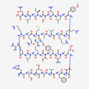
| CAS Number | 37221-79-7 |
|---|---|
| Molecular Formula | C147H237N43O43S |
| Molecular Weight | 3326.8 g/Mol |
| Purity | 99.8% |
| Lot Number | 25046 |
| Quantity | 10.08mg |
| Sequence | His-Ser-Asp-Ala-Val-Phe-Thr-Asp-Asn-Tyr-Thr-Arg-Leu-Arg-Lys-Gln-Met-Ala-Val-Lys-Lys-Tyr-Leu-Asn-Ser-Ile-Leu-Asn |
Cosmic Peptides Certificate of Analysis
Unable to load certificate. Download COA